
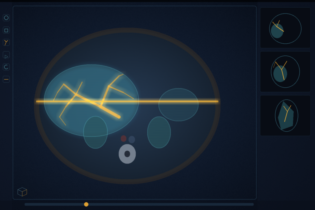
CoInS-Net takes two observed endpoint slices from the same CT or MRI volume and, in one forward pass, writes the image at an arbitrary in-between depth plus the segmentation overlay for that plane. The query is physical: relative depth t in (0, 1) and the through-plane spacing relative to in-plane pixel size. Sun, Que, Shi, Fu, Yang, Li, and Dai at Northeastern University (Shenyang) and Peking University submitted arXiv:2608.09391 on 10 August 2026. The paper is CoInS-Net: A Continuous Position-Aware Network for Joint Medical Image Interpolation and Segmentation. The same recipe is measured on MSD cardiac MRI, MSD pancreas CT, AMOS abdominal CT/MRI, and BraTS glioma MRI.
The preprint does not list a code URL. The backbone is a 2D Swin. It does not take a full 3D volume.
Shared Swin, physical t, two heads
Call the endpoints I0 and I1 at physical depths z0 and z1. The target depth is zt. Then t = (zt – z0) / (z1 – z0). A weight-shared 2D Swin Transformer encodes each endpoint on its own. The Physical Position-Aware Feature Generator builds a token from t, 1-t, t(1-t), log(1+ρ), and low-order sinusoids, where ρ is |z1 – z0| divided by mean in-plane spacing sxy. That token is why the same relative t is not treated as the same anatomical jump on a 2 mm series and an 8 mm series.
At every scale the generator starts from a linear blend of the two endpoint features, then adds a gated nonlinear correction. The gate is t(1-t), so at t = 0 and t = 1 the features recover the observed endpoints exactly. The peak of that gate is the midpoint, where linear blending misses organ appearance, disappearance, and bifurcation. Local attention looks at neighborhoods on both endpoints and biases the closer one. The authors argue 2D optical flow is the wrong model here: on thick-slice series structures pop in and out rather than translate.
Those position-aware features split into interpolation and segmentation adapters (1 x 1 conv, LayerNorm, GELU). Cross-Task Prototype Interaction then exchanges class-level summaries, not dense pixel-to-pixel attention. A coarse class map from the segmentation branch weights both a semantic prototype and an appearance prototype per class. Interpolation queries the semantic prototypes so tissue interfaces stay anatomically plausible. Segmentation queries the appearance prototypes so z-axis intensity and texture change help locate the organ. A dual-branch decoder shares a light cooperation feature at each upsample. Training loss is Charbonnier plus 0.15 SSIM on the image, Dice plus cross-entropy on the mask, and a warm-up spatial-affinity term with λsc up to 0.1. No adversarial or perceptual losses, on purpose, so the interpolator is not rewarded for inventing anatomy. Optimizer is AdamW at 1 x 10⁻⁴ with cosine annealing, mixed precision, up to 100 epochs on an RTX 4090. Parameter count is 42.98 M.
Four public volumes, patient-wise 7:1:2
Four public sets share the architecture. There are no anatomy-specific heads.
- MSD Task02 Heart: 21 cardiac MRI NIfTI volumes, single-class left atrium.
- MSD Task07 Pancreas: 282 abdominal CTs, dual-class pancreas and tumor.
- AMOS2022: abdominal CT or MRI, 15 structures. One modality per case. Per-organ numbers are reported for liver, stomach, spleen, and both kidneys.
- BraTS, t1n only. The text calls this BraTS 2024 post-treatment glioma. Footnote 3 points at a BraTS 2020 Kaggle page. Treat the cohort name as unverified beyond what the tables report.
Splits are patient-level 7:1:2. Training builds triples from a thinner reference volume, applies slice-profile blur along z, then downsamples, so the endpoints look like a thick-slice protocol rather than dropped slices. Endpoint intervals vary. Input order is swapped as (I0, I1, t) with (I1, I0, 1-t). Default spacing in the main tables is 4 mm. If you still mix the overlap coefficient with the training loss, read the note on Dice versus Dice loss.
One pass against two pipelines
The paper's comparison is a joint interpolator-segmenter versus single-task interpolators (best of those is SFCLI-Net) and single-task segmenters (best of those is SegMamba-V2), plus cascaded pairs of one interpolator plus one segmenter. Table 1 and Table 2 are the headline cells.
On interpolation, CoInS-Net PSNR is 37.88 dB on MSD Heart against SFCLI-Net 36.64, 37.01 against 35.70 on MSD Pancreas, 37.15 against 36.03 on AMOS, and 36.72 against 35.05 on the BraTS t1n split. SSIM follows the same order (0.975, 0.958, 0.958, 0.949 against 0.962, 0.945, 0.944, 0.933).
On segmentation, Dice is 0.968 against SegMamba-V2 0.947 on Heart, 0.974 against 0.956 on Pancreas, 0.867 against 0.838 on AMOS, and 0.842 against 0.819 on BraTS t1n. nnU-Net on the same table is 0.911, 0.922, 0.801, and 0.778. AMOS per-organ Dice for CoInS-Net is liver 0.937, spleen 0.892, right kidney 0.886, left kidney 0.870, stomach 0.808. Stomach is the hard cell.
On MSD Heart at 512 x 512, batch 4, one CoInS-Net pass is 37.70 ms (53.09 FPS), 118.23 G FLOPs, 408.92 MB. CycleINR plus SicTTA, the fastest cascaded pair in Table 13, is 72.64 ms. That is the 48.1% latency cut the abstract rounds to "nearly half." GPU for that timing is the RTX 4090 used for training.
Slice thickness and off-midpoint t
The 4 mm tables are not the whole protocol range. On MSD Pancreas and BraTS t1n the authors sweep 2, 3, 4, 6, 8, 10, 12, and 16 mm and compare against cascaded interpolator-plus-segmenter pairs. Dice on CoInS-Net drops 8.7 points on Pancreas and 9.8 points on BraTS from 2 mm to 16 mm. The paper's average drop for the cascaded baselines is 15.8 and 18.4 points on those two sets. At 16 mm, Pancreas Dice is still 0.901 and BraTS 0.781. Do not treat 16 mm as a protocol you would ship without a site check.
Relative position is not locked at the midpoint. At 4 mm, Pancreas Dice is 0.956 at t = 0.2, 0.972 at 0.33, 0.974 at 0.5, and 0.970 at 0.66. AMOS follows the same pattern, lowest at t = 0.2 (0.843) and highest at t = 0.5 (0.867). The t(1-t) gate is the paper's account of why the midpoint is easier. Five independent runs, Welch t-test, are how the asterisks on those tables were produced.
How this lands in a viewer
If you already hang AI in a DICOM viewer, treat this as a thick-slice helper: pick two axial endpoints, pass t and the Pixel Spacing / Slice Thickness ratio, and hang both the synthesized plane and its mask as an overlay. Keep a 3D organ model (nnU-Net, TotalSegmentator) on the native slices. Use CoInS-Net when the series is anisotropic and the reader needs an in-between plane that does not yet exist on disk. The 2D Swin never sees a full volume, so sagittal and coronal reconstructions of the filled stack are a post-step, not a native output. Small lesions are the paper's own weak cell. Public-set overlap is not a device claim.
RadYOLO is still the faster first-pass box. SAT3D is tumour plus uncertainty in Slicer. SLIP is click latency and undo. Ten-case MedSAM3 LoRA is a site adapter on a promptable SAM. CoM3eT is a frozen Swin plus a pyramid transformer. 3D-CurvSegFlow is three Euler steps on a vessel tree. This paper is one shared Swin, a physical t query, and two heads on the missing slice.
Rebuild from the protocol in arXiv:2608.09391. Do not wait on study weights. None were listed. The BraTS name versus the BraTS 2020 Kaggle footnote is unresolved in the preprint. Retrospective overlap on public NIfTI is not a scanner-vendor claim. The preprint is the source of record until a venue version exists.
Sources
- Sun, Y., Que, N., Shi, P., Fu, R., Yang, Y., Li, X., Dai, Y. CoInS-Net: A Continuous Position-Aware Network for Joint Medical Image Interpolation and Segmentation. arXiv:2608.09391. Submitted 10 August 2026. https://arxiv.org/abs/2608.09391 (HTML: https://arxiv.org/html/2608.09391). No code URL listed in the preprint. No public CoInS-Net repository found when checked 26 August 2026.
- Liu, Z., et al. Swin Transformer: Hierarchical Vision Transformer using Shifted Windows. ICCV 2021. arXiv:2103.14030
- Isensee, F., et al. nnU-Net: a self-configuring method for deep learning-based biomedical image segmentation. Nat. Methods 2021. DOI: 10.1038/s41592-020-01008-z
- Antonelli, M., et al. The Medical Segmentation Decathlon. Nat. Commun. 2022. http://medicaldecathlon.com/
- Ji, Y., et al. AMOS: A Large-Scale Abdominal Multi-Organ Benchmark. NeurIPS 2022 Datasets and Benchmarks. https://amos22.grand-challenge.org/
- Peng, C., Lin, W., Liao, H., Chellappa, R., Zhou, S. K. SAINT: Spatially Aware Interpolation Network for Medical Slice Synthesis. CVPR 2020.
- Song, H., Mao, X., Yu, J., Li, Q., Wang, Y. I3Net: Inter-Intra-slice Interpolation Network for Medical Slice Synthesis. IEEE TMI 2024.
- Xing, Z., et al. SegMamba-V2: Long-range Sequential Modeling Mamba for General 3D Medical Image Segmentation. IEEE TMI 2025. (best single-task segmentation row in Table 2)
- Li, W., et al. SFCLI-Net: Spatial-frequency collaborative learning interpolation network for computed tomography slice synthesis. Expert Syst. Appl. 2025. (best single-task interpolation row in Table 1)