
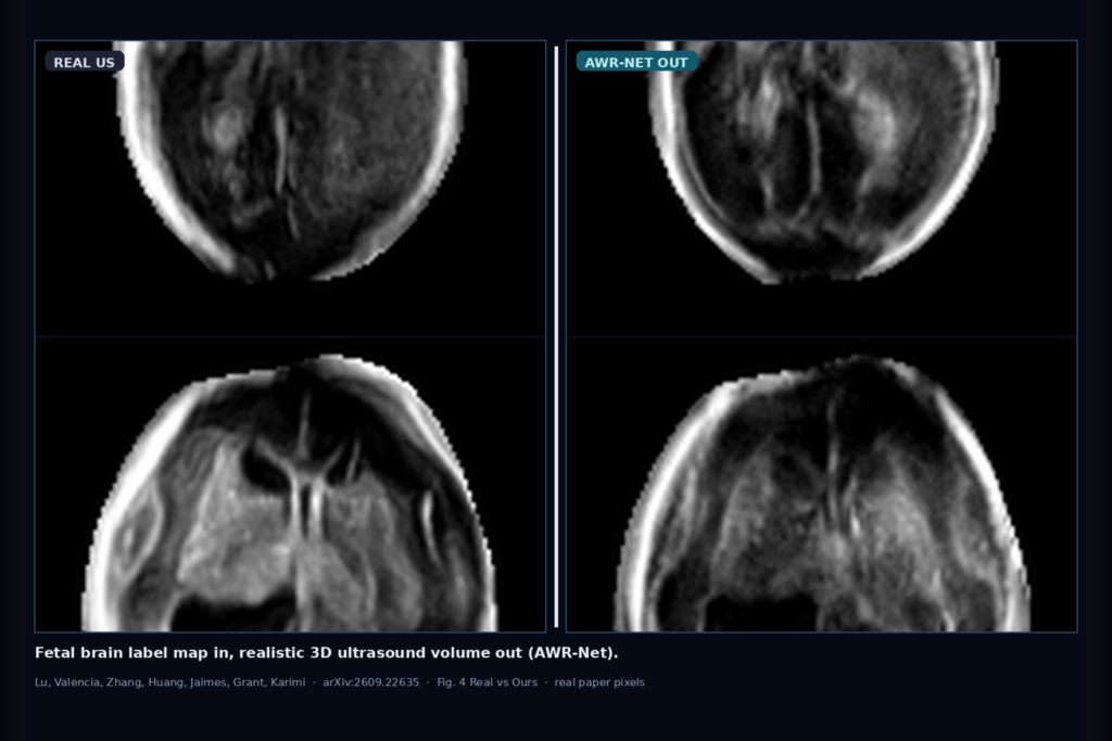
Hang a fetal brain anatomical label map into a 3D ultrasound viewer and the useful hang is a volume that looks like clinical US, not a smooth MRI-like render with anatomy painted on. Clinics have more MRI-derived labels and atlas annotations than paired MRI-US stacks. Synthesis that needs those pairs at inference stays stuck in the lab. Yuhuan Lu, Sergio Valencia, Yuanji Zhang, Yuhao Huang, Camilo Jaimes, P. Ellen Grant, and Davood Karimi take that gap seriously in arXiv:2609.22635, posted 18 September 2026. They call the method AWR-Net: Decoupling Anatomy and Appearance for 3D Fetal Brain Ultrasound Synthesis. The shop-facing idea is simple. Separate anatomy-conditioned wavelet diffusion from bounded appearance residual refinement, then at inference feed a label map and emit a realistic 3D fetal brain US volume. This is a paper walkthrough for practitioners, not a product claim.
Public code is at https://github.com/Luyuhuan/AWRNet. Volumes are resampled to 160×160×160 at 0.6 mm with a unified label space of background plus 13 structures.
What hangs on the viewer
Upstream is a 3D anatomical label map in that unified space, whether the labels came from fetal MRI (harmonized into the US label set) or from US annotation. Downstream is a synthetic 3D ultrasound volume with clinical-looking speckle and intensity layout. For a DICOM or 3D US viewer rail, the hangable pieces are: a label-to-US preview scrubber across axial, coronal, and sagittal, a path that accepts MRI-derived labels when paired MRI-US is missing at inference, and a model card that names the two stages (ACWD then AARL) instead of a single black-box generator.
The practical claim for a clinic AI shop is not a new probe. AWR-Net hands you a synthesis stack that learns anatomical correspondence on atlas pairs, then learns appearance residuals on real clinical US with the anatomy stage frozen. Inference still ends in a volume you can overlay against the input labels for QC.
How it works in plain words
Two stages matter. First, Anatomy-Conditioned Wavelet Diffusion (ACWD) works in the Haar wavelet domain on atlas ultrasound and label pairs. A 3D diffusion U-Net denoises wavelet coefficients while FiLM-style and mask conditioning keep the coarse volume aligned with the input anatomy. Atlas pairs give clean structural supervision before clinical clutter enters.
Second, Appearance-Adaptive Residual Learning (AARL) freezes ACWD and learns a bounded residual correction from real subject ultrasound. Residuals stay clipped so texture and probe-dependent appearance can move without rewriting the anatomical layout. Training mixes intensity and gradient matching in reliable regions, high-pass texture statistics, and a light PatchGAN term on brain ROI high-pass responses.
In shop language: labels drive anatomy in wavelet space, real clinical US teaches a small appearance residual, and inference does not require a paired MRI-US volume for the case you are synthesizing.
What the numbers say
US-Real is 91 clinical 3D fetal brain US volumes from Boston Children’s Hospital, split 48/8/35 train/val/test. MRI-Real adds 200 MRI volumes used for MRI-derived labels. On the US-Real test set (Table II), AWR-Net reports SSIM 0.470, PSNR 15.648, NCC 0.518, MAE 0.124, Grad-NCC 0.399, and FetalCLIP FID 8.905. Those sit above the listed 3D baselines including cWDM and MOTFM on the paper’s image-domain and FetalCLIP feature metrics.
Downstream, mixing synthetic US into nnU-Net training helps segmentation when real abnormal data are scarce (Fig. 2). A blinded reader study (Table V, Fig. 3) scores Ours above cWDM, MOTFM, and ACWD-only on realism, structure visibility, and label-map consistency (average 4.27 on a 1–5 scale in the paper’s reporting). Figure 4 shows qualitative US-Real normal and severe abnormal strips; Figure 5 shows the MRI-derived label path. Treat panels and metrics as paper evidence, not as a promise on your local vendor protocol.
Where it fails and what not to trust
This is research software on in-house and atlas fetal brain cohorts, not a cleared medical device. Rebuild and validate on your own acquisition protocol, gestational-age mix, and abnormality distribution before you hang synthetic volumes next to reportable reads. ACWD trains on normal atlas pairs; abnormal synthesis rides on label maps plus the residual stage, so bad or incomplete labels will still steer the volume.
Do not treat SSIM 0.470 or FetalCLIP FID 8.905 as a guarantee on a different scanner stack. Do not skip a local holdout with clinical review of ventricles, CSP, and skull boundary against the input labels. And do not hide that generator training still used real US for AARL even though inference from a label map does not need a paired MRI-US case.
For a viewer or clinic AI shop
Wire anatomical label map in; hang a realistic 3D fetal brain US volume out. Prefer AWR-Net when you want label-conditioned synthesis without paired MRI-US at inference: ACWD for anatomy in wavelet space, AARL for bounded appearance residuals, unified 13-structure labels at 160³ / 0.6 mm. Keep a QC scrubber so support can compare synthetic slices to the label overlay and to any available real US. Log label source, gestational age band, and the exact ACWD/AARL checkpoints.
Validate on at least one internal cohort before you claim synthetic fetal US hang in production. Start from the public repo and the preprint. Rebuild from arXiv:2609.22635. PDF: https://arxiv.org/pdf/2609.22635. Code: https://github.com/Luyuhuan/AWRNet.
Sources
- Lu, Y., Valencia, S., Zhang, Y., Huang, Y., Jaimes, C., Grant, P.E., Karimi, D. AWR-Net: Decoupling Anatomy and Appearance for 3D Fetal Brain Ultrasound Synthesis. arXiv:2609.22635, 2026. https://arxiv.org/abs/2609.22635. PDF: https://arxiv.org/pdf/2609.22635. Code: https://github.com/Luyuhuan/AWRNet.
- Table II US-Real test: Ours SSIM 0.470, PSNR 15.648, NCC 0.518, MAE 0.124, Grad-NCC 0.399, FetalCLIP FID 8.905.
- US-Real 91 volumes (BCH), split 48/8/35; MRI-Real 200 volumes; volumes 160³ at 0.6 mm; unified label space background + 13 structures.
- Fig. 4 qualitative US-Real normal + severe abnormal; Fig. 5 MRI-derived labels; Table V / Fig. 3 reader study favors Ours over listed synth baselines.