
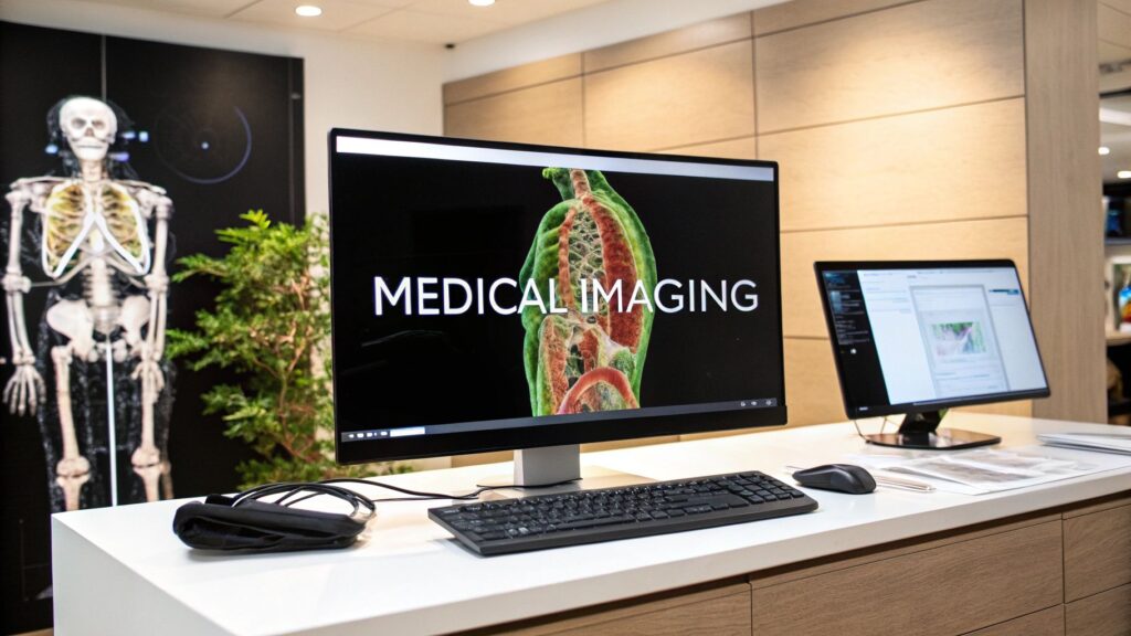
Medical imaging software is the hospital stack that takes a scan from the machine to a report: store it, find it, show it, and attach it to the right order. It is not one product. The four jobs people mean are a PACS (archive), a RIS (scheduling / billing / worklist), a viewer (window-level, cine, MPR), and an archive that can outlive one vendor (often a VNA).
If you meant software that measures (segment, register, count), that is image analysis software. If you meant the algorithms (enhancement, segmentation, registration), that is medical image processing. If you meant 3D reconstruction (MPR / MIP / volume rendering), that is 3D imaging software. If you meant how to build one of these systems, that is medical imaging software development. If you meant “open this DICOM / NIfTI and paint a mask,” that is the Slicer + ITK-SNAP how-to. PACS as its own topic sits on PACS and DICOM.
Three modules, not one box
The old “software in medical imaging” page was this three-box picture with more adjectives. Same job as this page.
| Module | What it does | Analogy |
|---|---|---|
| PACS | Store, retrieve, and display studies. DICOM in, DICOM out. | The digital library |
| RIS | Orders, scheduling, worklist, billing, report status. | The command center |
| Viewer / processing | Window-level, cine, MPR, measure. Sometimes a bolt-on analysis module. | The analyst’s toolkit |
A toolkit that cannot talk to the archive is a side tool. A PACS that cannot talk to the RIS is a pile of unbilled studies. The stack is the product, not any one logo.
What the software actually does
| Job | What it is | What you get |
|---|---|---|
| Visualization | Render the series; zoom, window-level, cine, MPR / 3D | A study you can read, not a folder of JPEGs |
| Analysis | Measure, segment, compare visits | A number or a mask a report can use. Deep analysis is a different page. |
| PACS archive | Store and retrieve the study, keep the history | Last year’s CT without a film room |
| Workflow | Hook to RIS / EHR so the order, the images, and the report stay one thing | A closed loop, not a ZIP on a share |
PACS, RIS, viewer, archive
PACS — the digital library
A Picture Archiving and Communication System stores and retrieves studies. Query/retrieve (C-FIND / C-MOVE, or DICOMweb) is the job. Without it, people export ZIP. With it, a radiologist opens last year’s CT from any authorized workstation. That is the archive, not the measurement product.
RIS — the command center
A Radiology Information System does not hold the pixels. It holds the work around them: schedule the slot, attach the right patient, track the study through the department, bill it, send the report. PACS shows the image. RIS closes the order.
Viewer and advanced visualization
A routine PACS viewer window-levels and scrolls. Advanced visualization is MPR, 3D / 4D, a model a surgeon can rotate before the case. That is still “show me the anatomy.” Reconstruction software as its own job is 3D imaging software.
PACS vs VNA
A PACS is usually one department’s library (radiology, sometimes cardiology). A Vendor Neutral Archive is the city library: many departments, many vendors, one long-term store that does not die when you swap the viewing client. You can run a PACS viewer on top of a VNA. Do not buy a second archive because the first one is branded.
The same stack, different scanners
The 301’d “medical imaging softwares” page organized this by modality. Same stack. Different pixels.
| Modality | Primary use | What the software must do | Limit |
|---|---|---|---|
| CT | Trauma, bone, tumor location | Fast volume load, MPR, 3D | Dose; huge series |
| MRI | Brain, spine, soft tissue | Multi-series hanging, good window presets | Slow acquire; protocol soup |
| Ultrasound | OB, echo, live guidance | Cine, Doppler, measurements on a moving loop | Operator-dependent |
| X-ray | Bone, chest, dental | Fast 2D, annotation, simple calipers | No depth |
| PET (often PET-CT) | Uptake, oncology | Fusion onto CT / MRI, SUV tools | Needs a registered anatomy series |
Multi-modality support means the same patient, two scanners, one hanging protocol — not two logins.
Must-haves (the boring ones that actually ship)
- DICOM. The image language. A GE series has to open on a Siemens workstation. Non-standard tags will still show up; the software has to not drop them.
- HL7 / FHIR. The order and the report language. RIS → scanner demographics, report → EHR. Without this, someone retypes.
- MPR / 3D. Axial-only is 1998. Coronal, sagittal, a custom oblique, a volume you can spin.
- Security. Encryption at rest and in transit, role-based access, an audit trail, a BAA. HIPAA is the floor in the US, not a feature bullet.
How to choose, then how to install
Do not start with a feature matrix. Start with the worklist you already have.
- Integration first. Live query/retrieve against your PACS. Images inside the EHR, not a second app. Push new studies back without duplicates. The 301’d “medical images software” page was this checklist.
- Clearance. A viewer that stores PHI is a medical device conversation. An AI that flags a bleed is a SaMD conversation. Research Slicer is not a purchase order for diagnosis. How you classify that is on the development page.
- Who clicks it. A tool the fellow will not open after week two is a sunk license.
- Migrate in batches. Clean duplicates and names first. Validate each department or modality. Set a kill date for the old store so new studies stop landing there.
FAQ
Is this the same as a DICOM viewer?
A viewer is one piece. The stack is PACS + RIS + viewer + archive. Viewer lists live elsewhere. This page is the hospital system around the viewer.
Do I need a VNA if I already have a PACS?
Only if you have more than one archive (or you are about to swap vendors and do not want to migrate twice). One department, one vendor, one PACS is often enough.
Where does AI sit?
As a module on the viewer or as a sidecar that writes a result back to the study. It is not a fourth archive. Triage (push the bleed to the top of the worklist) is the use that actually ships. Do not buy “AI” as a synonym for the stack.
PYCAD builds the imaging side of this when the viewer or the archive hook has to live in a clinic app. Case studies.
Keep Reading
Related Articles
Explore the full DICOM HubAll services, tools, and guides on this topic — in one place.
Visit Hub →