
One number a radiologist reports from an MRCP is the maximum width of the main pancreatic duct, and getting it by hand is slow. In this study a duct of 5 mm or more counts as dilated. Dilation is the most common indirect sign of a pancreatic tumour, and it also shows up with intraductal papillary mucinous neoplasms (IPMN). Measuring it means scrolling a heavily T2-weighted 3D volume, finding the widest point of a thin, curving tube and dropping calipers on it. Two experienced readers rarely land on the same value. Zipei Zhang and colleagues describe an automatic pipeline for this job in arXiv:2610.40073. The authors are Zipei Zhang, Jaakko Sahlsten, Mikko Saukkoriipi and Kimmo Kaski from the Department of Computer Science at Aalto University, and Johanna Ronkainen, Mona Lehto, Akseli Aittoniemi and Irina Rinta-Kiikka from the Department of Radiology at Tampere University Hospital and the Faculty of Medicine and Health Technology at Tampere University, Finland.
Why the duct is hard to segment
Most published work on automatic duct analysis uses CT. MRCP is the better picture of a fluid-filled duct and uses no ionising radiation, but annotated 3D MRCP datasets are rare. The duct is also tiny relative to the scan. In this dataset it fills about 0.019% of the volume, so a segmentation network trained on whole volumes sees almost nothing but background.
The data comes from the pancreatic cyst registry of Tampere University Hospital: 256 MRCP volumes from 69 patients. Two clinical researchers drew voxel-wise duct masks in 3D Slicer, and two abdominal radiologists with more than 20 years of MRI experience reviewed every mask. Volumes were resampled to 0.9 x 0.625 x 0.625 mm. The split is by patient: 201 volumes for development and 55 volumes from 14 patients for testing. Several test patients have repeat scans taken years apart, with a mean gap of 2.89 years between the first and last.
How the pipeline works
The authors did not design a new network. They took standard 3D models from MONAI and arranged them for a thin tubular target.
- Coarse localisation. A 3D U-Net sees the whole MRCP volume downsampled four times and predicts a rough duct mask. It is tuned for recall, so that almost no duct voxels fall outside its output.
- Crop. The bounding box of that rough mask, widened by a fixed safety margin, is cut from the full-resolution volume. The crop is about four times smaller than the scan.
- Fine segmentation. A 3D SegResNet segments the duct inside the crop at full resolution, and the result is pasted back into scan coordinates.
- Ensemble and clean-up. Five coarse-plus-fine model pairs, one per cross-validation fold, run on every scan. Their probability maps are averaged and thresholded at 0.5, and only the largest connected component is kept.
Both stages train on weighted cross-entropy plus Dice loss, with the duct class weighted up. The team also tried 3D U-Net and Swin UNETR as the fine model, and DynUNet (MONAI’s nnU-Net-style model) as a one-stage reference on the full volume. The U-Net then SegResNet pairing came out best, with a test Dice of 0.634. Swin UNETR came last, which the authors put down to the small training set.
The review flag
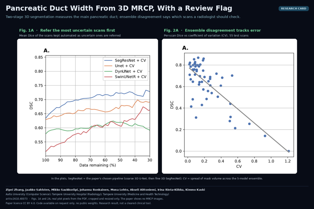
The five ensemble members also give an uncertainty score per scan. The authors tested three: mean voxel entropy over the whole volume, entropy over the predicted duct only, and the coefficient of variation (CV) of the predicted duct volume across the five models. CV is the simplest of the three and worked best. If the five models disagree on how big the duct is, the mask is probably poor. Per scan, CV correlated strongly with Dice and with the radiologists’ grade of segmentation quality.
That makes a referral rule possible. Sort scans by CV, send the most uncertain ones to a human, and keep the rest automatic. In the paper’s referral curve (Fig. 1A), mean Dice on the scans kept automatic climbs as the uncertain ones are removed, and stays above the other pipelines across the whole range.
What it hangs on a scan
For each MRCP series you get a 3D mask of the main pancreatic duct, a maximum width read from that mask, a dilated or not call at the 5 mm threshold, and one CV number. In the study the width was not computed by an algorithm. Two radiologists measured it in 3D Slicer on both the reference mask and the predicted mask, and their mean was used. Against the reference, the predicted masks gave a width error of 0.355 mm on average, which is smaller than the 0.618 mm the two radiologists differed by when measuring the reference masks.
Why a viewer or clinic shop would care
A duct measurement is an easy feature to put in front of a reader because it maps onto something they already do. A plausible workflow in a DICOM viewer or reporting platform:
- Detect the 3D MRCP series when the abdominal MRI study arrives from PACS.
- Run the coarse and fine models with the five-fold ensemble in the background.
- Store the duct mask as a DICOM SEG object linked to the source series, and show it as an overlay on the MIP and on the source slices.
- Place a proposed caliper at the widest point and put the value into a structured report field that the radiologist confirms or edits.
- Show the CV score next to the measurement, and mark high-CV cases as needing a manual check.
For pancreatic cyst surveillance, where the same patient comes back every year or two, the reader will want the measurement history across visits. The test set here already has patients with repeat scans, so the authors could report how much each patient’s results varied from visit to visit.
Where it fails
- Masks are rough. On a five-point usability scale, both radiologists graded the predicted masks below the middle grade on average, which on their scale means large errors or missing detail. Some predictions spill outside the lumen, and the authors list duct continuity as something to fix.
- Dilation calls are noisy. Sensitivity for a dilated duct was 75.0%, but precision was 45.0%, so more than half of the dilated calls were wrong. The authors suggest using it for triage, with every positive reviewed.
- A miss gives a confident wrong number. In one test scan the model found no duct at all, which produced a misleading measurement. The authors found no consistent pattern behind the errors.
- The flag tracks mask quality better than width error. CV correlated well with Dice and with segmentation grades, but its correlation with width error was weak and not statistically significant. A low CV does not promise a correct width.
- Small, single-site data. One hospital, 14 test patients, and dilation in this cohort comes only from main-duct or mixed-type IPMN. Other scanners, protocols and causes of dilation, such as chronic pancreatitis or an obstructing cancer, are untested.
- Manual width step. Widths were read by radiologists on the masks. A product would need its own automatic width method. The authors name cross-sectional analysis orthogonal to the centreline as future work.
Code, data and licence
The code is not public. The paper says it is available from the corresponding author on reasonable request, and the method section is detailed enough to rebuild the pipeline from MONAI components. There are no released weights. The patient data cannot be shared under Finnish data protection law and GDPR, but researchers who meet the access criteria can request it from the Wellbeing Services County of Pirkanmaa. The preprint is licensed CC BY 4.0. Funding came from Business Finland through the Medical AI and Immersion project. This is a research result and not a cleared clinical tool.
My take
The segmentation is not good enough to show a radiologist as a finished mask, and the authors say so. The part I would reuse is the combination of a recall-first crop for a thin structure and a five-model ensemble whose volume disagreement tells you which cases to hand back. That costs nothing beyond the cross-validation models you already trained. Before shipping anything like this I would add automatic width measurement along the centreline, check that the flag also catches bad widths, and test on MRCP from other scanners and from patients with chronic pancreatitis.