
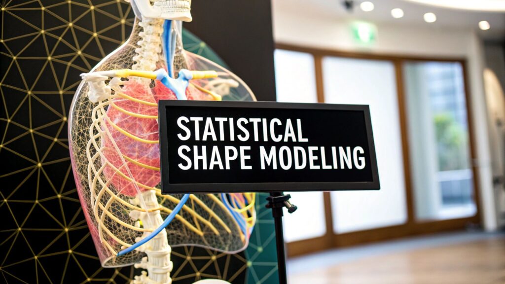
Statistical shape modeling (SSM) is a population model of anatomy: a mean shape plus a few modes of variation learned from corresponding landmarks (or particles) on many examples. It is not a segmentation network. It is not a 3D print. It is not a PYCAD shape product.
The slug ends in -3. Live search found no /blog/statistical-shape-modeling/ and no -2. This is the only SSM URL. If you meant drawing a mask → medical image segmentation. If you meant a patient mesh you can print → 3D lung model. If you meant how the scanner made the volume → acquisition of images.
PYCAD builds custom web DICOM viewers and imaging models. It does not ship an SSM toolkit, an implant designer, or a “precision-medicine shape engine.”
What you actually build
A shape here is not a volume and not a screenshot. It is a set of points that mean the same anatomy on every subject — the same vertex on the femoral head, the same landmark on the left atrium. Once those correspondences exist, you can treat each shape as a long vector and ask how the population moves.
| Step | What it is | What breaks it |
|---|---|---|
| Correspondences | The same anatomical point on every training shape (manual landmarks, or automatic particles) | A missing point, a swapped left/right, a tumor that has no twin on the atlas |
| Alignment | Procrustes (or similar): take out translation, rotation, and usually scale so pose is not “shape” | Leaving pose in. PCA then spends its first modes on “the patient was tilted” |
| PCA | Mean shape + orthogonal modes ranked by variance. A new shape ≈ mean + Σ bi · modei | Too few examples for the number of points. Modes that describe noise, not anatomy |
| Use | Fit a new scan (ASM / AAM), compare a patient to the cloud, or constrain a segmentation | Treating ±3σ as a diagnosis. The model only knows the training set |
The hard step is correspondences, not the eigen-decomposition. Manual landmarks are honest and slow. Automatic methods (SPHARM-PDM, entropy-based particles) scale; they still fail when the anatomy is missing or cut.
PCA is the compressor, not the diagnosis
Stack the aligned coordinates. Subtract the mean. The first principal component is the direction in that high-dimensional space where the training set varies most — often “how tall the ventricle is,” not “disease.” Later modes are smaller, more local, and easier to overfit.
A new shape is then a short list of coefficients b. People bound those (commonly about ±3 standard deviations along each mode) so the reconstruction stays inside the training cloud. That bound is a regularizer, not a clinical cutoff.
The old page shipped a table of “hand 90% / heart 95% / brain 98% variance” with no dataset. Dropped. How many modes you need is a property of your correspondences and your cohort, not a number you copy from a listicle.
ASM and AAM
Active Shape Models (Cootes, Taylor and colleagues, 1990s — the usual citation is Cootes et al., Computer Vision and Image Understanding, 1995) add a search: move each landmark toward an edge along the surface normal, then project the deformed wireframe back onto the PCA subspace. The model can only take shapes the training set allows. That is the point.
Active Appearance Models add texture (intensities inside the mesh), not only the contour. Useful when the edge is weak and the interior pattern is the cue. Heavier, easier to overfit, still a 1990s linear model — not a U-Net.
Neither is “AI that sees like a doctor.” An ASM is a constrained template. A U-Net is a learned mask. You can use an SSM as a prior around a net; that is a paper, not this URL, and not a PYCAD SKU.
Where it actually shows up
- Population morphometrics. What “normal” looks like, and how a cohort (age, a disease, a surgical state) sits off the mean. Research, not a signed report.
- A prior for segmentation or registration. The fit cannot leave the subspace. Good when the organ is a smooth closed surface. Bad when the thing you care about is a resection cavity the atlas never saw.
- Implant / guide design. A statistical bone atlas can size a standard implant or start a patient-specific plan. The surgeon still checks the metal. PYCAD does not design implants.
A named, maintained toolkit for correspondence-based SSM is ShapeWorks (SCI Institute, University of Utah; source). 3D Slicer is a viewer and a module host, not an SSM product by itself. scikit-learn will do the PCA once you already have the matrix; it will not place landmarks on a femur.
What this page is not
- Not a second segmentation methods article with an “SSM” costume. Threshold / U-Net / atlas sit there.
- Not a claim that SSM “detects Alzheimer’s or lung cancer before traditional methods.” The old page said that without a trial. Dropped.
- Not a drug-response oracle, a CAGR, or a covariance-matrix calculator affiliate. Dropped.
- Not a PYCAD statistical-shape product. The closer is a viewer or a model if the mesh has to live in a clinic app — not an SSM engine.
If the missing piece is hanging the volume or training a mask, that is the imaging piece. Case studies.