
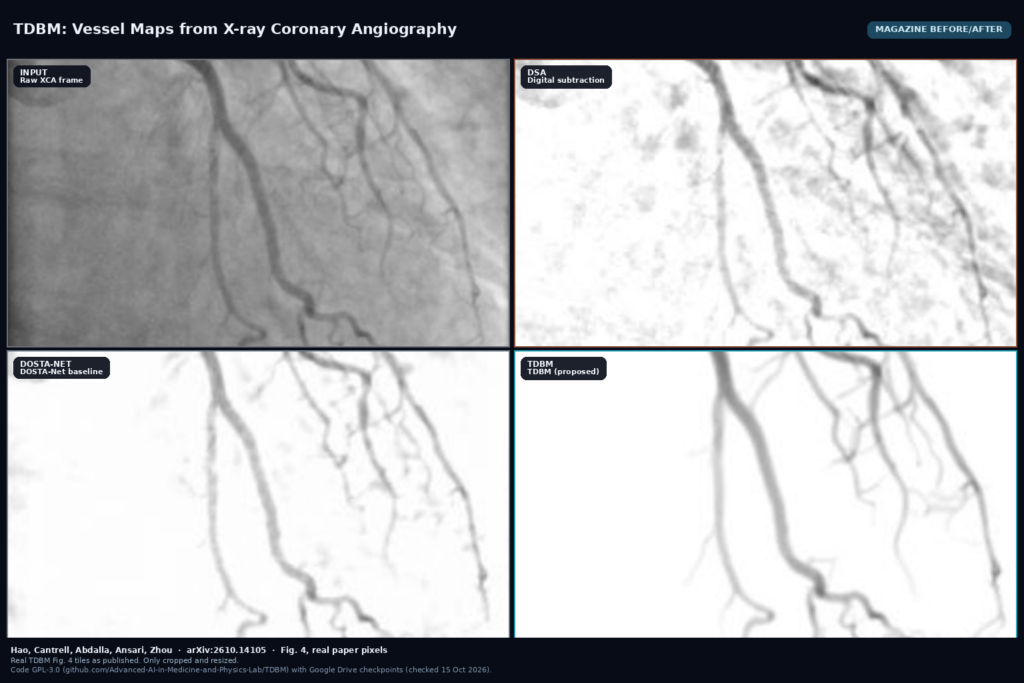
Cath-lab X-ray coronary angiography (XCA) still fights bone and soft-tissue overlay, and classic digital subtraction angiography (DSA) breaks when the heart or diaphragm moves between frames. Jinkui Hao, Donald R. Cantrell, Ramez Abdalla, Sameer A. Ansari and Bo Zhou (Department of Radiology, Northwestern University) posted arXiv:2610.14105 on 8 October 2026 with TDBM, a temporal diffusion bridge that recovers vessels as a contrast-preserving residual instead of a binary mask. The model trains on physics-informed synthetic XCA pairs, so it never needs hand-drawn vessel labels. Code is public under GPL-3.0 with Google Drive checkpoints.
What goes in and what comes out
The input is a short window of contrast-filled XCA frames (three neighboring frames works best in their ablations). At inference you point inference.py at one directory per sequence of PNG frames, plus a config and a checkpoint.
The output is a vessel image that keeps intra-arterial contrast intensity, not only a 0/1 mask. For Dice-style scoring they binarize with a fixed global threshold, but the continuous map is what a reader would overlay in a viewer for stenosis review or path planning. The same checkpoint is also tested zero-shot on retinal fundus vessels (DRIVE) and fluorescence microscopy vessels (VessMAP).
How it works
TDBM treats vessel extraction as image translation: predict the contrast-free frame from the contrast-filled sequence, then take the residual as the vessel map. Training pairs come from a physics-based synthesis engine that grows 3D arterial trees with a Lindenmayer system under Murray’s law, simulates contrast bolus flow, renders digitally reconstructed radiographs, and composites them onto real contrast-free fluoroscopic backgrounds with Poisson noise.
The network is an attention U-Net inside a Brownian bridge between the two domains, conditioned on the temporal window so cardiac and respiratory motion do not wipe thin branches. A vessel-aware contrastive loss (VCL) uses background-invariant synthetic pairs to push vessel features apart from background. An annealing schedule mixes a small amount of imperfect real data during training without requiring vessel annotations on those real frames.
What the numbers say
On their internal labeled set, TDBM reaches DSC about 0.867 and accuracy about 0.986, ahead of DOSTA-Net at DSC about 0.789 (Table 2; Wilcoxon p < 0.001 vs the next-best method on the external set). On the external XCA30 set it stays ahead at DSC about 0.755 versus DOSTA-Net at about 0.716. With DDIM acceleration, 50 sampling steps cut latency from about 20 s/frame to about 1.0 s/frame at essentially the same DSC; 10 steps land near 0.20 s/frame with DSC about 0.753.
Three board-certified radiologists ranked DSA, DOSTA-Net and TDBM on internal and external real sequences. TDBM took Rank 1 on most internal cases; on the external set it kept the top rank while DOSTA-Net often fell behind conventional DSA. Zero-shot on VessMAP, TDBM reports DSC about 0.773 against MedSegX at about 0.591, still short of a supervised nnU-Net trained on that domain (about 0.919).
What the pictures show
The featured image is cut from Fig. 4 (a) of the paper. Left to right on one internal sequence frame: the raw XCA input, DSA, DOSTA-Net, and TDBM. DSA still shows motion residue and broken edges. DOSTA-Net cleans the background but thins or drops some distal branches. TDBM keeps the tree continuous on a quiet background while preserving contrast along the lumen. A viewer can scrub that overlay locked to the cine without pretending the vessels are binary paint.
Where it falls short
Diffusion sampling is slower than single-pass networks such as pix2pix or nnU-Net even after DDIM; about 1 s/frame at 50 steps is usable offline, not yet cheap enough for every live fluoro frame. The model still needs a trained checkpoint and a fixed neighbor count that must match training. Annotation-free training does not erase domain shift: external DSC is lower than internal, and zero-shot microscopy trails a supervised in-domain nnU-Net. Challenging interventional hardware (catheters, wires) is shown qualitatively in Fig. 5, but the paper is not a full failure atlas for every device. Synthetic backgrounds are drawn from a real fluoro pool, so coverage of rare C-arm geometries and dose settings is still finite.
Code, weights, and data
The repo is github.com/Advanced-AI-in-Medicine-and-Physics-Lab/TDBM, GNU GPL-3.0 (checked 15 October 2026; default branch main, license file present, last push around 5 October 2026). Install with conda/pip from requirements.txt; the data engine has its own extras. Entry points: train.py, inference.py, and evaluate.py. Pretrained weights are linked from the README as a Google Drive file (“TDBM Checkpoints”); they are not vendored inside the git tree, so download the .pth and pass --checkpoint.
Public evaluation sets named in the paper include DCA1 and XCA30 for XCA, plus DRIVE and VessMAP for zero-shot vessel segmentation. The Northwestern internal reader-study sequences are institutional.
How we would wire it into a viewer
We would treat TDBM as a server-side derived series. Read the fluoro or XA cine, export a short PNG or NIfTI window of three frames centered on the current index, run inference.py with the released checkpoint at 50 DDIM steps, and store the continuous vessel map as a secondary DICOM series or overlay object that never overwrites the original pixels. In the viewport, scrub the vessel map locked to the cine, offer a threshold slider for a binary mask when a centerline or QCA tool needs one, and flag frames where residual background energy is high so a reader can fall back to raw or DSA.
Before shipping we would time 10-step and 50-step settings on our matrix sizes, compare a labeled subset against DOSTA-Net and DSA with the same radiologist ranking rubric, and keep neighbor count and checkpoint path in config. For related work on this site, see our posts on 3D coronary trees from angiogram masks and numbered rib centerlines from thoracic CT.
Sources
- Hao, J., Cantrell, D.R., Abdalla, R., Ansari, S.A., Zhou, B. Temporal Diffusion Bridge Model for Vessel Extraction Using Physics-informed Synthetic Data. arXiv:2610.14105, 2026. https://arxiv.org/abs/2610.14105. PDF: https://arxiv.org/pdf/2610.14105.
- Code: https://github.com/Advanced-AI-in-Medicine-and-Physics-Lab/TDBM (GPL-3.0, inference/train CLI, checked 15 October 2026).
- Weights: Google Drive checkpoint linked from the README as “TDBM Checkpoints” (download separately; not in git).
- Related baselines cited in the paper: DSA, ETRPCA, DDSA, DOSTA-Net, AngioNet, pix2pix, nnU-Net, MedSegX; datasets DCA1, XCA30, DRIVE, VessMAP.
- Featured image: Fig. 4 (a) of arXiv:2610.14105 (Input, DSA, DOSTA-Net, TDBM on an internal XCA sequence), as published.