
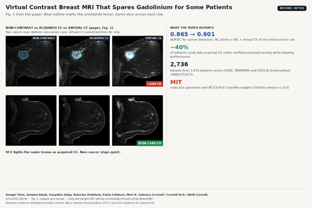
Contrast-enhanced breast MRI shows tumor vascularity that non-contrast sequences often miss, but every exam means an IV line, gadolinium, extra time on the table, and cost. In arXiv:2611.00140, Hongxi Chen, Sunwoo Kwak, Sanyukta Adap, Katerina Dodelzon, Paula Feldman and Mert R. Sabuncu (Cornell, Cornell Tech and Weill Cornell Medicine) ask whether a virtual contrast-enhanced (VCE) image, predicted from the non-contrast (NC) volume alone, can add enough signal that some patients skip the acquired contrast-enhanced (CE) phase without losing cancer detection.
They do not claim to replace gadolinium. They propose confidence-based routing. A 3D U-Net builds the VCE image. Medical Slice Transformer classifiers score NC, CE and VCE. On the held-out test set, NC alone reached an AUROC of 0.865. Fusing NC with VCE predictions lifted that to 0.901. Acquired CE still led at 0.945. In a simulation that sends the most confident NC cases down an NC+VCE path, about 40% of patients could avoid acquired CE while diagnostic performance stayed close to the full-CE baseline.
How it works
The generator is a five-stage 3D U-Net. It takes the fat-suppressed NC volume and predicts an enhancement residual. The VCE image is the NC volume plus that residual. Training losses weigh reconstruction inside the breast, enhancement fidelity, missed enhancement and excess enhancement, so the network is pushed to light the right regions without inventing bright spots elsewhere.
Cancer detection uses three independent Medical Slice Transformer (MST) classifiers, one each for NC, acquired CE and VCE. The authors also run VCE through the CE-trained classifier. Fusion is a simple average of classifier logits. That keeps the pipeline easy to reason about in a viewer: one score from the raw NC series, one from the synthetic CE series, then combine them.
The cohort mixes paired NC and first-phase CE exams from DUKE, MAMAMIA and ODELIA. After removing DUKE duplicates that overlap MAMAMIA, each bilateral volume is split into left and right breasts with BreastDivider, resampled, and padded to a fixed size. The final set has 2,736 breasts from 1,410 patients, split patient-wise into 1,888 / 375 / 473 for train / val / test.
What the numbers show
Standalone VCE matched NC (AUROC 0.866 vs 0.865) and did better when scored by a classifier trained on virtual images (0.883). The useful jump came from fusion: NC + VCE reached 0.901 with the CE-trained head and 0.896 with the VCE-trained head, closing nearly half the gap between NC and acquired CE (0.945). Gains held on all three source datasets, though ODELIA remained the hardest.
The featured image is Fig. 1 of the paper: NC, acquired CE and virtual CE on a cancer case and a non-cancer case, with blue outlines on the annotated lesion. On the cancer row the virtual image lights the same region as the acquired CE. On the non-cancer row both CE and VCE stay quiet.
For routing, patients were ranked by NC prediction confidence, defined as max(p, 1-p) and averaged across a patient’s breasts so both sides stay on the same pathway. Confidence-based replacement beat random replacement across matched rates. At roughly 60% of patients on the NC+VCE path (about 40% still getting acquired CE), AUROC and AUPRC stayed near the full-CE curve in their simulation.
Why a viewer or clinic team should care
If you ship breast MRI tools, the decision layer matters as much as the synthetic series:
- Hang NC and VCE side by side. A DICOM viewer can show the acquired NC series next to a derived VCE series the same way it hangs a subtracted DCE phase. The reader sees where the model thinks enhancement should appear, with the NC anatomy still in view.
- Surface a confidence score before injection. Because NC is acquired first, a triage score can be computed while the patient is still on the table. High-confidence negatives (or clear positives) could finish without contrast; uncertain cases proceed to CE as usual.
- Keep acquired CE for characterization. The authors are explicit that acquired CE remains the standard for lesion characterization, disease extent and biopsy guidance. A product that frames VCE as a routing aid, not a CE replacement, matches the paper’s claim.
Wiring looks familiar if you have already hung synthetic series. You need a NIfTI (or in-memory volume) path into the generator, then a derived series back into the study. We covered related plumbing for synthetic contrast in brain MRI with MoFD, and for breast MRI segmentation overlays with CoM3eT.
Where it falls short
- VCE is not a substitute for acquired CE. The authors state this directly. Detection improves when VCE is added to NC, but acquired CE still wins, and characterization work stays on real contrast.
- Cohorts are cancer-enriched and preoperative, not screening. Most exams in screening will be negative. The paper flags that the datasets do not match that mix, and that overall sample size was limited.
- Lesion-level fidelity is imperfect. Prior synthesis work already showed poor overlap between synthetic enhancement and lesions. This study measures downstream detection, not pixel-perfect enhancement maps, so do not treat VCE as a quantitative perfusion substitute.
- ODELIA is harder. Dataset-specific AUROCs drop on ODELIA relative to DUKE and MAMAMIA, which is a warning for multi-site deployment.
- No prospective reader study yet. Future work they list includes radiologist performance with synthetic images and real-time routing. Those results are not in this preprint.
Code, weights and license
Code is at github.com/XiGabe/VirtualContrast-BreastMRI under the MIT license. A scripts/reproduce.sh path regenerates Table 1 and Figure 2 from included prediction files without downloading MRI. Generator and NC/CE/VCE classifier weights (three seeds) ship as GitHub release v1.0.0 via scripts/download_weights.py. Third-party notices cover DINOv2, BreastDivider and the source datasets (DUKE, MAMAMIA, ODELIA), which you still obtain from their own pages. The preprint is under arXiv’s standard non-exclusive license. Credit: Hongxi Chen, Sunwoo Kwak, Sanyukta Adap, Katerina Dodelzon, Paula Feldman and Mert R. Sabuncu.
My take
I would trial this as a pre-injection triage experiment, not as a CE replacement. Run NC through the generator, hang VCE next to NC, log the fused score, and measure how often a confident NC+VCE call would have matched the CE read. The MIT weights make that local test cheap. Keep acquired CE for anything that needs margin, kinetics or biopsy planning, which is where the paper itself draws the line.