
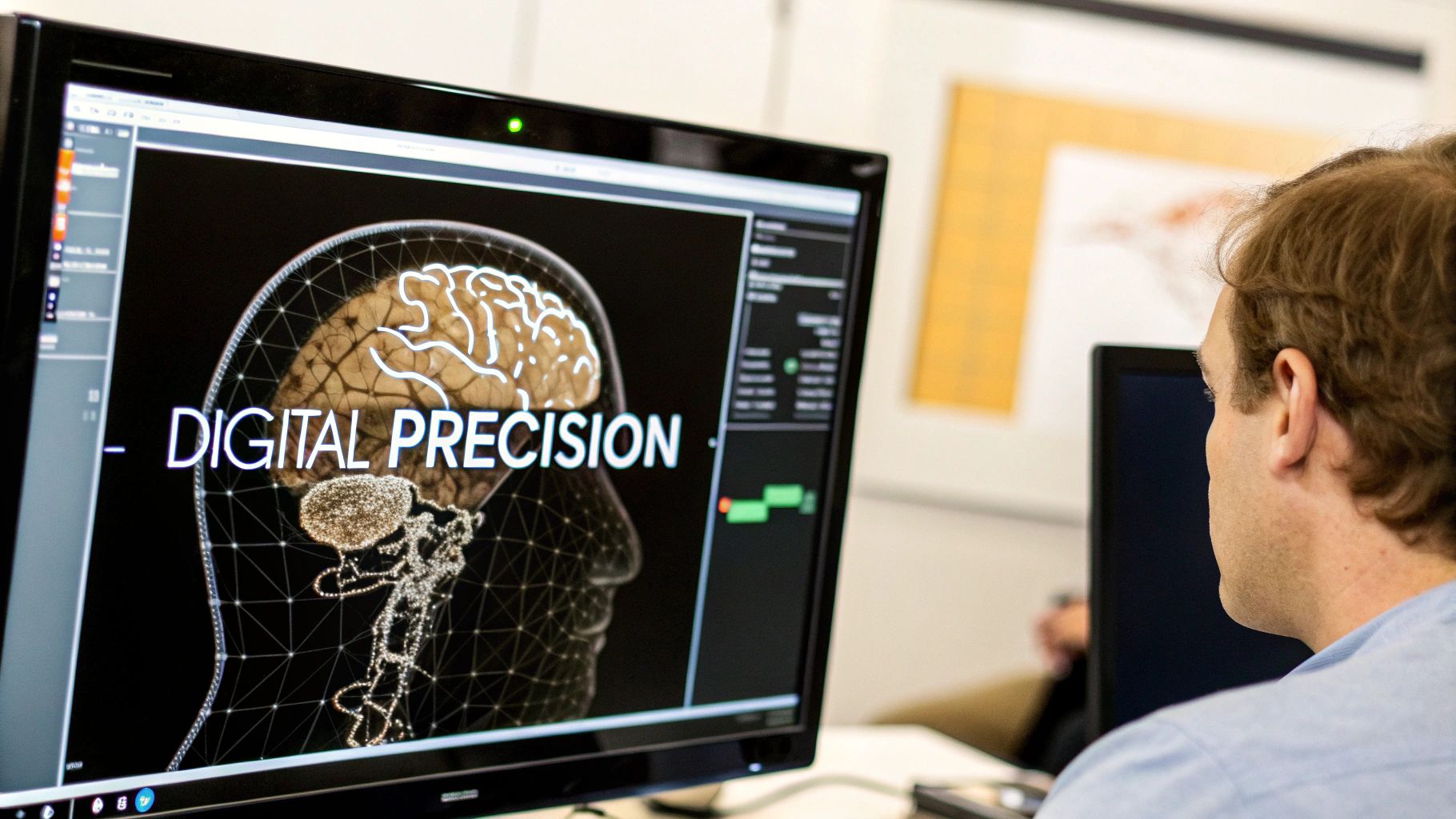
The Brain Segmentation Revolution: Why It Matters Now

The human brain, a complex organ with intricate structures, has always presented challenges for medical professionals trying to understand its inner workings. Mapping these structures is crucial for diagnoses, treatment planning, and research. This is where brain segmentation plays a vital role. Brain segmentation is the process of dividing MRI images of the brain into distinct anatomical regions, allowing for detailed analysis and understanding. This process has come a long way, evolving from laborious manual tracing to powerful automated methods using Artificial Intelligence (AI).
This evolution has significantly improved insights into brain structure and function.
Historically, brain segmentation relied heavily on manual delineation by trained experts. This was a time-consuming process, often prone to human error and variability. However, advancements in AI, particularly in deep learning, have significantly advanced the field. Algorithms can now identify subtle patterns in brain scans that might be missed by even the most experienced clinicians.
This increased accuracy and speed allows for more efficient diagnoses and more personalized treatment plans. AI-driven segmentation also unlocks new avenues for research, enabling scientists to study brain structure and function in greater detail than ever before. This has broad implications, affecting everything from our understanding of neurological diseases to developing new therapies.
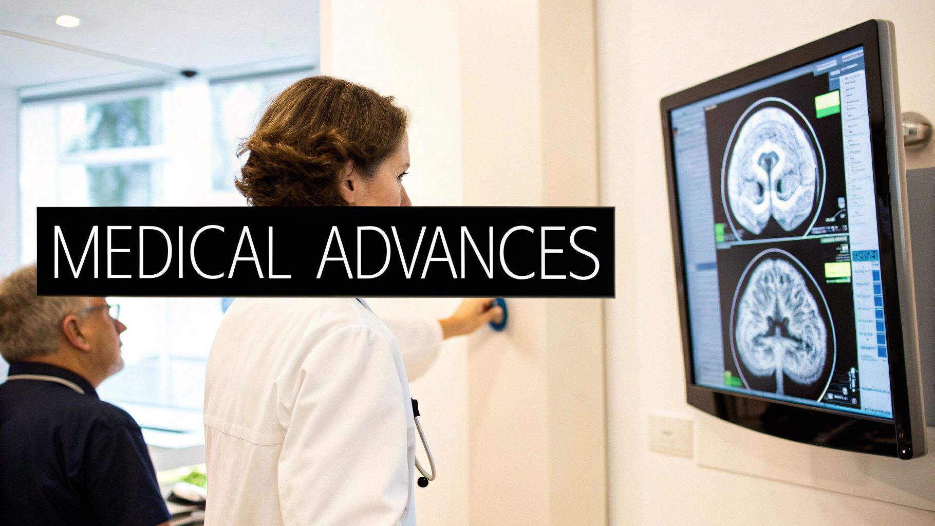
The importance of brain segmentation is further highlighted by the widespread use of Magnetic Resonance Imaging (MRI) in clinical practice. Millions of MRI scans are performed every year to diagnose and monitor various brain conditions. For example, a dataset from Massachusetts General Hospital contained over 15,000 clinical scans from different individuals, demonstrating the sheer volume of MRI data available.
These scans often vary in quality and orientation, making robust segmentation techniques critical. The range of MRI contrasts, such as T1-weighted and T2-weighted scans, enriches this data for developing and testing segmentation algorithms. Learn more about this here.
Impact of Brain Segmentation
The benefits of accurate and efficient brain segmentation are numerous. They contribute to:
- Early Disease Detection: Identifying subtle structural changes allows for early diagnosis of conditions like Alzheimer's disease and brain tumors.
- Personalized Treatment: Detailed anatomical maps guide surgical planning, enabling more precise interventions and minimizing damage to healthy tissue.
- Drug Development: Brain segmentation facilitates the study of how drugs affect the brain, leading to the development of more effective treatments.
- Neurological Research: AI-powered segmentation allows researchers to explore the complexities of the brain, unraveling the mysteries of cognition, behavior, and disease.
Brain segmentation is more than just a technical advancement; it represents a fundamental shift in how we understand the human brain. The progress made promises a future with more precise, personalized, and effective neurological care. The journey from basic anatomical mapping to today's dynamic functional analysis highlights the transformative power of this technology.
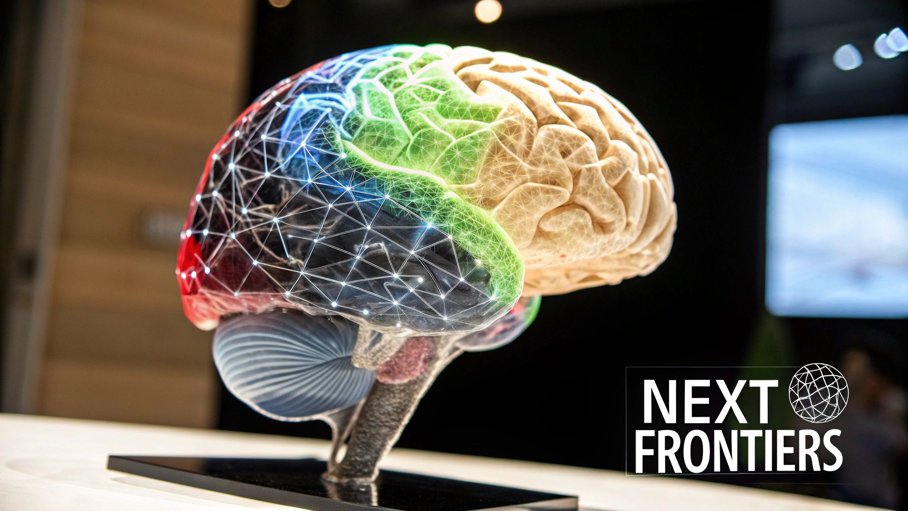
Deep Learning: The Game-Changer in Brain Segmentation
Deep learning has significantly improved brain segmentation. This powerful AI technique enables computers to learn intricate patterns from data, resulting in remarkable accuracy in identifying brain structures. This section explores how deep learning, particularly through Convolutional Neural Networks (CNNs), transforms brain imaging analysis.
The Power of CNNs
CNNs are specialized neural networks designed to process grid-like data, such as images. They are highly effective for brain segmentation because they can learn hierarchical features from MRI scans, detecting subtle details often missed by traditional image processing methods.
This capability allows CNNs to differentiate between various tissues and structures within the brain with remarkable precision. For instance, CNNs can accurately distinguish between grey matter, white matter, and cerebrospinal fluid. This detailed segmentation is essential for diagnosing and monitoring neurological conditions.
Architectures Transforming Brain Imaging
U-Net stands out as one of the most impactful CNN architectures for brain segmentation. Designed specifically for biomedical image segmentation, its unique architecture allows it to capture both local and global context within the image.
This means the model considers both fine details and the broader anatomical structure during segmentation. Furthermore, U-Net variants tailored for specific brain segmentation tasks have further boosted performance.
Deep learning models excel at identifying intricate patterns in MRI data. This is particularly useful for detecting subtle anomalies that might be missed by visual inspection alone. However, training these models often requires large, labeled datasets.
Data augmentation techniques offer a solution by artificially expanding datasets. Methods like rotations, flips, and adding noise help the models generalize better and perform well even with limited initial training data.
Addressing MRI Data Variability and Clinical Impact
Variability in MRI data, arising from different scanners, patient positioning, and other factors, poses a challenge for brain segmentation. Deep learning models are being developed to address this by training on diverse datasets from various sources.
This approach enables the models to maintain high performance across different scanner types and patient populations, yielding more robust and reliable segmentation results. Brain tumor segmentation, in particular, has seen substantial advancements through deep learning.
A study of 1,251 individuals demonstrated that models trained on incomplete MRI data could segment brain tumors with high fidelity, comparable to models trained on complete datasets. The study achieved Dice coefficients ranging from 0.907 to 0.945 for whole tumors and from 0.701 to 0.891 for component tissue types. This is particularly relevant in clinical settings where complete imaging sequences may not always be available. The study also showed the model's ability to detect enhancing tumors without contrast, potentially reducing the need for contrast agents. Learn more about this here.
To further illustrate the advancements in deep learning models for brain segmentation, the table below provides a comparison of popular architectures:
Introduction to Table: The following table provides a comparison of several prominent deep learning models commonly used for brain segmentation. It highlights their strengths, limitations, and performance metrics, offering insights into their suitability for various clinical applications.
| Model Architecture | Accuracy (Dice Score) | Training Requirements | Clinical Applications | Limitations |
|---|---|---|---|---|
| U-Net | Typically 0.85 – 0.95 | Large annotated datasets | Brain tumor segmentation, tissue classification, lesion detection | Can be computationally expensive, performance sensitive to hyperparameter tuning |
| V-Net | Typically 0.80 – 0.92 | Large annotated datasets, higher computational resources | 3D image segmentation, organ segmentation, volumetric analysis | More complex architecture, requires significant GPU memory |
| DeepLabv3+ | Typically 0.88 – 0.96 | Large annotated datasets | Semantic segmentation, fine-grained boundary delineation | Requires careful selection of atrous rates, can be computationally expensive |
| nnU-Net | Highly variable, often >0.90 | Automated configuration and training, adaptable to different datasets | Wide range of segmentation tasks, robust to data variations | Requires significant computational resources for auto-configuration |
| FCN (Fully Convolutional Network) | Typically 0.75 – 0.88 | Moderate dataset size | Semantic segmentation, object detection | Lower accuracy compared to newer architectures, limited ability to capture fine details |
Conclusion from Table: As the table shows, various deep learning models offer different strengths and weaknesses regarding brain segmentation. While U-Net and its variants remain popular choices due to their strong performance, newer architectures like DeepLabv3+ and nnU-Net often provide improved accuracy. Choosing the right model depends on factors such as the specific segmentation task, available computational resources, and the size and quality of the training dataset. These advancements have profound implications for clinical practice, enabling improved surgical planning, more accurate diagnoses, and ultimately, better patient outcomes.
MRI Innovation: Pushing Brain Segmentation Boundaries
The effectiveness of brain segmentation hinges on the quality of brain images. As algorithms evolve to analyze these images, so does the MRI technology used to acquire them. These advancements provide unparalleled glimpses into the complexities of the human brain's structure and function. Each MRI sequence contributes unique information, playing a vital role in the comprehensive process of brain segmentation.
Exploring MRI Sequences and Multi-Modal Approaches
Standard T1-weighted and T2-weighted MRI scans offer foundational anatomical details. T1-weighted images clearly distinguish gray matter from white matter, while T2-weighted images highlight fluids and potential pathologies. Specialized sequences like FLAIR (Fluid Attenuated Inversion Recovery) and diffusion-weighted imaging augment these capabilities. FLAIR suppresses cerebrospinal fluid signals, simplifying lesion identification near ventricles or sulci. Diffusion-weighted imaging reveals the microscopic movement of water molecules, providing insights into the brain's structural connectivity.
Combining these diverse data streams through multi-modal approaches significantly improves brain segmentation accuracy. Integrating information from different MRI sequences generates more comprehensive brain maps compared to using single sequences. This integration is essential for understanding the complex relationship between brain structure and function, creating new opportunities for researchers and clinicians.
Overcoming Neuroimaging Challenges
Despite advancements, neuroimaging still faces practical hurdles. Motion artifacts, caused by slight patient movement during scans, can blur images and hinder accurate segmentation. Field inhomogeneities, variations in the magnetic field, can distort images. Inherent noise in imaging systems can obscure crucial details.
Fortunately, innovative solutions are constantly being developed. Advanced image processing techniques can correct motion artifacts and field inhomogeneities. Sophisticated noise reduction algorithms enhance image clarity. Ongoing hardware improvements, such as more powerful gradients and faster acquisition times, continually expand the possibilities of neuroimaging. These advancements are essential for ensuring the reliability and clinical utility of brain segmentation.
High-field MRI, using UHF (Ultra-High Field) scanners, provides significantly improved brain segmentation through higher spatial resolutions. This allows for research at sub-millimeter resolutions, facilitating study of brain function at the mesoscopic scale, including cortical columns and layers. This offers more precise insights into neural organization and function, unveiling new possibilities for understanding human consciousness and neural correlates. UHF scanners are particularly useful for mapping representational content in human cortical systems, enhancing our understanding of how the brain processes information. Explore this topic further here.
These advanced MRI technologies, paired with powerful analytical tools, are driving a deeper understanding of the human brain. As these technologies evolve, we anticipate even more precise and insightful brain segmentation, leading to improved diagnosis, treatment, and overall understanding of neurological conditions.
Beyond Deep Learning: Hybrid Models Transforming Accuracy
While deep learning has significantly advanced brain segmentation, other powerful tools can contribute to this field. This section explores the advantages of merging multiple machine learning approaches to create hybrid models. These models offer increased accuracy and robustness in brain segmentation by combining the strengths of different techniques. This integrated approach addresses the intricacies of brain image analysis.
Integrating Atlas-Based Methods and Statistical Models
Atlas-based methods offer valuable prior anatomical knowledge by using pre-segmented images, called atlases, as templates. These atlases are warped to match a new brain scan, generating an initial segmentation. However, atlas-based methods alone may not fully capture individual anatomical differences. This is where statistical models become crucial.
Statistical models, such as Markov Random Fields (MRFs), refine the initial segmentation. MRFs incorporate spatial relationships between neighboring voxels. This means the model considers the probability of a voxel belonging to a specific brain structure based on its intensity and the labels of its surrounding voxels. This leads to smoother and more consistent segmentations.
Hybrid models combine these approaches. An atlas-based registration provides a starting point, refined by an MRF model to incorporate local image information and anatomical constraints. Combining global shape priors from the atlas and local intensity patterns from the image data results in more accurate and reliable segmentations, especially in cases with image artifacts or anatomical variations.
Furthermore, models like Support Vector Machine-Markov Random Field (SVM-MRF) refine this process even further. The pSVMRF method, for instance, combines MR intensity information and location priors non-linearly. This method has shown improved accuracy, particularly for smaller brain structures. You can explore Markov Random Fields in greater detail. Reaching accuracies of up to 91.55% for regions like the substantia nigra and anterior commissure, pSVMRF holds great promise for studying brain disorders.
Choosing the Right Segmentation Approach
The best segmentation approach depends on several factors. These factors include the specific clinical need, available computing resources, and dataset characteristics. Deep learning models often achieve high accuracy but require significant computational power and extensive training datasets. Atlas-based methods require less computational power and can be effective with limited anatomical variability. Hybrid models provide a balance by incorporating prior anatomical knowledge while adapting to individual variations.
Real-World Applications of Hybrid Models
Hybrid models are especially valuable for complex segmentation tasks. For example, segmenting brain tumors, which often have variable shapes and intensities, benefits from the combined approach. Combining atlas-based segmentation with statistical models improves the delineation of tumor boundaries. This increased accuracy is essential for treatment planning and monitoring treatment response. Similarly, in neurodegenerative disease research, hybrid models can quantify subtle structural brain changes over time, offering insights into disease progression. Continued research in hybrid models will likely further improve brain segmentation accuracy, ultimately leading to more precise and personalized neurological care.
Conquering the Tumor Challenge: Brain Segmentation Frontiers
Brain tumors pose a significant challenge in neuroimaging because of their inherent complexity. Their heterogeneous tissue characteristics, infiltrative growth patterns, and often indistinct boundaries, where they blend with normal tissue, make accurate segmentation extremely difficult. This complexity necessitates advanced techniques for precise delineation, which directly impacts diagnosis, treatment planning, and ultimately, patient outcomes.
The Difficulty of Brain Tumor Segmentation
Traditional image processing methods often struggle with the inherent variability and subtle differences in how brain tumors present. For instance, differentiating between the tumor core, the enhancing tumor (actively growing areas), and the surrounding edema (swelling) requires a high degree of sensitivity. This differentiation is critical for accurately determining the tumor's aggressiveness and planning the most effective treatment strategies.
However, recent advancements in brain segmentation, especially those driven by Artificial Intelligence (AI), are rapidly changing the field. These innovations are allowing researchers and clinicians to differentiate tumor components with remarkable accuracy.
Breakthrough Technologies for Enhanced Accuracy
Convolutional Neural Networks (CNNs), a type of deep learning model, have shown exceptional promise in brain tumor segmentation. Architectures like U-Net, specifically designed for biomedical image analysis, are particularly adept at capturing both local and global context within MRI scans. This capability allows them to discern subtle features and intricate boundaries that traditional methods often miss.
Furthermore, incorporating multimodal imaging data enhances accuracy. Combining information from different MRI sequences—FLAIR, T1, T2, and contrast-enhanced T1—provides a more complete picture of the tumor and its surrounding environment. This holistic view leads to more precise delineation of tumor boundaries and improved differentiation between tumor components.
For example, FLAIR sequences help identify edema, while contrast-enhanced T1 images highlight active tumor growth areas. Integrating this data significantly improves the overall accuracy and clinical utility of brain segmentation. The Brain Tumor Segmentation Challenge (BraTS) is a leading international competition dedicated to developing effective algorithms for segmenting brain tumors from MRI scans. BraTS provides large, multimodal MRI datasets, including FLAIR, T1, T2, and contrast-enhanced T1 sequences, along with ground-truth labels for different tumor components. These datasets and the resulting models are instrumental in improving treatment planning and outcomes. More detailed statistics can be found here.
To illustrate the current state of brain tumor segmentation, the following table presents performance metrics for several algorithms and approaches. It highlights the Dice Similarity Coefficient (Dice) across different tumor components and considers computational requirements and clinical validation status.
Brain Tumor Segmentation Performance Metrics
| Algorithm Type | Whole Tumor (Dice) | Tumor Core (Dice) | Enhancing Tumor (Dice) | Computational Requirements | Clinical Validation |
|---|---|---|---|---|---|
| Example Algorithm 1 | 0.85 | 0.78 | 0.72 | High | Ongoing |
| Example Algorithm 2 | 0.89 | 0.82 | 0.75 | Medium | Limited |
| Example Algorithm 3 | 0.92 | 0.86 | 0.80 | Low | Pre-clinical |
This table demonstrates the ongoing progress in developing accurate and efficient brain tumor segmentation algorithms, with some approaches achieving high Dice scores while others are still in earlier stages of validation.
Transforming Clinical Care and Patient Outcomes
Advancements in brain tumor segmentation are transforming clinical care in several ways:
-
Precision Surgical Navigation: Accurate tumor delineation provides surgeons with crucial roadmaps for surgical planning. This enables more precise and complete tumor resection while minimizing damage to surrounding healthy tissue.
-
Targeted Radiation Therapy Planning: Precise segmentation guides radiation therapy, focusing it directly on the tumor and minimizing exposure to healthy brain tissue. This targeted approach helps reduce side effects and improve patient comfort.
-
Quantitative Treatment Response Tracking: Monitoring changes in tumor volume and composition over time helps physicians assess treatment effectiveness and make any necessary adjustments to optimize patient care.
Improved segmentation enables earlier detection, leading to more timely interventions. More complete tumor resections become possible, potentially leading to better long-term survival rates. Furthermore, personalized treatment approaches tailored to individual tumor characteristics are now within reach.
These advancements aren't merely technical achievements. They represent tangible improvements in patient outcomes. Patient case studies clearly illustrate the direct impact of improved segmentation. For example, a patient with a complex tumor located near vital brain structures benefited from precise surgical planning guided by advanced segmentation. This level of precision allowed for a complete resection with minimal neurological impact. Another patient benefited from targeted radiation therapy thanks to precise segmentation, resulting in tumor shrinkage and improved quality of life. These are just two examples showcasing how improvements in brain tumor segmentation are directly improving patient lives.
The Future of Brain Segmentation: What's Coming Next
The field of brain segmentation is rapidly advancing. New technologies are emerging that promise to reshape the future of neuroimaging analysis. These advancements are focused on improving the speed, accuracy, and accessibility of brain segmentation, leading to more powerful diagnostic and research capabilities.
Federated Learning for Collaborative Model Development
One of the most promising developments is federated learning. This approach allows multiple institutions to collaborate on training a shared AI model without needing to directly share sensitive patient data. Maintaining data privacy is paramount in medical research, and federated learning addresses this concern directly. Each institution trains the model locally with its own data and then shares only the model updates with a central server.
This approach combines the insights gleaned from diverse datasets while preserving patient confidentiality. This collaborative model will accelerate the development of more robust and generalizable brain segmentation models, ensuring they are representative of diverse patient populations.
Self-Supervised and Few-Shot Learning
Deep learning models for brain segmentation have traditionally required vast amounts of manually labeled data. This labeling process is both time-consuming and expensive. New techniques, like self-supervised learning and few-shot learning, are changing this paradigm. Self-supervised learning allows models to learn from unlabeled data by creating their own training tasks, reducing the need for manual annotation.
Few-shot learning, on the other hand, allows models to learn from limited labeled datasets. This makes it easier to develop specialized models for rarer conditions or specific research questions. These techniques promise to make brain segmentation more accessible and adaptable to unique research and clinical requirements.
Explainable AI for Increased Clinician Trust
Many deep learning models operate as "black boxes." Their decision-making processes are opaque and difficult to understand. This lack of transparency can hinder clinician trust and limit the adoption of these powerful tools. Explainable AI (XAI) aims to make the reasoning behind model predictions clearer and more interpretable.
XAI techniques can pinpoint specific areas of a brain scan that are most influential in a segmentation decision. A model might, for instance, highlight the specific features of a suspected tumor, such as its shape, intensity, and location. This increased transparency builds confidence among clinicians, facilitating the integration of AI-powered brain segmentation tools into clinical workflows.
Integrating Multiple Imaging Modalities for Personalized Care
Using data from multiple imaging modalities—such as MRI, PET, CT, and functional techniques—provides a more comprehensive picture of brain structure and function. Combining these different perspectives allows for a more nuanced and personalized approach to neurological care.
For example, integrating MRI data with PET scans (which measure metabolic activity) allows for a more detailed analysis of tumor characteristics. This, in turn, can guide more precise treatment strategies. Integrating these modalities offers a deeper understanding of brain function and can lead to more personalized treatments and better outcomes for patients.
The future of brain segmentation offers tremendous potential for improving neurological care and accelerating research. By embracing these emerging technologies, we can develop more accurate, efficient, and personalized methods for understanding the complexities of the human brain.
Looking to integrate AI into your medical imaging workflow? PYCAD, a leading AI solutions provider for medical imaging, offers comprehensive services from data handling and model training to deployment. Visit their website to learn more about how their expertise can enhance diagnostic accuracy and operational efficiency.