The Future of Healthcare is Here: Seeing Beyond the Surface
This listicle reveals eight key applications of computer vision in healthcare, demonstrating how this technology improves diagnoses, treatments, and patient care. Learn how computer vision is transforming medical imaging analysis, surgical robotics, pathology, dermatology, ophthalmology, patient monitoring, drug discovery, and even medical documentation. These advancements offer increased accuracy, efficiency, and personalized medicine, ultimately leading to better patient outcomes. Explore these transformative applications and see how computer vision is shaping the future of healthcare.
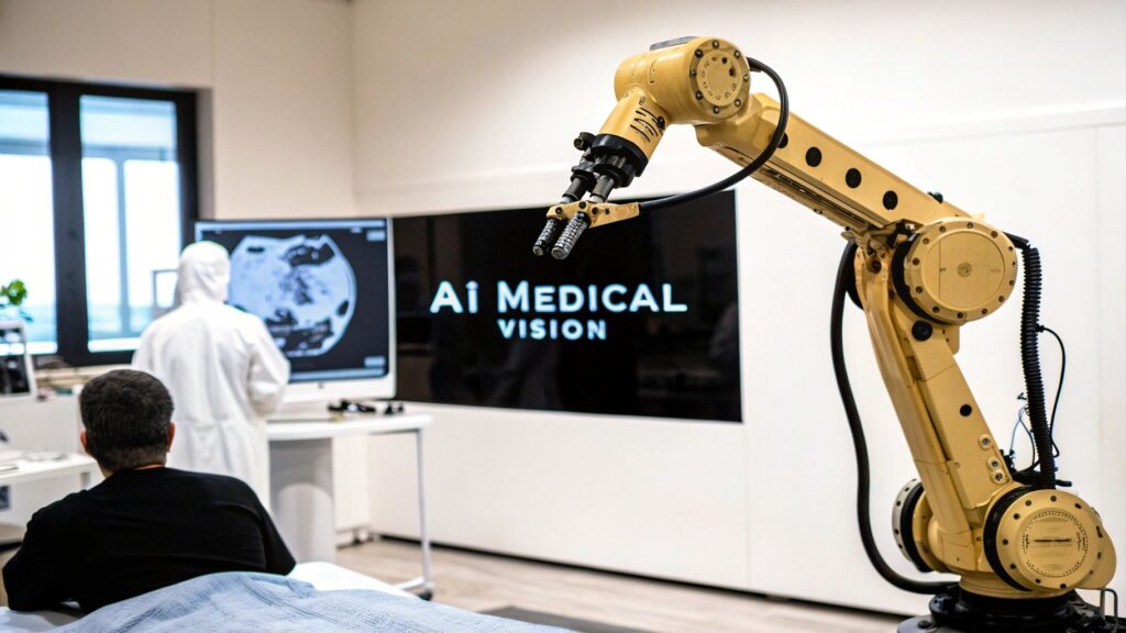
1. Medical Imaging Analysis and Diagnosis
Medical imaging analysis and diagnosis represents a revolutionary application of computer vision in healthcare. This technology leverages the power of artificial intelligence, specifically deep learning algorithms, to analyze medical images such as X-rays, CT scans, MRIs, mammograms, and ultrasounds. By training these algorithms on vast datasets of labeled medical images, computer vision systems can identify intricate patterns and anomalies often imperceptible to the human eye, assisting radiologists in detecting a range of conditions including tumors, fractures, and other pathological indicators. These systems are capable of providing both primary diagnoses and crucial second opinions, contributing significantly to improved patient care.
Computer vision achieves this remarkable feat through several key features: deep learning-based pattern recognition, support for various imaging modalities (X-ray, CT, MRI, ultrasound), automated lesion detection and segmentation, quantitative analysis and measurement tools, and seamless integration with Picture Archiving and Communication Systems (PACS). This integration with existing hospital infrastructure ensures smooth workflows and efficient data management.
The benefits of incorporating computer vision into medical imaging analysis are multifaceted. Increased diagnostic accuracy and consistency minimize human error and subjectivity, leading to more reliable results. Faster image interpretation and reporting significantly expedite diagnosis and treatment planning, which is particularly crucial in time-sensitive situations. Furthermore, this technology enables the early detection of diseases like cancer, potentially improving patient outcomes and survival rates. By automating routine tasks, computer vision reduces radiologist workload and burnout, allowing them to focus on complex cases and patient interaction. Finally, the 24/7 availability of these systems ensures prompt image analysis for emergency cases, even during off-peak hours.
However, integrating computer vision into medical imaging also presents challenges. The initial implementation costs can be substantial, including hardware, software, and training expenses. Regulatory approval processes are rigorous and time-consuming. These systems require extensive, high-quality training data to achieve optimal performance, and the potential for false positives and negatives remains a concern. Furthermore, integrating new technology can sometimes meet with resistance from medical professionals accustomed to traditional workflows.
Several examples highlight the successful implementation of computer vision in medical imaging. Google's AI has demonstrated its proficiency in detecting diabetic retinopathy in eye scans, while IBM Watson for Oncology analyzes cancer images to assist oncologists in developing personalized treatment plans. Zebra Medical Vision's AI1 platform offers automated chest X-ray analysis, and Aidoc's AI solution streamlines emergency radiology triage. These advancements demonstrate the real-world impact of computer vision in enhancing healthcare delivery.
For healthcare providers looking to integrate computer vision into their practices, several actionable tips can facilitate a smoother transition. Starting with pilot programs in specific departments allows for controlled testing and refinement before widespread deployment. Ensuring robust data privacy and security measures is paramount for patient confidentiality. Actively involving radiologists in the validation process fosters acceptance and ensures the technology aligns with clinical needs. Establishing clear workflows for AI-assisted diagnosis promotes efficient integration into existing practices. Crucially, maintaining human oversight and final decision-making ensures that medical expertise remains at the center of patient care.
This advancement in medical imaging analysis is a significant reason for its inclusion on this list of impactful computer vision applications in healthcare. Its transformative potential stems from its ability to address critical bottlenecks in medical imaging workflows, ultimately leading to more accurate, efficient, and accessible diagnostic services.
The following infographic visualizes key data points related to the growing influence of computer vision in medical image analysis: the substantial volume of medical images used for training these algorithms and the projected market size of this rapidly expanding field.
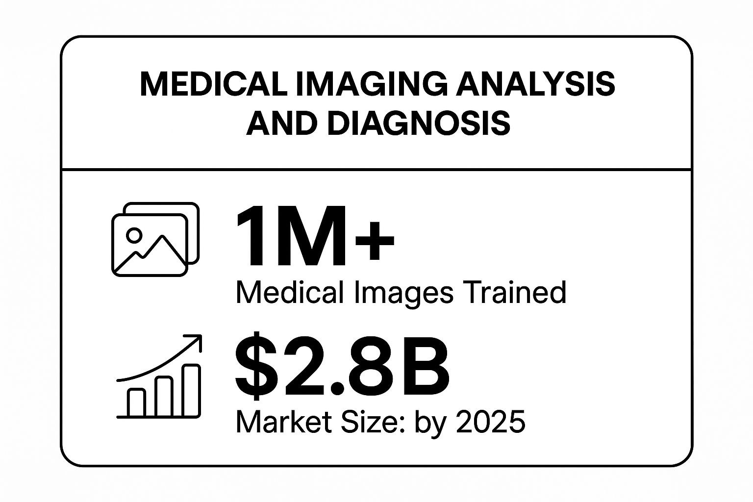
The infographic reveals the impressive scale of data utilized ("1M+ Medical Images Trained") and the significant market growth projected ("Market Size: $2.8B by 2025"). This underscores the increasing investment and adoption of computer vision in medical imaging, highlighting its potential to revolutionize diagnostic practices and improve patient care globally. This growth is driven by the need for faster, more accurate diagnoses, and the increasing availability of large medical image datasets, coupled with advances in deep learning algorithms. The rising market size reflects the growing recognition of the value of computer vision in addressing critical challenges in healthcare.
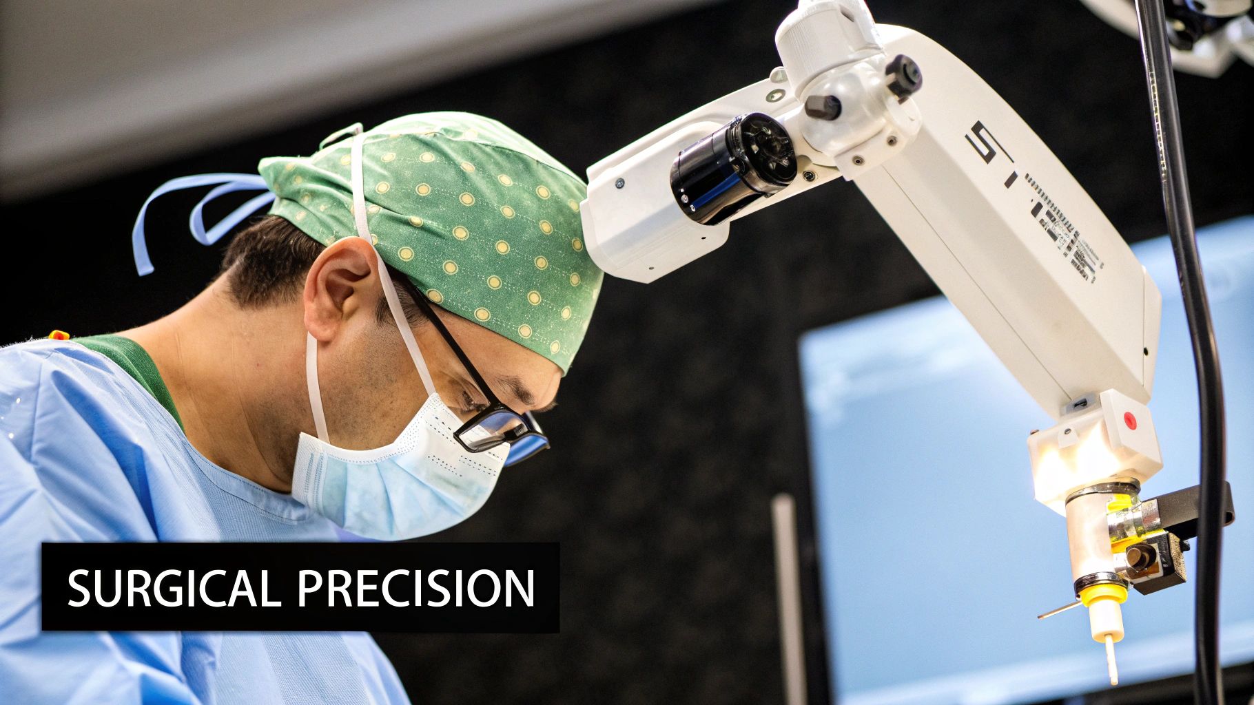
2. Surgical Navigation and Robotics
Computer vision in healthcare is revolutionizing surgical procedures through advanced navigation and robotics systems. These systems leverage computer vision algorithms to provide surgeons with real-time visualization, instrument tracking, and detailed anatomical mapping, ultimately enhancing precision, minimizing invasiveness, and improving patient outcomes. By integrating with robotic surgical platforms, computer vision empowers surgeons with augmented reality overlays and 3D reconstructions, transforming the operating room into a high-tech, data-driven environment.
This technology works by utilizing cameras and sensors to capture real-time images of the surgical field. These images are then processed by sophisticated computer vision algorithms that identify and track surgical instruments, create 3D models of the patient's anatomy, and overlay this information onto the surgeon's view. This augmented reality provides a comprehensive understanding of the surgical site, allowing for more precise and controlled movements. The integration with robotic systems further amplifies these benefits, enabling surgeons to perform complex procedures with enhanced dexterity and stability.
Several successful implementations demonstrate the transformative potential of computer vision in surgical navigation and robotics. The da Vinci Surgical System by Intuitive Surgical is a prime example, offering minimally invasive surgery across various specialties. Stryker's Mako robotic arm, specifically designed for orthopedic surgery, utilizes computer vision to enhance precision in joint replacement procedures. Medtronic's StealthStation navigation system provides real-time tracking and guidance during neurosurgical and spinal surgeries. Furthermore, platforms like TouchSurgery's AR surgical planning platform allow surgeons to visualize and rehearse procedures before entering the operating room.
This approach is particularly valuable in complex procedures requiring high precision and minimal invasiveness. Examples include minimally invasive cardiac surgery, neurosurgery where accuracy is paramount, and orthopedic procedures requiring precise bone cutting and implant placement. The benefits are multifaceted, including enhanced surgical precision and accuracy, reduced complications and recovery time, and the ability to perform minimally invasive procedures that were previously impossible. Furthermore, computer vision-assisted surgery provides improved training opportunities for surgical residents and facilitates better documentation of surgical procedures.
However, it's crucial to acknowledge the challenges associated with this technology. The high equipment costs can be a significant barrier to adoption, especially for smaller hospitals. Surgeons face a steep learning curve to master these systems, requiring comprehensive training programs. The technical complexity of these systems necessitates specialized maintenance and support, adding to the overall cost. Furthermore, the potential for system failures during surgery, although rare, is a critical concern. Traditional surgical capabilities must be maintained as a backup in case of technical issues.
Tips for successful implementation:
- Invest in comprehensive surgeon training programs: Ensure surgeons are thoroughly trained on the specific systems being implemented.
- Start with less complex procedures: Gradually introduce the technology into the surgical workflow, beginning with less complex cases.
- Ensure redundant safety systems: Implement backup systems and protocols to mitigate the risk of system failures during surgery.
- Maintain traditional surgical capabilities as backup: Be prepared to revert to traditional surgical techniques if necessary.
- Regular system calibration and maintenance: Adhere to a strict maintenance schedule to ensure optimal system performance and reliability.
Despite these challenges, the potential of computer vision in surgical navigation and robotics is undeniable. It represents a significant advancement in surgical care, offering tangible benefits for both surgeons and patients. By addressing the current limitations and continuing to refine the technology, computer vision promises to further transform the operating room and redefine the future of surgery. This revolutionary approach firmly secures its place on the list of transformative applications of computer vision in healthcare, paving the way for a new era of precision and minimally invasive surgery.
3. Pathology and Histopathology Analysis
Computer vision in healthcare is revolutionizing numerous fields, and pathology and histopathology analysis is a prime example. This application leverages AI-powered microscopy systems to analyze tissue samples, blood smears, and cellular structures, enabling faster and more accurate diagnoses of various diseases, including cancer. This technology holds immense potential for improving patient outcomes and transforming the field of pathology.
Traditional pathology relies heavily on manual microscopic examination of stained tissue sections by pathologists. This process, while effective, can be time-consuming, subjective, and prone to inter-observer variability. Computer vision offers a solution by automating and standardizing the analysis process. Digitized slides are analyzed by machine learning algorithms, which are trained to recognize patterns and anomalies indicative of specific diseases. This approach offers several significant advantages.
How it Works:
The process begins with whole slide imaging (WSI), where glass slides are digitized using high-resolution scanners, creating a digital representation of the entire tissue sample. These digital slides can then be accessed and analyzed remotely, eliminating the need for physical slides and enabling faster consultations. AI algorithms then process these images, using computer vision to identify and classify cells, detect and measure morphological changes, and quantify biomarkers. These algorithms are trained on vast datasets of labeled images, allowing them to identify subtle patterns that might be missed by the human eye. The output can include information about the presence and type of disease, tumor grading and staging, predicted treatment response, and other clinically relevant information.
Features and Benefits:
- Whole Slide Imaging and Digitization: Facilitates remote access and collaboration, streamlines workflow, and enables quantitative analysis.
- Automated Cell Counting and Classification: Provides accurate and rapid cell counts, differentiating various cell types and identifying abnormal cells.
- Cancer Grading and Staging Assistance: Supports pathologists in determining the aggressiveness of cancers and the extent of disease spread, crucial for treatment planning.
- Morphological Analysis and Measurement: Quantifies cellular and tissue features like size, shape, and texture, aiding in the identification of subtle abnormalities.
- Integration with Laboratory Information Systems (LIS): Seamlessly integrates with existing laboratory infrastructure, enabling efficient data management and reporting.
Examples of Successful Implementation:
Several organizations are at the forefront of applying computer vision to pathology and histopathology:
- Google AI Healthcare: Has developed AI models capable of detecting metastatic breast cancer in lymph node biopsies with high accuracy.
- PathAI: Offers AI-powered solutions for tumor detection, grading, and molecular profiling, aiming to improve diagnostic accuracy and efficiency. (https://www.pathai.com/)
- Paige.AI: Developed a comprehensive cancer detection platform that uses machine learning to analyze digitized pathology slides, aiding pathologists in identifying cancerous tissue. (https://www.paige.ai/)
- Proscia: Provides a digital pathology workflow platform that incorporates AI tools for image analysis, collaboration, and reporting. (https://proscia.com/)
Pros and Cons:
Pros:
- Consistent and Objective Analysis: Reduces inter-observer variability and improves diagnostic consistency.
- Faster Turnaround Times for Results: Automates time-consuming tasks, enabling faster diagnosis and treatment initiation.
- Enhanced Detection of Rare Conditions: AI algorithms can be trained to identify subtle features associated with rare diseases that might be missed by human observers.
- Remote Pathology Consultation Capabilities: Facilitates consultations with specialists regardless of location.
Cons:
- High-Quality Imaging Equipment Required: Requires investment in specialized scanners for digitizing slides.
- Challenges with Tissue Preparation Variability: Variations in tissue preparation can impact the accuracy of AI analysis.
- Need for Pathologist Validation: AI algorithms should be used as a tool to assist pathologists, not replace them entirely. Pathologist review and validation are essential.
- Limited Performance on Rare Diseases: Training data for rare diseases may be limited, impacting the accuracy of AI algorithms in these cases.
- Storage Requirements for Digital Slides: Large digital image files require significant storage capacity.
Actionable Tips for Implementation:
- Standardize Tissue Preparation Protocols: Minimize variability in tissue preparation to improve the consistency of digital images and AI analysis.
- Implement Quality Control Measures: Regularly assess the performance of AI algorithms and imaging equipment to ensure accuracy and reliability.
- Train Pathologists on Digital Workflows: Provide adequate training to pathologists on the use of digital pathology tools and the interpretation of AI-generated results.
- Establish Clear Reporting Standards: Develop standardized reporting formats for AI-assisted pathology results to ensure clarity and consistency.
- Ensure Adequate IT Infrastructure: Invest in robust IT infrastructure to support the storage, processing, and retrieval of large digital image files.
Computer vision-powered pathology and histopathology analysis holds significant promise for improving the accuracy, efficiency, and accessibility of diagnostic services. By addressing the challenges and following the tips outlined above, healthcare organizations can effectively integrate this transformative technology into their workflows, ultimately benefiting both patients and healthcare professionals.
4. Dermatology and Skin Cancer Detection
Computer vision is revolutionizing healthcare, and one of its most promising applications lies in dermatology and skin cancer detection. This technology empowers medical professionals and patients alike to analyze skin lesions, moles, and rashes with increased accuracy and accessibility. By leveraging the power of algorithms trained on vast datasets of dermatological images, computer vision systems can differentiate between benign and malignant skin conditions, track changes over time, and provide crucial early warning signs for melanoma and other skin cancers. This capability significantly enhances the diagnostic process and improves patient outcomes, solidifying its position as a vital component of modern dermatological practice.
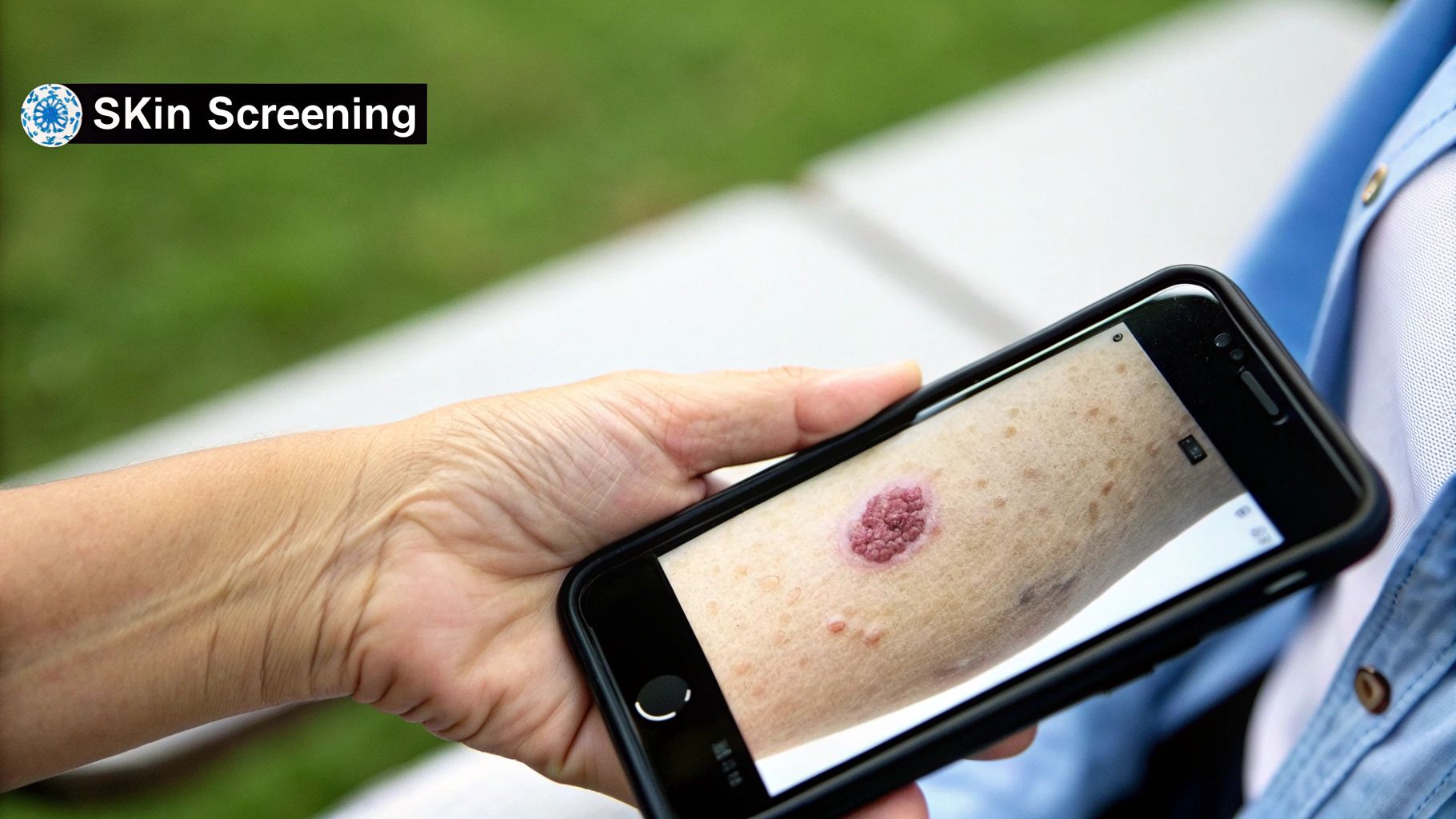
These computer vision systems operate by processing images captured through various sources, including smartphone cameras and specialized dermoscopy devices. Dermoscopy, a non-invasive technique using a handheld device to magnify and illuminate the skin, allows for detailed visualization of skin lesions. When combined with computer vision, dermoscopy images can be analyzed for specific patterns and features indicative of malignancy. The algorithms employed in these systems are trained on extensive datasets of labeled images, enabling them to recognize subtle variations in color, texture, and shape that may escape the human eye. This automated analysis provides objective assessments of skin lesions, assisting dermatologists in making more informed diagnostic and treatment decisions.
Several key features contribute to the effectiveness of computer vision in dermatology: automated lesion detection and segmentation precisely identify and isolate areas of interest within an image; melanoma risk assessment scoring provides a quantitative measure of malignancy risk, aiding in prioritization of suspicious lesions; comparison with dermatological databases allows for cross-referencing with known cases, enhancing diagnostic accuracy; temporal tracking of skin changes monitors lesion evolution over time, providing valuable insights into disease progression; and integration with smartphone cameras increases accessibility and facilitates patient self-monitoring.
Examples of successful implementations of this technology include the SkinVision app, which allows users to capture images of their skin lesions and receive a risk assessment for melanoma. DermEngine, developed by MetaOptima, provides AI-powered dermoscopy image analysis for healthcare professionals. 3Gen integrates its DermLite camera with computer vision algorithms for enhanced lesion visualization and analysis. MoleMap offers a comprehensive mole tracking system, facilitating early detection and intervention for skin cancer.
Pros of using computer vision in dermatology and skin cancer detection:
- Early detection of skin cancer: Enables timely diagnosis and treatment, leading to improved patient outcomes.
- Accessible screening for remote areas: Extends the reach of dermatological expertise to underserved populations.
- Cost-effective mass screening: Offers a scalable and affordable solution for population-level skin cancer screening.
- Patient self-monitoring capabilities: Empowers patients to take an active role in their skin health.
- Reduced need for unnecessary biopsies: Minimizes invasive procedures and associated costs and patient discomfort.
Cons and limitations to consider:
- Variable image quality from smartphones: Can affect the accuracy of analysis and requires careful image acquisition.
- Limited effectiveness on darker skin tones: Existing algorithms may exhibit bias due to underrepresentation of diverse skin tones in training datasets. Active research and development are addressing this critical limitation.
- Cannot replace professional dermatological exam: Serves as a valuable tool to support, but not replace, clinical examination by a dermatologist.
- Potential for patient anxiety from false alarms: Requires careful communication of results and appropriate disclaimers to manage patient expectations.
- Regulatory challenges for consumer apps: Navigating regulatory landscapes for medical devices and software requires careful consideration.
Tips for effective implementation:
- Use consistent lighting and image quality: Standardized image acquisition protocols improve the reliability of analysis.
- Combine with regular dermatologist visits: Computer vision should complement, not replace, professional dermatological examinations.
- Train on diverse skin tone datasets: Addressing algorithmic bias requires inclusive training data.
- Implement clear user guidance for image capture: Clear instructions improve image quality and consistency.
- Provide appropriate disclaimers about limitations: Transparency about the technology’s capabilities and limitations manages patient expectations.
The convergence of computer vision and dermatology holds immense potential to transform skin cancer detection and management. By addressing current limitations and continuing to refine these technologies, we can further enhance the accuracy, accessibility, and effectiveness of skin cancer screening and improve patient outcomes worldwide. This application of computer vision in healthcare exemplifies the innovative power of technology to address pressing medical needs.
5. Ophthalmology and Retinal Disease Detection
Computer vision in healthcare is revolutionizing various medical specialties, and ophthalmology stands as a prime example. This technology offers powerful tools for detecting and monitoring a range of retinal diseases, contributing significantly to preventative care and improved patient outcomes. This section explores how computer vision is transforming ophthalmology and retinal disease detection, detailing its functionalities, benefits, limitations, and implementation considerations.
Computer vision algorithms, trained on vast datasets of retinal images, can analyze photographs and Optical Coherence Tomography (OCT) scans to detect subtle changes indicative of diseases like diabetic retinopathy, age-related macular degeneration, glaucoma, and other eye conditions. These systems work by identifying patterns and anomalies within the images, often imperceptible to the human eye, enabling early detection and timely intervention. The process typically involves several stages: image acquisition, pre-processing to enhance image quality, feature extraction to identify relevant characteristics, and classification based on the learned patterns.
How it Works:
The core of this technology lies in its ability to analyze medical images. Fundus photography, a common ophthalmic imaging technique, captures images of the retina, optic disc, and blood vessels. Computer vision algorithms analyze these images, searching for signs of abnormalities like microaneurysms, hemorrhages, and exudates, which can indicate diabetic retinopathy. Similarly, OCT scans provide cross-sectional images of the retina, allowing the algorithms to assess retinal thickness and identify structural changes associated with macular degeneration and glaucoma. The algorithms are trained using thousands of labeled images, enabling them to differentiate between healthy and diseased retinas with remarkable accuracy.
Successful Implementations:
Several real-world applications highlight the impact of computer vision in ophthalmology:
- Google's AI for Diabetic Retinopathy Screening: Google has developed an AI system that can detect diabetic retinopathy with accuracy comparable to ophthalmologists. This system has been deployed in several countries, particularly in areas with limited access to specialists.
- IDx-DR: This FDA-approved autonomous AI diagnostic system analyzes retinal images and provides a diagnosis of diabetic retinopathy without requiring clinician oversight, enabling wider access to screening.
- Topcon's Harmony AI platform: This platform integrates AI-powered image analysis tools for various ophthalmic conditions, streamlining workflow and enhancing diagnostic capabilities.
- Nidek's retinal analysis software: This software uses AI to analyze retinal images and OCT scans, providing detailed reports that assist ophthalmologists in diagnosis and treatment planning.
Features and Benefits:
- Automated Fundus Photography Analysis and OCT Scan Interpretation: Reduces the manual workload on ophthalmologists and accelerates the diagnostic process.
- Diabetic Retinopathy Grading and Glaucoma Progression Monitoring: Enables objective and consistent assessment of disease severity and progression.
- Integration with Electronic Health Records: Facilitates seamless data sharing and improves patient management.
- Early Detection Prevents Vision Loss: By identifying diseases in their early stages, computer vision enables timely intervention, preserving vision and improving patient quality of life.
- Scalable Screening Programs: Cost-effective screening solutions can be implemented in underserved areas, reaching a larger population and improving public health outcomes.
- Reduced Need for Specialist Referrals: AI-powered systems can triage patients, reducing the burden on specialists and prioritizing those requiring immediate attention.
Pros and Cons:
Pros: Early detection, scalable screening, reduced specialist referrals, objective disease grading, cost-effective population health screening.
Cons: Requires high-quality imaging equipment, performance limitations on poor-quality images, need for ophthalmologist oversight (in some cases), challenges with image standardization, potential for technology dependence.
Actionable Tips for Implementation:
- Ensure Proper Camera Calibration and Maintenance: High-quality images are crucial for accurate analysis.
- Train Technicians on Image Quality Standards: Consistent image acquisition protocols are essential for reliable results.
- Establish Clear Referral Pathways: Develop protocols for referring patients identified with potential diseases for further evaluation and treatment.
- Monitor System Performance Regularly: Ongoing monitoring ensures the accuracy and reliability of the AI system.
- Integrate with Existing Workflow Systems: Seamless integration with existing EHR systems optimizes workflow and data management.
Computer vision in ophthalmology is transforming eye care, providing powerful tools for early detection, diagnosis, and monitoring of retinal diseases. By leveraging this technology, healthcare providers can improve patient outcomes, enhance accessibility to care, and contribute to more effective population health management. While challenges remain, the continued development and refinement of computer vision algorithms promise even greater advancements in ophthalmology and beyond.
6. Patient Monitoring and Vital Signs Detection
Computer vision in healthcare is revolutionizing patient care, and one of its most impactful applications is in patient monitoring and vital signs detection. This technology leverages cameras and sensors, coupled with sophisticated algorithms, to continuously monitor patients and detect subtle changes in their condition, offering a contactless and often more comprehensive approach than traditional methods. This makes it a crucial component of the evolving landscape of healthcare technology.
How it Works:
Computer vision systems for patient monitoring work by analyzing visual data captured by cameras. This data can range from simple movements and posture changes to subtle variations in skin color and even micro-movements that are imperceptible to the human eye. Advanced algorithms process these visual cues to extract vital signs like heart rate, respiratory rate, and oxygen saturation. This contactless approach eliminates the need for cumbersome wires and sensors attached to the patient, significantly improving comfort and reducing the risk of skin irritation, particularly beneficial for long-term monitoring. Furthermore, some systems can detect and analyze facial expressions to assess pain levels, offering a valuable tool for managing patient discomfort, especially for those unable to communicate verbally. The data collected is then relayed to healthcare providers in real-time, allowing for immediate intervention if necessary.
Successful Implementations:
Several companies have pioneered this technology, demonstrating its effectiveness in various healthcare settings. EarlySense, for example, offers a contactless monitoring platform that tracks heart rate, respiratory rate, and movement in hospital beds and at home. Philips Healthcare’s IntelliVue patient monitoring system integrates computer vision to enhance its comprehensive suite of patient monitoring tools. GE Healthcare also utilizes video-based monitoring for patient observation and fall detection. SafelyYou focuses specifically on fall prevention in elderly care facilities, employing computer vision to analyze video feeds and alert staff to potential falls. These real-world applications showcase the versatility and potential of computer vision in improving patient outcomes and safety.
Actionable Tips for Implementation:
- Address Privacy Concerns Proactively: Transparency and clear communication with patients about data collection and usage are crucial. Implement strict data security protocols and ensure compliance with relevant regulations, such as HIPAA.
- Calibrate Systems for Different Environments: Lighting conditions, camera angles, and patient characteristics can affect accuracy. Regular calibration and validation are essential to maintain optimal performance.
- Train Staff on Alert Interpretation: Healthcare providers need to understand how to interpret the data and alerts generated by the system. Comprehensive training is vital to ensure appropriate responses and prevent alarm fatigue.
- Implement Gradual Deployment Strategies: Introduce the technology incrementally, starting with pilot programs in specific units or patient populations. This allows for iterative feedback and adjustments before widespread deployment.
- Maintain Backup Monitoring Methods: While computer vision offers significant advantages, relying solely on it can be risky. Maintain traditional monitoring methods as a backup in case of technical issues or limitations of the computer vision system.
When and Why to Use This Approach:
Computer vision-based patient monitoring is particularly valuable in situations requiring continuous observation, such as:
- ICUs: Closely monitoring critically ill patients for subtle changes in vital signs can be life-saving.
- Elderly Care Facilities: Fall detection and prevention are critical for this vulnerable population. Computer vision can significantly reduce fall-related injuries.
- Post-Surgical Care: Monitoring patients remotely after surgery can improve recovery and reduce hospital readmissions.
- Home Healthcare: Remote monitoring allows patients to remain in the comfort of their homes while still receiving continuous observation.
- Patients Requiring Continuous Observation: Patients with neurological conditions or those at risk of seizures can benefit from constant monitoring without the discomfort of traditional sensors.
Benefits and Drawbacks:
The benefits of computer vision in patient monitoring are numerous, including continuous monitoring without patient discomfort, early detection of patient deterioration, reduced need for physical contact, improved patient safety and fall prevention, and enhanced staff efficiency. However, there are also challenges. Privacy concerns related to camera monitoring must be addressed carefully. Environmental factors can affect the accuracy of the system, and performance may be limited in low-light conditions. The potential for false alarms also exists, and patient acceptance and comfort with being monitored by cameras need to be considered.
Computer vision in patient monitoring represents a significant advancement in healthcare technology. By offering continuous, contactless monitoring, it empowers healthcare providers to deliver more proactive and personalized care, ultimately leading to improved patient outcomes and a safer healthcare environment. As technology continues to evolve and mature, computer vision will undoubtedly play an increasingly important role in shaping the future of healthcare.
7. Drug Discovery and Development
Computer vision is revolutionizing healthcare, and its impact on drug discovery and development is particularly profound. This cutting-edge technology accelerates pharmaceutical research by enabling scientists to analyze cellular images, protein structures, and drug interactions at the molecular level with unprecedented speed and accuracy. This application of computer vision in healthcare significantly reduces the time and cost associated with traditional drug development processes while simultaneously increasing the likelihood of identifying successful drug candidates.
Traditional drug discovery is a laborious and expensive process, often taking years and billions of dollars to bring a single new drug to market. Computer vision offers a powerful alternative by automating many of the traditionally manual tasks involved. High-content screening analysis, a key feature of this technology, allows researchers to rapidly analyze thousands of cells treated with different compounds, identifying potential drug candidates with much greater efficiency. By automating this process, computer vision drastically reduces the time required for initial screening and allows researchers to focus on the most promising leads.
Furthermore, computer vision facilitates a deeper understanding of protein structures, which are crucial for drug development. Specialized software can visualize and analyze complex 3D protein structures, enabling researchers to identify potential drug binding sites and predict drug efficacy. This ability to understand drug-target interactions at a molecular level is critical for designing effective therapies. Tools like DeepMind's AlphaFold, for instance, have demonstrated remarkable success in predicting protein structures, opening up new possibilities for drug design and development.
Computer vision also excels at assessing cell morphology and behavior. By analyzing microscopic images of cells, these systems can detect subtle changes in cell shape, size, and movement in response to different drug treatments. This allows for a more nuanced understanding of drug mechanisms and potential side effects. This capability is particularly useful in areas like oncology, where understanding how cancer cells respond to treatment is crucial for developing effective therapies.
Several companies are pioneering the use of computer vision in drug discovery. Atomwise, for instance, utilizes its AI drug discovery platform to identify potential drug candidates for a variety of diseases. BenevolentAI leverages AI to analyze vast amounts of biomedical data, accelerating drug development. Insilico Medicine focuses on molecular design using AI and deep learning, while Recursion Pharmaceuticals employs cellular analysis powered by computer vision to discover new drugs. These examples demonstrate the transformative potential of computer vision in healthcare and specifically in pharmaceutical research.
Pros of using computer vision in drug discovery:
- Accelerated drug discovery timelines: Automating tasks like high-throughput screening significantly reduces the time it takes to identify promising drug candidates.
- Reduced research and development costs: By streamlining the discovery process, computer vision minimizes the need for expensive and time-consuming laboratory experiments.
- Improved hit identification rates: The enhanced analysis capabilities of computer vision increase the likelihood of finding effective drug candidates.
- Enhanced understanding of drug mechanisms: Analyzing cellular images and protein structures provides valuable insights into how drugs interact with their targets.
- Automated analysis of large datasets: Computer vision systems can efficiently process massive amounts of data, enabling researchers to analyze complex datasets that would be impossible to analyze manually.
Cons of using computer vision in drug discovery:
- High computational requirements: Processing large image datasets and complex algorithms requires significant computing power.
- Complex data interpretation challenges: Analyzing the vast amount of data generated by computer vision systems can be challenging and requires specialized expertise.
- Need for specialized expertise: Implementing and utilizing computer vision systems effectively requires skilled personnel with expertise in both computer science and biology.
- Validation requirements for regulatory approval: Rigorous validation is necessary to ensure the accuracy and reliability of computer vision-based findings for regulatory approval.
- Limited applicability to some drug types: While promising, computer vision approaches may not be applicable to all types of drug discovery, particularly those involving complex biological systems.
Tips for implementing computer vision in drug discovery:
- Invest in high-quality imaging infrastructure: High-resolution images are essential for accurate analysis.
- Collaborate with AI and computer vision experts: Partnering with specialists ensures the effective implementation and utilization of these technologies.
- Establish robust data management systems: Efficient data management is crucial for handling the large datasets generated by computer vision systems.
- Validate findings through traditional methods: Confirming results using established experimental techniques is essential for building confidence in the findings.
- Consider partnerships with tech companies: Collaborating with technology companies specializing in computer vision can provide access to cutting-edge tools and expertise.
Computer vision is poised to transform drug discovery and development, offering a faster, more efficient, and cost-effective approach to identifying and developing new therapies. For organizations involved in pharmaceutical research, embracing computer vision in healthcare is no longer a luxury but a necessity for remaining competitive and bringing life-saving drugs to market faster.
8. Medical Documentation and Workflow Automation
Computer vision in healthcare is revolutionizing numerous aspects of the industry, and one area where its impact is particularly profound is medical documentation and workflow automation. This application of computer vision tackles the traditionally labor-intensive and error-prone processes of documentation, coding, billing, and even physical asset tracking, freeing up valuable time for healthcare professionals to focus on patient care. This technology deserves its place on this list due to its potential to significantly improve efficiency, accuracy, and ultimately, patient outcomes.
This innovative approach leverages computer vision algorithms to interpret and extract information from various medical data sources. These sources range from structured data like printed forms and prescriptions to unstructured data like handwritten clinical notes and medical images. Optical Character Recognition (OCR) plays a key role, allowing the system to "read" and digitize text, converting it into machine-readable formats for further processing and analysis. Beyond simple text extraction, computer vision can also interpret more complex visual information, such as identifying specific medical instruments in images or analyzing patient flow patterns within a hospital.
How it works:
The core of medical documentation automation lies in training computer vision models to recognize and understand the relevant information within medical documents. This training involves feeding the system a vast dataset of labeled examples, teaching it to identify key elements like patient names, diagnoses, medications, and other crucial details. Once trained, the system can accurately extract this information from new, unseen documents, significantly reducing the need for manual data entry.
Workflow automation expands on this foundation by integrating these extracted data points into existing hospital systems. For instance, information extracted from a patient's admission form can automatically populate electronic health records (EHRs), streamlining the admission process and eliminating redundant data entry. Similarly, computer vision can track the location and usage of medical equipment, optimizing resource allocation and minimizing downtime.
Examples of Successful Implementation:
Several leading healthcare technology companies have already implemented computer vision for documentation and workflow automation:
- Epic's AI-powered documentation tools: Epic Systems, a major EHR provider, utilizes computer vision to assist with tasks like automated chart review and coding suggestions.
- Cerner's workflow optimization systems: Cerner Corporation offers solutions that use computer vision to analyze patient flow patterns, helping hospitals optimize staffing and resource allocation.
- Nuance's Dragon Medical speech recognition: While not strictly computer vision, Nuance's speech recognition software complements computer vision systems by allowing clinicians to dictate notes directly into the EHR, which can then be further processed by computer vision algorithms.
- Zebra Technologies' healthcare asset tracking: Zebra Technologies provides solutions that leverage computer vision and RFID tags to track medical equipment and supplies, ensuring efficient inventory management.
Features and Benefits:
- Optical character recognition for medical documents: Digitizes handwritten and printed text for seamless integration with EHRs.
- Prescription and form digitization: Automates data entry and reduces errors in medication management.
- Equipment and asset tracking: Improves resource utilization and minimizes equipment downtime.
- Patient flow analysis and optimization: Enhances operational efficiency and patient experience.
- Automated billing and coding assistance: Reduces administrative burden and improves accuracy.
Pros:
- Reduced administrative burden on staff
- Improved accuracy in documentation
- Enhanced operational efficiency
- Better resource allocation and planning
- Reduced manual data entry errors
Cons:
- Challenges with handwritten text recognition (especially poor handwriting)
- Need for system integration with existing hospital infrastructure
- Initial setup and training requirements for the computer vision models
- Potential resistance to workflow changes from staff
- Data security and compliance concerns necessitate robust security measures
Tips for Implementation:
- Start with high-volume, repetitive tasks: Target areas where automation can provide the most immediate impact.
- Ensure compliance with healthcare regulations: Adhere to HIPAA and other relevant data privacy regulations.
- Provide comprehensive staff training: Ensure staff are comfortable using the new systems and understand the benefits.
- Implement gradual rollout strategies: Avoid overwhelming staff with sudden changes; introduce the technology incrementally.
- Monitor and optimize system performance: Continuously evaluate the system's effectiveness and make adjustments as needed.
When considering implementing computer vision for medical documentation and workflow automation, it's crucial to carefully evaluate the specific needs of your organization. This technology offers a powerful tool to streamline processes, improve accuracy, and enhance efficiency, ultimately leading to better patient care and optimized resource allocation. By carefully addressing the potential challenges and following best practices for implementation, healthcare providers can fully realize the transformative potential of computer vision in this crucial area.
Comparative Overview of 8 Healthcare CV Applications
| Application Area | Implementation Complexity 🔄 | Resource Requirements ⚡ | Expected Outcomes 📊 | Ideal Use Cases 💡 | Key Advantages ⭐ |
|---|---|---|---|---|---|
| Medical Imaging Analysis and Diagnosis | High: deep learning, regulatory hurdles | High: extensive training data, costly setup | Increased diagnostic accuracy, faster reporting | Radiology departments, emergency diagnostics | Early disease detection, reduced workload |
| Surgical Navigation and Robotics | Very High: real-time tracking, robotics integration | Very High: expensive equipment, maintenance | Enhanced surgical precision, reduced recovery time | Complex surgeries needing precision guidance | Minimally invasive procedures, improved training |
| Pathology and Histopathology Analysis | High: digitization and ML model training | Moderate: quality imaging and storage | Faster, consistent tissue analysis, reduced errors | Cancer diagnosis, lab pathology workflows | Objective grading, remote consultations |
| Dermatology and Skin Cancer Detection | Moderate: smartphone integration, image variability | Moderate: dermoscopy devices or smartphone cameras | Early detection, accessible screening | Remote screening, patient self-monitoring | Cost-effective, reduces unnecessary biopsies |
| Ophthalmology and Retinal Disease Detection | Moderate-High: imaging standards and AI grading | Moderate: specialized imaging equipment | Early detection of retinal diseases, scalable screening | Population health screening, remote diagnostics | Cost savings, reduces specialist referrals |
| Patient Monitoring and Vital Signs Detection | Moderate: sensor calibration, privacy management | Moderate: cameras, sensors, IT infrastructure | Continuous monitoring, early deterioration alerts | ICU, elderly care, fall risk management | Contactless, improves patient safety |
| Drug Discovery and Development | High: complex molecular analysis, data handling | High: computational power, specialized expertise | Accelerated drug discovery, cost reductions | Pharmaceutical R&D, compound screening | Faster timelines, improved hit identification |
| Medical Documentation and Workflow Automation | Moderate: OCR and system integration challenges | Moderate: software and staff training | Reduced admin time, improved accuracy | Hospital admin, billing, inventory management | Efficiency gains, reduced manual errors |
Embracing the Vision: Computer Vision's Impact on Healthcare's Future
From revolutionizing medical imaging analysis and diagnosis to streamlining surgical navigation and robotics, the transformative potential of computer vision in healthcare is undeniable. This article explored eight key applications, including pathology analysis, dermatological and ophthalmological diagnostics, patient monitoring, drug discovery, and even workflow automation. These advancements are not merely incremental improvements, but represent a paradigm shift towards more efficient, effective, and patient-centered care. Mastering these applications of computer vision empowers healthcare professionals to enhance diagnostic accuracy, personalize treatment plans, and accelerate research, ultimately leading to improved patient outcomes and a more sustainable healthcare system. As we move forward, the convergence of human expertise and artificial intelligence through computer vision will continue to redefine healthcare delivery. The future promises even more sophisticated applications, building upon the foundations laid by the innovations we see today.
Looking to implement cutting-edge computer vision solutions in your healthcare projects? PYCAD offers advanced tools and expertise to help you leverage the power of computer vision. Visit PYCAD to explore how we can assist you in developing innovative solutions for a healthier future.


