
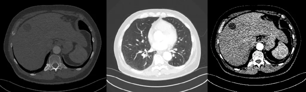
CT window settings in this page are the two numbers you pass to a converter: window center (WC) and window width (WW). The job is to apply them to a DICOM CT series in Python so a liver, lung, tumor, or bone window is a file you can open — not a slider lecture.
If you meant WW vs WL as a concept → window width vs window level. If you meant the HU scale → what Hounsfield units are. If you meant windowing as a preprocess step before a model → what is image preprocessing.
This URL is also the landing for the leftover /dicommriwindowing/ and /niftictwindowing/ paths. Same apply-windowing job. Not a new post.
WC and WW
Window center (WC) is the midpoint of the HU range you display. Raise it and the image goes darker; lower it and the image goes brighter. Same slider 635 calls window level.
Window Width (WW) is the width of that range. Narrow WW = high contrast (tissues near the center separate). Wide WW = more greys at once (survey view, less punch).
A monitor shows about 256 greys. A CT stores a few thousand HU. Windowing is the clip that makes one organ readable. The numbers themselves live on the Hounsfield scale.
Apply it in Python
The converter is DicomCTWindowing from pycad-medic. Point it at a DICOM series folder. It writes a windowed output.
from pycad.preprocessing import DicomCTWindowing

dicom_dir = "path/to/dicom/series"
output_dir = "path/to/output"
window_center = 40
window_width = 400
converter = DicomCTWindowing(window_center, window_width, visualize=True)
converter.convert(dicom_dir, output_dir)
window_center and window_width are the two knobs. visualize=True opens a preview so you can see the preset before you batch. Change the pair, re-run. That is the whole how-to.
Presets already in the examples
| Target | WC (HU) | WW (HU) | Why that pair |
|---|---|---|---|
| Soft-tissue default (script above) | +40 | 400 | Midpoint on soft tissue; a usable first pass |
| Liver | +60 | 150 | Narrow width so liver texture and a lesion in it separate |
| Lungs | −600 to −700 | 1500 to 2000 | Center on air-filled lung; wide enough for vessels and a nodule |
| Tumor (against nearby soft tissue) | Slightly above the host organ | Moderate | Raise WC so the mass is not the same grey as the bed it sits in |
| Bone | +400 to +700 | 1500 to 2000 | Center on cortex; wide enough for marrow vs cortex |
These numbers are the ones the original examples used. They are starting points, not a protocol Bible. A PE study still wants the lung pair; a hairline fracture still wants the bone pair. If you need the clinical why for those three named windows, that is window width vs window level.
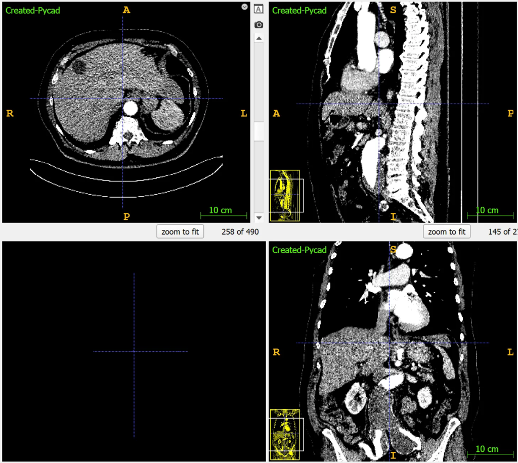
Liver
WC about +60 HU, WW 150 HU. Liver texture and a density change inside it show up; a wide soft-tissue window washes that out.
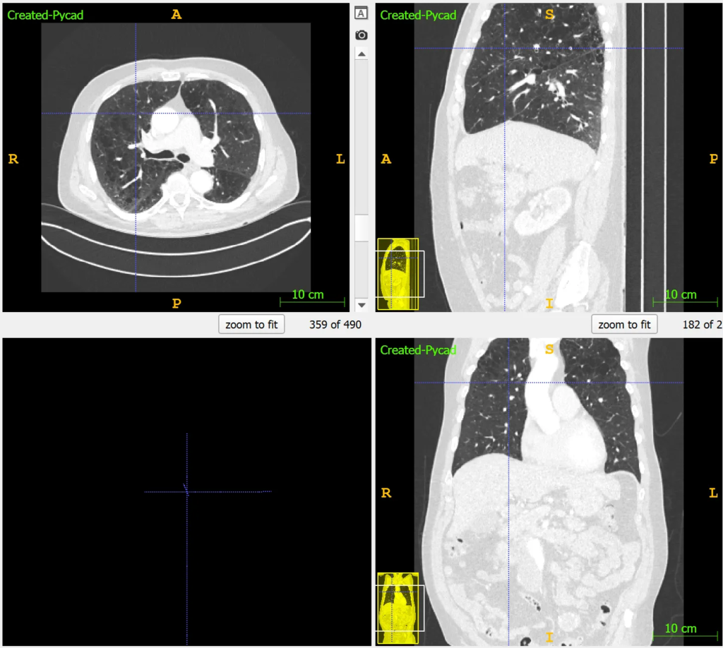
Lungs
WC −600 to −700 HU, WW 1500 to 2000 HU. Air is the bed; vessels and a nodule stay visible because the width is wide.
Tumors
Location decides the pair. A slightly higher WC than the host organ, and a moderate WW, is the usual move so the mass is not the same grey as the tissue around it.
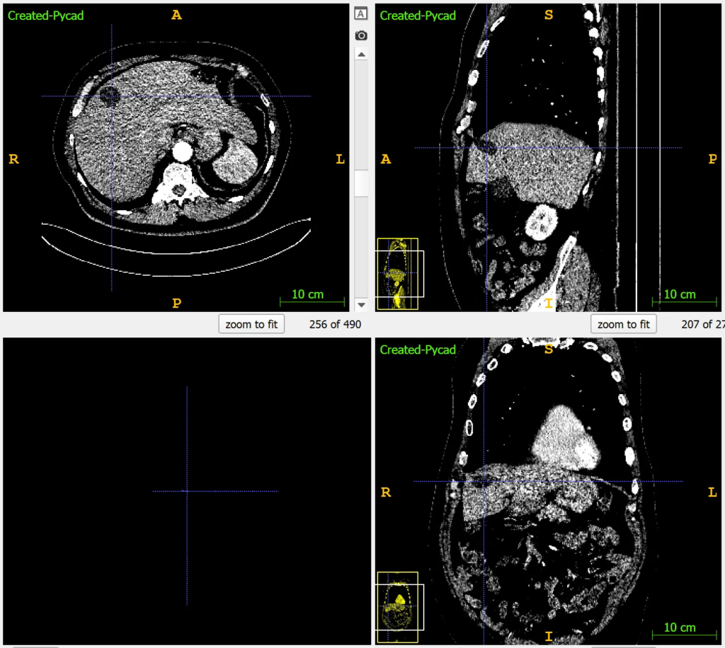
Bones
WC +400 to +700 HU, WW 1500 to 2000 HU. Cortex stays bright; a fracture line is a dark break, not a smear.
What this page is not
It is not a comparison of the two sliders — that is 635. It is not the HU table — that is 622. MRI intensity has no Hounsfield scale; do not paste these presets onto an MRI series and expect the same picture.
PYCAD builds the imaging side of products that have to window a series for real. Case studies.