Skip to content
  • Home
  • Services
    • All Services
    • Custom DICOM Viewer
    • Case Studies
  • Products
    • All Products
    • PYCAD DICOM Viewer
    • PYCAD Dental Viewer
    • PYCAD SSR Engine
  • Case Studies
  • Blog
  • About us
  • Home
  • Services
    • All Services
    • Custom DICOM Viewer
    • Case Studies
  • Products
    • All Products
    • PYCAD DICOM Viewer
    • PYCAD Dental Viewer
    • PYCAD SSR Engine
  • Case Studies
  • Blog
  • About us
Get in Touch

Category: machine learning

KIR-NO: 3D MRI Reconstruction Trained Small, Deployed Large
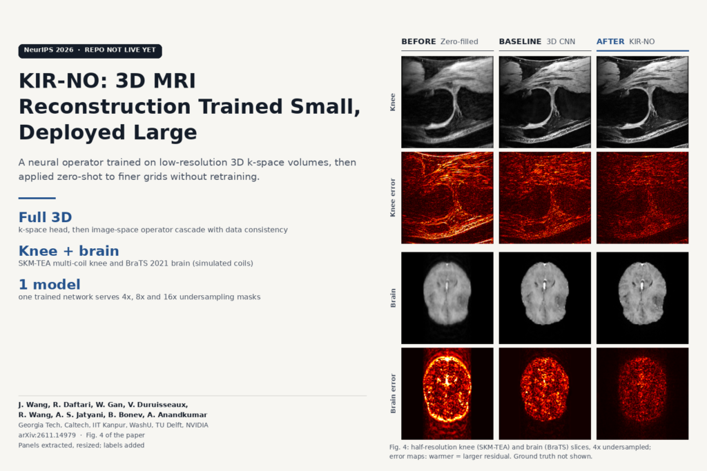
Fig. 4 of arXiv:2611.14979 by Wang, Daftari and colleagues: half-resolution knee and brain MRI slices reconstructed by zero-filling, a 3D CNN and KIR-NO, each with an error map, the KIR-NO error maps showing the smallest residuals.

A neural operator trains full 3D MRI recon on downsampled k-space, then runs the same weights on finer grids. Repo not public yet.

N2N-iMF: Fast Self-Supervised CT Denoising Without Clean Scans
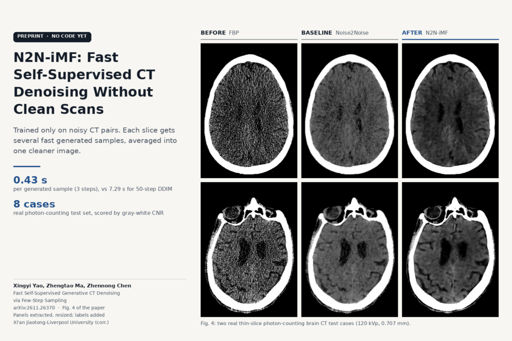
Fig. 4 of arXiv:2611.26370 by Yao, Ma and Chen: two real thin-slice photon-counting brain CT slices shown as noisy FBP, Noise2Noise output and N2N-iMF output, with the N2N-iMF images less grainy and cortical sulci still visible.

Trained on noisy CT pairs only, it averages fast generated samples per slice. Tested on real photon-counting brain CT. No code yet.

Geometry-Bridge Flow Matching: Fixing Head Motion in CBCT
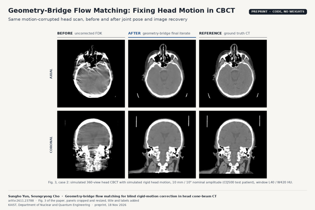
Fig. 3, case 2 of arXiv:2611.23788 by Yun and Cho: axial and coronal head CBCT slices of a simulated motion-corrupted scan, uncorrected FDK with streaks and doubled bone edges, the geometry-bridge final reconstruction, and the ground truth CT.

A KAIST method recovers head pose for every CBCT projection from the raw data, then rebuilds the scan. Code is public; weights are not.

VoxForge: One Frozen 3D Encoder for MRI, CT and PET
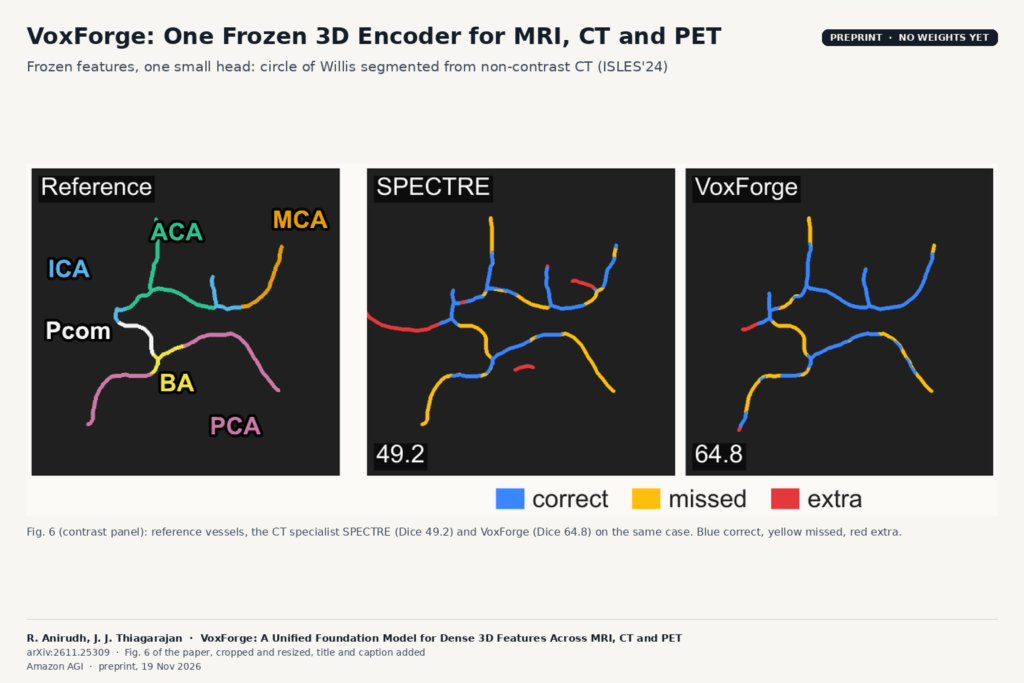
Fig. 6 of arXiv:2611.25309 by Anirudh and Thiagarajan: reference circle of Willis vessels, the SPECTRE result (Dice 49.2) and the VoxForge result (Dice 64.8) segmented from non-contrast CT, with correct, missed and extra vessel segments colour coded.

Amazon AGI’s 388M-parameter ViT, trained on 235K commercial-use volumes, segments, classifies and registers scans without fine-tuning.

MedImgAnnotator: Batch Annotation for Large CT and MRI Cohorts
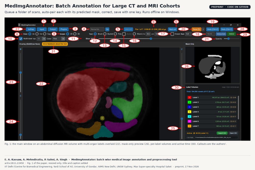
Fig. 1 of arXiv:2611.22450 by Kassaw et al.: the MedImgAnnotator main window showing an abdominal diffusion MR slice with multi-organ labels overlaid, a mask-only preview and a per-label volume and annotation time panel.

A Windows tool that queues folders of CT and MRI, pairs each scan with its predicted mask, and cuts the time spent per case.

SWIMSS: An Open MS Lesion Segmenter Trained on Seven Swiss Centres
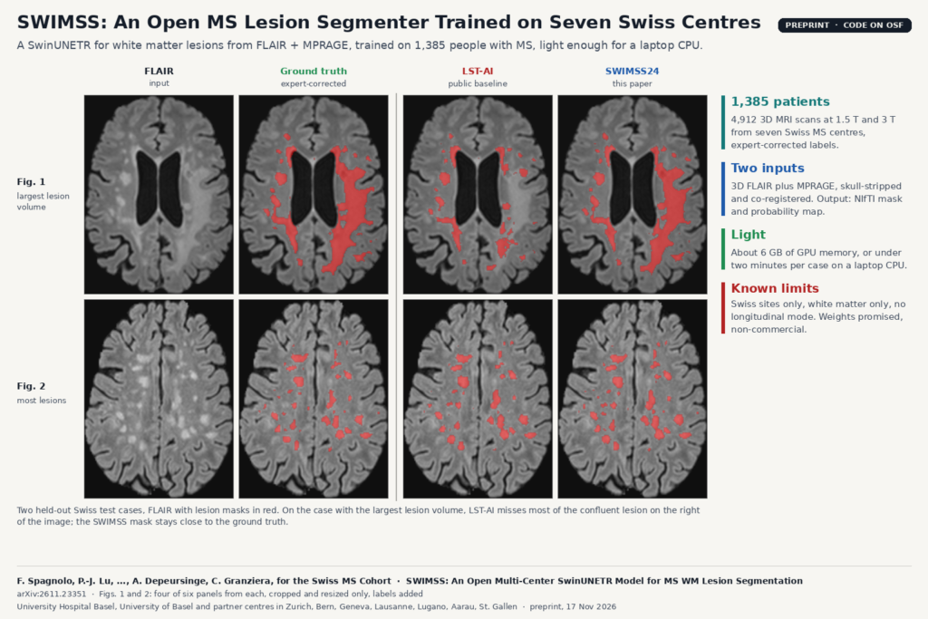
Figs. 1 and 2 of arXiv:2611.23351 by Spagnolo et al.: two axial FLAIR brain MRI test cases with multiple sclerosis, showing the input, the expert-corrected ground-truth lesion mask, the LST-AI mask and the SWIMSS24 mask in red.

A SwinUNETR trained on 1,385 people with MS from seven Swiss centres segments white matter lesions from FLAIR and MPRAGE, even on a CPU.

GRACE: Fixing Segmentation Masks With Retrieved Verified Cases
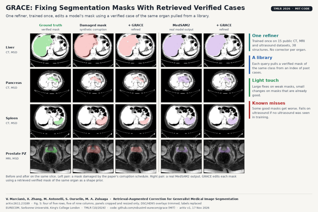
Fig. 5 of arXiv:2611.23189 by Marcianò et al.: liver, pancreas and spleen CT and prostate peripheral zone MRI slices showing the ground-truth mask, a synthetically damaged mask and its GRACE correction, and a MedSAM2 mask and its GRACE correction.

One refiner, trained once on CT, MRI and ultrasound, corrects a model’s mask using a verified case of the same organ from a library.

PathoDraw: One Zero-Shot Lesion Detector for Any Brain MRI
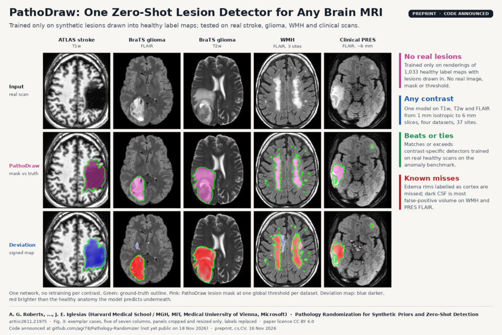
Fig. 3 of arXiv:2611.21975 by Roberts et al.: axial brain MRI from ATLAS T1w, BraTS FLAIR and T2w, WMH FLAIR and a clinical PRES FLAIR scan, with the input image, ground-truth outline in green with the PathoDraw lesion mask in pink, and the signed deviation map in blue and red.

Trained only on synthetic lesions, one network flags stroke, glioma and white matter lesions on T1w, T2w or FLAIR brain MRI.

Plug-and-Play Prior vs Tuned TV: Sparse-View CT Beyond PSNR
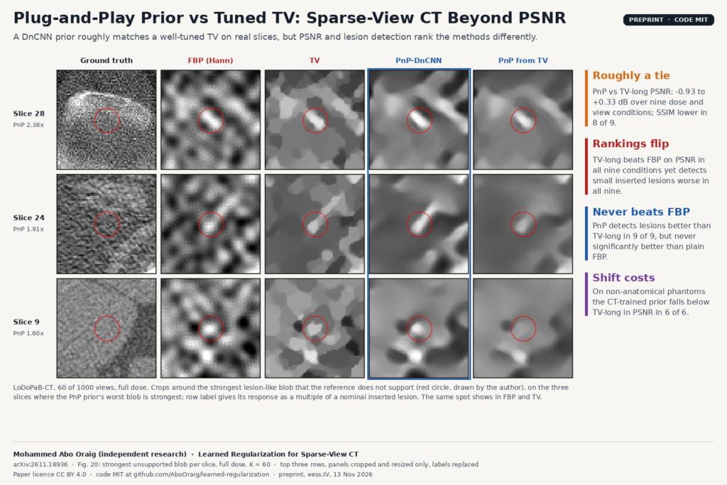
Fig. 20 of arXiv:2611.18936 by Mohammed Abo Oraig: crops of three LoDoPaB-CT slices reconstructed from 60 views at full dose with Hann-filtered FBP, total variation, a Plug-and-Play DnCNN prior and PnP started from TV, red circles marking the strongest lesion-like blob not supported by the reference image.

A DnCNN prior roughly matches a well-tuned TV on real sparse-view CT, but PSNR and lesion detection rank the methods differently.

Scribble Once: Echo Cine Segmentation From One Scribbled Frame
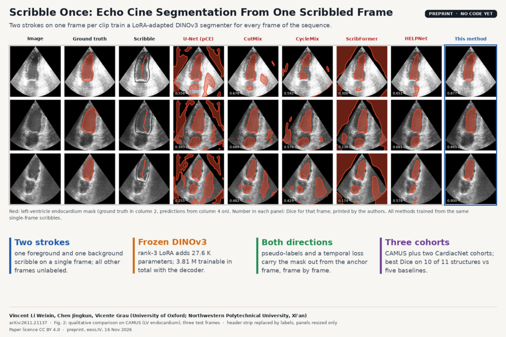
Fig. 2 of arXiv:2611.21137 by Vincent Li Weixin, Chen Jingkun and Vicente Grau (University of Oxford, Northwestern Polytechnical University): CAMUS two-chamber echo frames with left-ventricle endocardium masks from U-Net, CutMix, CycleMix, ScribFormer, HELPNet and the single-scribble method, per-frame Dice printed in each panel.

Two scribbles on one frame per clip train a LoRA-adapted DINOv3 model to segment heart chambers across the whole echo sequence.

Next →