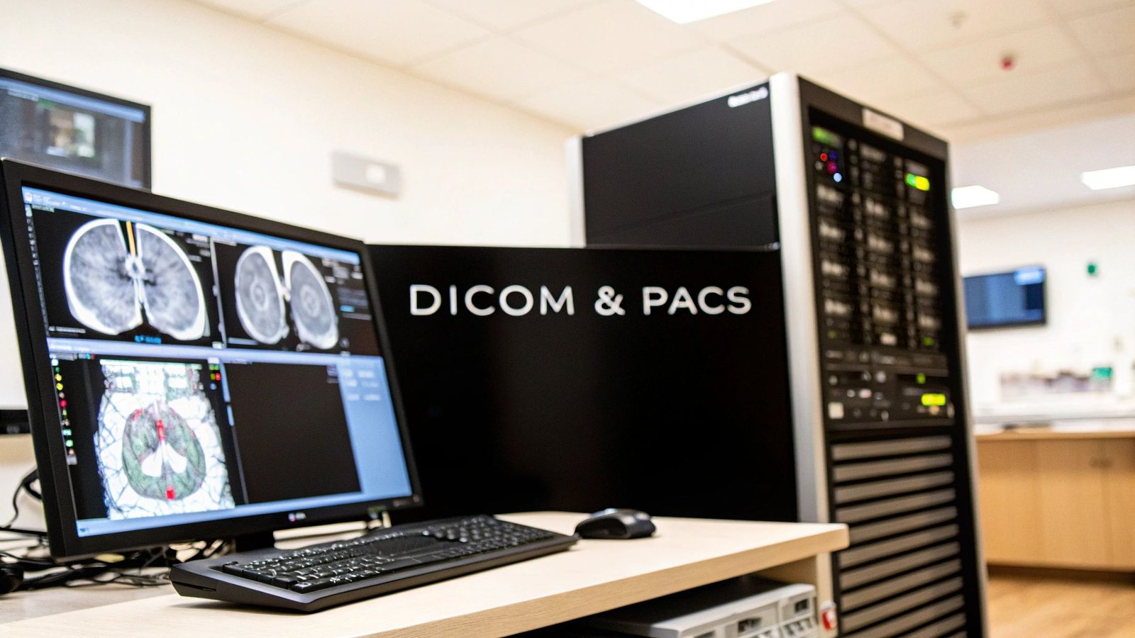
Welcome to the operational core of modern medicine. In here, DICOM and PACS aren't just technical acronyms; they are the digital backbone that makes modern diagnostics possible.
Imagine DICOM as a universal translator, a common language that allows every piece of medical imaging equipment—from MRI machines to CT scanners—to speak to each other perfectly. Now, picture PACS as a vast, incredibly organized digital library, built to securely store, manage, and instantly retrieve every single one of those medical images. One is the language, the other is the library.
The Heartbeat of Modern Medical Imaging
This guide is for the innovators—the medical device creators and healthcare IT leaders who are building the future. We're going to break down everything you need to know, from the basic building blocks of this technology to its most sophisticated uses. Mastering DICOM and PACS isn't just a technical skill; it's the key to unlocking the next generation of patient care.
These two systems are completely intertwined. DICOM standardizes the format for the images and all the critical patient data attached to them. PACS provides the robust infrastructure to handle these enormous datasets. Without this partnership, the journey of a diagnostic image from a scanner in a hospital basement to a specialist’s screen across the country would grind to a halt.
What Makes It All Work?
At its core, the goal is surprisingly straightforward: ensure a patient's X-ray, MRI, or ultrasound is captured, stored, and viewed with perfect accuracy and security. It doesn't matter who made the equipment or where the doctor is located.
This foundational structure is what allows for incredible leaps forward in diagnostics and treatment.
The market for this technology is massive. The global Picture Archiving and Communication Systems (PACS) market hit a valuation of around USD 9.7 billion in 2024. Within that, radiology PACS makes up the largest slice of the pie at 45.8%, which makes sense given it handles the bulk of diagnostic imaging. For a deeper dive into the numbers, you can explore detailed insights about the PACS market to see just how big this field is.
To quickly grasp their distinct roles, let's break it down.
DICOM vs PACS At a Glance
| Concept | Role of DICOM (The Language) | Role of PACS (The Library) |
|---|---|---|
| Primary Function | A universal standard for transmitting, storing, and formatting medical images and related data. | A system for archiving, retrieving, distributing, and presenting medical images. |
| What It Is | A communication protocol and file format. Think of it like a .JPEG or .PDF, but for medicine. | A technology infrastructure, including servers, storage, and workstations. |
| Key Action | Defines how a CT scanner and a radiologist's workstation talk to each other. | Stores the images from that CT scan and makes them available to the workstation. |
| Analogy | The rules of grammar and vocabulary for medical imaging conversations. | The bookshelf, catalog, and retrieval system for all the books written in that language. |
This table highlights the symbiotic nature of the two: DICOM provides the standardized content, and PACS provides the system to manage it all.
The Real Power Is in Connection
The true magic happens when this imaging ecosystem connects with other clinical systems. When you can seamlessly integrate imaging data with a patient's Electronic Health Record (EHR), you give clinicians a complete, holistic view of their patient's health story.
For anyone building medical devices or medtech software, this isn't just a technical footnote—it's a strategic necessity. A tightly integrated system doesn't just make workflows more efficient. It cuts down on diagnostic errors and opens the door for incredible innovations like teleradiology and AI-powered analysis.
At PYCAD, this is exactly what we live and breathe. We build custom web DICOM viewers and integrate them into medical imaging web platforms, creating the very connected solutions that push healthcare forward. You can see some of our work in action by visiting our portfolio page.
Decoding DICOM: The Universal Language of Medical Images
Ever wonder how an image from a GE scanner can pop up flawlessly on a Siemens workstation halfway across the hospital? The magic behind that is DICOM. It’s the invisible handshake, the universal translator that makes sure every piece of imaging equipment speaks the same language.
This isn't just about file types—it’s a dynamic communication protocol that forms the very foundation of modern medical imaging.
Think of a DICOM file like a highly detailed package. The image is the precious cargo inside, but it's the DICOM header—the attached shipping label—that tells you everything you need to know. This header is packed with a goldmine of metadata.
This structured data is what breathes life into a simple image. It includes crucial context like:
- Patient Information: Name, ID, date of birth.
- Study Details: What kind of scan it was (CT, MRI), when it happened, and why.
- Equipment Parameters: Details on the machine used, down to the specific settings.
- Image Specifics: Resolution, orientation, and other technical data.
Without this rich context, a medical image is just a picture. With it, it becomes a complete clinical record, ready for a radiologist to make a diagnosis.
The Rules of the Road for Image Communication
DICOM goes beyond just defining the file structure; it sets the rules for how imaging devices talk to each other. This communication is managed through a set of services that dictate how data gets from point A to point B. In any DICOM and PACS environment, two of the most common commands you'll hear about are C-STORE and C-MOVE.
At its core, DICOM is a promise. It’s a promise that a pixel isn't just a dot of light, but a precise data point, full of context, that a clinician can trust—no matter what machine or software they're using. That trust is everything in diagnostics.
For example, C-STORE is the "push" command. When an MRI machine finishes a scan, it uses C-STORE to send the new DICOM files over to the PACS server for safekeeping.
On the other hand, C-MOVE is the "pull" command. A radiologist at their workstation uses C-MOVE to request a specific patient's studies from the PACS, bringing them to their local machine for review.
DICOM's Impact on the Healthcare Market
This universal standard isn't just a technical nicety; it's a massive economic engine. The market for acquiring and managing DICOM content makes up a huge slice of the North American PACS pie—around 35-40%. Radiology applications are still the king, holding nearly 45% of the market, driven largely by the constant need for imaging in chronic disease management.
This common language is what allows the entire ecosystem to innovate. At PYCAD, for instance, we build custom web DICOM viewers that plug directly into our clients' medical imaging platforms. That entire process hinges on the structured, predictable nature of DICOM to give doctors secure access to images wherever they are. You can check out our work on our portfolio page.
For organizations looking to bridge different systems, exploring tools like DICOM Gateway solutions can be a game-changer. Ultimately, having this shared language empowers companies like ours to build the tools that help clinicians save lives.
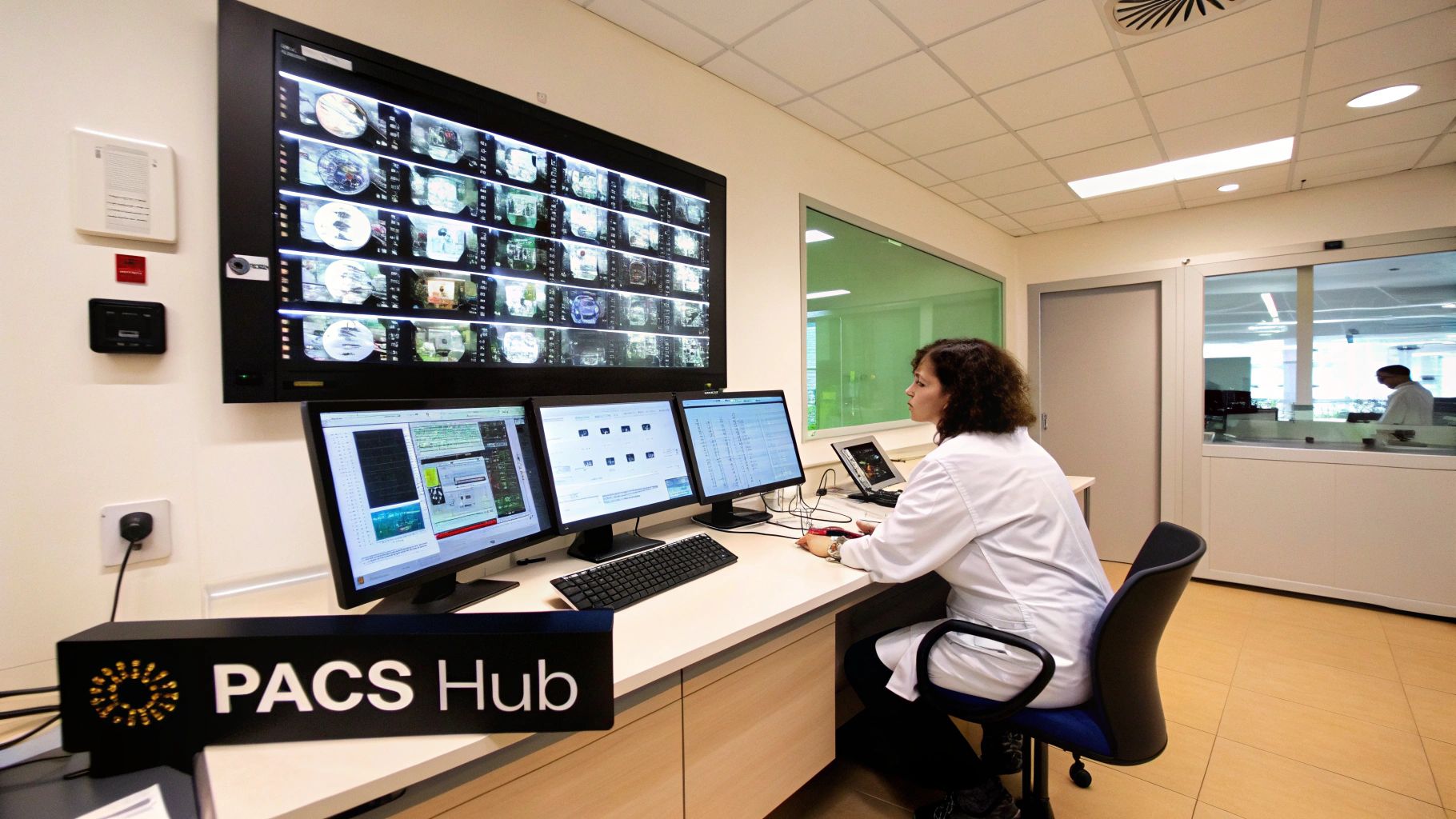
Mastering PACS: The Secure Hub for Medical Images
If DICOM is the universal language of medical imaging, think of a PACS (Picture Archiving and Communication System) as the central nervous system that manages the entire conversation. It's so much more than a digital filing cabinet. It's the operational heart of modern radiology, the command center that orchestrates the entire journey of a medical image, from its creation to the final diagnostic report.
Imagine a patient has just finished an MRI. The moment that scan is complete, the images—packaged neatly in the DICOM format with all their crucial metadata—are whisked away to the PACS. This is where the real work begins. The system doesn't just dump the file into a folder; it intelligently indexes, securely archives, and makes it instantly available to the right clinicians when they need it.
Without a PACS, that life-saving image could easily get lost in a digital haystack. It’s the engine that drives efficiency, collaboration, and ultimately, better patient care.
Tracing the Image Journey Through PACS
To really get a feel for how a DICOM and PACS ecosystem functions, let’s follow that MRI scan through its typical workflow. It's a beautifully coordinated dance, designed for speed and unerring accuracy.
- Acquisition: The MRI machine—what we call a modality in the industry—captures the images. As soon as the study is finished, it sends the DICOM files over a secure network connection straight to the PACS server.
- Archiving: The PACS receives the study and immediately files it away in its archive. Most modern systems are smart about this, using a tiered approach. Recent and frequently needed studies live on lightning-fast storage, while older, less-accessed images are moved to more economical long-term archives.
- Distribution: A radiologist gets an alert: a new study is ready for reading. From their specialized viewing workstation, they send a quick query to the PACS. Almost instantly, the system finds and delivers the requested images.
- Viewing and Reporting: Now the expertise comes in. The radiologist examines the images on a high-resolution diagnostic monitor, using sophisticated tools to zoom, pan, measure, and manipulate the views. They document their findings in a report, which is then linked directly back to the study within the PACS, closing the loop.
This entire sequence, often completed in just a matter of minutes, is a testament to the seamless integration between the imaging equipment and the central PACS hub.
Departmental vs. Enterprise PACS Architectures
Of course, not all PACS are built the same. Their design usually depends on the scale of the healthcare organization they serve, which leads to two primary models.
- Departmental PACS: These systems are built for the unique needs of a single clinical department, like radiology, cardiology, or even ophthalmology. They're fine-tuned for the specific workflows and image types generated by that specialty.
- Enterprise PACS: This is the big-picture approach. A single, unified PACS serves the entire healthcare system, breaking down the traditional data silos between departments. The result is one comprehensive imaging record for every patient.
While departmental systems offer powerful, specialized tools, the industry is clearly shifting toward enterprise solutions. Why? Because they provide a more complete, holistic view of a patient's health journey.
A well-designed PACS doesn't just manage data; it unlocks potential. By centralizing imaging information, it provides the foundation for advanced analytics, enterprise-wide collaboration, and the integration of powerful new technologies like AI.
The Rise of Cloud-Based PACS
For years, PACS meant on-premise systems—huge investments in servers, storage hardware, and the IT staff needed to maintain it all. But the cloud has completely changed the game. Cloud-based PACS deliver incredible advantages in scalability, accessibility, and cost-effectiveness. Instead of buying and managing physical servers, a hospital can tap into cloud infrastructure that grows and shrinks with their needs.
This shift is more than just a trend; it's essential for enabling modern healthcare. It powers things like teleradiology and remote consultations, allowing specialists to contribute from anywhere.
This forward-thinking approach is exactly where we at PYCAD live and breathe. We build custom web DICOM viewers and integrate them into medical imaging web platforms, giving clinicians secure, browser-based access to patient images from anywhere in the world. This kind of flexibility isn't a luxury anymore—it's a necessity. To see how we empower healthcare innovators, check out some of our projects on our portfolio page.
Unlocking Access with Custom Web DICOM Viewers
For decades, traditional PACS workstations have been the command centers of radiology. They're powerful, specialized, and reliable—but they're also typically anchored to a desk inside the hospital. The future, however, is all about collaboration and flexibility, and that requires a much more agile way of working. This is where web-based DICOM viewers are making a massive impact.
These modern viewers tear down the hospital walls. By giving clinicians secure access to medical images from any device with a web browser, they open up a world of possibilities. It’s a "zero-footprint" approach, which means no software to install or maintain on the user's computer. That’s a huge win for IT departments and makes access instant and universal.
A New Era of Collaboration
This technology is what makes modern teleradiology possible, letting specialists read scans from anywhere on the globe. It completely changes how multi-disciplinary teams meet—now, surgeons, oncologists, and radiologists can review the exact same images together, in real-time, from entirely different locations.
The benefits even trickle down to the patient. Providers can now build patient portals that grant people secure access to their own imaging studies. This small step empowers patients to become active participants in their own care, leading to better conversations and deeper understanding.
Web-Based vs. Traditional PACS: A Look Under the Hood
The real difference between a classic PACS setup and a modern web viewer is in the architecture. Traditional systems are built on thick-client applications—heavy software installed on dedicated workstations, all wired into on-premise servers. It’s a model that’s powerful but often rigid and a headache to scale.
Web-based viewers, on the other hand, operate on a much nimbler model:
- Cloud-Native Foundation: Most are built on cloud infrastructure (like AWS or Azure), which allows for nearly infinite, on-demand scalability.
- Zero-Footprint Client: All the user needs is a browser. This eliminates local installs and makes updates a breeze for everyone.
- Decoupled Architecture: The viewing layer is completely separate from the image archive, opening up far more options for integration and deployment.
This modern structure means that as an organization's imaging volume explodes, the system can grow right along with it—without massive upfront costs for new hardware. For any medtech company exploring their options, a great place to start is understanding what tools like the OHIF Viewer can do.
The PYCAD Approach: Beyond Off-the-Shelf
Off-the-shelf viewers are great, but they rarely fit the unique workflows of a specific medical device or healthcare platform like a glove. This is where custom development becomes a game-changer.
The true power of a web viewer is unlocked when it stops being just a tool and becomes an integrated part of a clinical ecosystem. It should feel like a natural extension of your platform, designed for your users.
At PYCAD, this is precisely what we do. We build custom web DICOM viewers and integrate them directly into medical imaging platforms. Our entire focus is on creating seamless, intuitive, and secure experiences that actually help clinicians, not hinder them. We get that every workflow is unique, and our solutions are built to match.
By creating custom solutions, we help our clients build a real competitive advantage, drive user adoption, and future-proof their platforms for a more collaborative era of healthcare. We’re passionate about transforming clinical work with smart, human-centered technology. You can see how we've helped others do just that on our portfolio page.
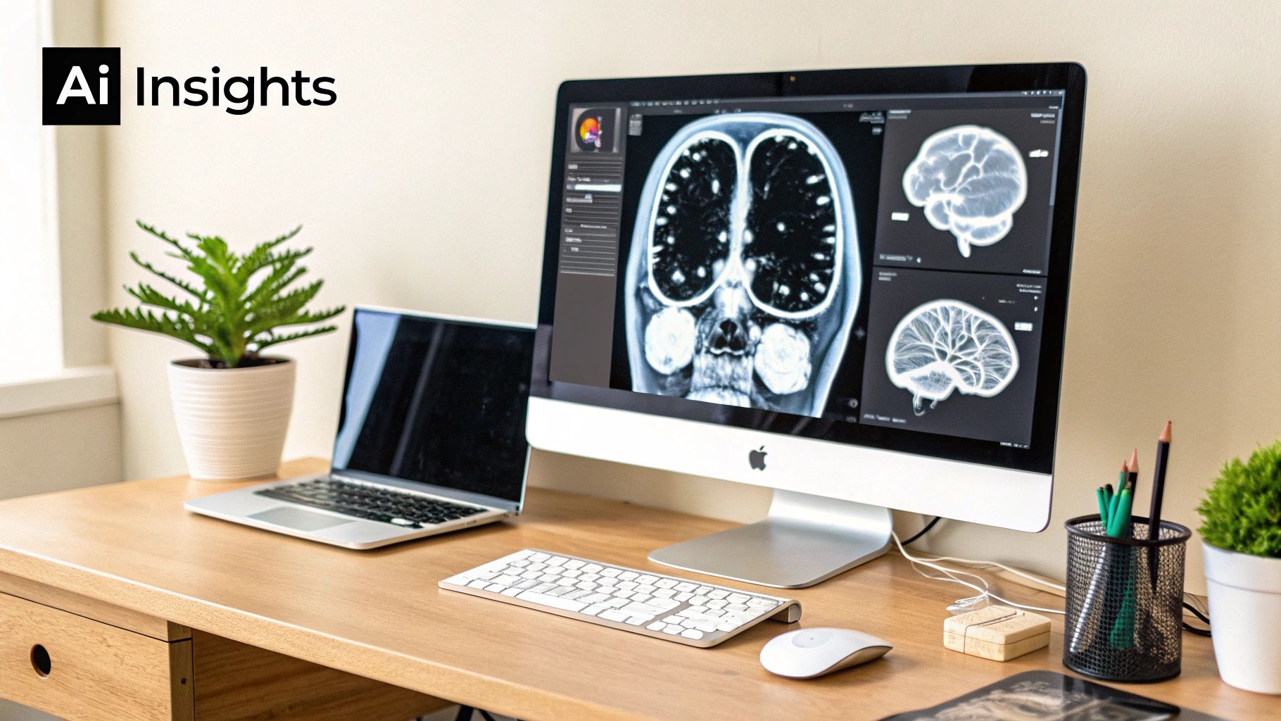
Weaving AI into the Fabric of Diagnostics
This is where medical imaging gets truly exciting. When you combine the highly structured world of DICOM and PACS with the predictive power of Artificial Intelligence, you're not just connecting systems—you're creating the future of diagnostics. This partnership gives clinicians a powerful new ally in their mission to deliver faster, more accurate care.
Think about it: every DICOM file is more than just an image. It’s a complete story, packed with rich, organized metadata—patient details, study parameters, and crucial clinical context. This structured data is the perfect fuel for training and validating diagnostic AI models, allowing algorithms to learn from huge datasets with incredible precision.
Once you unlock that potential, the impact is immediate. You can have AI algorithms working quietly within the PACS workflow, analyzing new studies the moment they arrive and flagging potential abnormalities for a radiologist's immediate review.
A Force Multiplier for Clinicians
Let's be clear: this isn't about replacing the seasoned expertise of a clinician. It's about augmenting it. AI acts as a tireless assistant, handling repetitive tasks and pointing out subtle details, which frees up specialists to focus their incredible skills where they matter most. The goal is to elevate care, not just automate it.
Here's how it's already playing out in hospitals today:
- Smarter Triage: An AI can scan a long queue of chest X-rays, instantly prioritizing studies that show signs of a collapsed lung. This ensures the most critical patients get seen first, without delay.
- A Second Set of Eyes: In a busy mammography clinic, an algorithm can highlight suspicious microcalcifications that are incredibly subtle to the human eye, acting as a vigilant second reader.
- Objective Measurements: Instead of manual measurements, an AI tool can precisely calculate tumor volume from a series of CT scans, giving oncologists objective data to track how well a treatment is working.
This isn't science fiction; it's happening right now. Integrating AI into DICOM and PACS workflows is helping doctors catch diseases earlier and speed up the entire diagnostic journey. We dive much deeper into this in our guide on the role of machine learning in medical imaging, which really unpacks these applications.
AI's Impact in Specialized Medicine
The effect is especially profound in specialized fields. Take ophthalmology, where AI is making incredible strides. An algorithm can now scan a retinal image and instantly spot the earliest signs of diabetic retinopathy, giving doctors a critical head start to prevent vision loss.
This fusion of structured data and intelligent algorithms represents a monumental shift. It transforms medical imaging from a static record of the past into a dynamic, predictive tool that can help shape a patient's future.
The market trends tell the same story. Ophthalmology PACS is one of the fastest-growing segments, with its global market valued at about USD 164 million in 2024 and projected to climb to nearly USD 333 million by 2034. This explosion is fueled by AI-powered screening programs that prove just how vital specialty PACS are for improving diagnostic accuracy. You can learn more about the rapid growth in ophthalmology PACS technology and the waves it's making.
At PYCAD, we live and breathe this evolution. We don't just build software; we architect intelligent ecosystems. We specialize in creating custom web DICOM viewers and integrating them into medical imaging web platforms, which become the perfect launchpad for these game-changing AI tools. To see how we’re helping innovators bring smarter diagnostic solutions to life, take a look at our portfolio page.
Navigating Critical Security and Compliance Challenges
In medical imaging, trust isn't a feature—it's the foundation. For any DICOM and PACS system, security and compliance are the non-negotiable promises that safeguard patient privacy and uphold the integrity of clinical data. This goes far beyond just avoiding hefty fines; it’s about honoring the ethical duty at the very heart of healthcare.
When we talk about regulations like HIPAA or GDPR, we're not just throwing acronyms around. These are the frameworks that provide the critical rules of the road for handling sensitive patient information. They dictate how Protected Health Information (PHI) is stored, transmitted, and accessed, ensuring every digital touchpoint is secure, justified, and, above all, trustworthy.
Building a Fortress Around Patient Data
Securing a modern medical imaging environment demands a defense-in-depth strategy. It all starts with encryption, which essentially scrambles data so it's completely unreadable to anyone without the right key. This is a must-have for data "at rest" (sitting on a server) and data "in transit" (zipping across a network).
But encryption alone isn't enough. Granular access controls are absolutely paramount. This means designing a system where people can only see or interact with the specific data they are authorized for, based on their role. Think of it as the digital equivalent of making sure only the right doctor can open the right patient's chart.
And to tie it all together, you need meticulous audit trails. These logs create an unchangeable record of every single action taken within the system: who accessed what, when they did it, and from where. This level of accountability is non-negotiable for both real-time security monitoring and proving compliance.
Core Pillars of a Secure Imaging System
A truly secure system is built on a few key principles that work together to protect sensitive information from every possible angle.
- End-to-End Encryption: Data must be encrypted from the moment it leaves the CT scanner until it lands on the radiologist's screen, leaving zero vulnerable gaps in between.
- Role-Based Access Control (RBAC): This is what prevents a surgeon from accessing psychiatric notes or an administrator from viewing clinical images without a legitimate reason. Access is strictly on a need-to-know basis.
- Comprehensive Audit Logging: Every login, every query, and every image view is recorded. This digital footprint is vital for investigating potential incidents and demonstrating compliance.
- Secure Authentication: Strong passwords, multi-factor authentication (MFA), and secure sign-on (SSO) protocols are your first line of defense against unauthorized entry.
Think of compliance not as a restrictive hurdle, but as a strategic enabler. It’s what builds the patient trust and institutional confidence needed to innovate responsibly and ethically in healthcare.
Compliance as a Blueprint for Trust
Achieving compliance is an ongoing commitment, not a one-time project. It requires a deep and practical knowledge of the regulatory landscape and proactive measures to manage risk. A great starting point is understanding compliance risk assessment and how it applies to your specific environment.
This commitment has to permeate every part of the system, from the PACS server right down to the viewing software on a clinician's device. For healthcare technology companies, ensuring every component is compliant is a foundational responsibility.
At PYCAD, we know that security is the bedrock of innovation. That’s why we build custom web DICOM viewers and integrate them into medical imaging web platforms with security and compliance baked in from day one. We engineer solutions that are not only powerful and intuitive but are fundamentally secure. To see our commitment to robust and secure design in action, you can view our portfolio page.
Common Questions About DICOM and PACS
Diving into the world of medical imaging technology always sparks a few questions. For innovators and developers, getting a grip on the practical side of DICOM and PACS is the key to building tools that actually work in the real world—tools that are reliable, secure, and ready to grow. Let's tackle some of the most common things people ask when they start working with these foundational systems.
A big one is always performance. How does a classic, on-premise PACS stack up against a modern, web-based DICOM viewer? A local PACS can be lightning-fast inside the hospital’s own network, but that speed often vanishes the moment a clinician tries to log in from home. Web-based viewers, on the other hand, are built for this. They use cloud infrastructure to deliver images quickly and efficiently, no matter where you are.
Is One System More Secure Than the Other?
Security is another hot topic. There's a common belief that keeping data on-premise is automatically safer because it's "in-house." But this thinking puts the entire weight of security—firewalls, intrusion detection, you name it—squarely on the shoulders of the institution's IT department.
In reality, a well-designed cloud system often comes out ahead on security. The top cloud providers pour billions into their infrastructure, giving you access to advanced threat detection, sophisticated identity management, and compliance controls that most individual organizations simply can't match.
This lets your development team stop worrying about the foundation and focus on what they do best: building great features and securing the application itself, all while standing on an incredibly secure base.
What Is the Biggest Challenge in Implementation?
If there’s one hurdle that trips up almost everyone, it’s interoperability. DICOM is a standard, yes, but vendors can interpret that standard in slightly different ways. This is where the real headaches begin, like when a scanner from one company just won't talk to a PACS from another.
Fixing these communication gaps takes deep technical know-how. Sometimes it even requires a go-between, like a DICOM router, to translate the "dialects" spoken by different systems. It’s a powerful reminder to partner with vendors who are true to the DICOM standard and have the integration battle scars to prove it.
This is exactly the kind of puzzle we live to solve. At PYCAD, we don't just write code; we navigate these complex integration challenges every day. We build custom web DICOM viewers and integrate them into medical imaging web platforms, creating a seamless flow of information across your entire tech stack. Our experience is your guarantee that everything will just work, no matter what equipment you're using. You can learn more about our projects by visiting our portfolio page.
At PYCAD, we build secure, scalable, and intelligent medical imaging solutions that help healthcare innovators bring their ideas to life. To see how our custom platforms can accelerate your vision, check out our work on our portfolio page.


