
Learning to read a head MRI is all about becoming fluent in the visual language of different imaging sequences. It’s not just about looking at pictures; it’s about systematically analyzing sequences like T1, T2, FLAIR, and DWI to understand how different tissues and fluids behave. This process of decoding patterns of light and dark signals is how you build a complete clinical picture, one slice at a time.
Decoding the Language of Head MRI Sequences
Diving into head MRI interpretation feels a lot like learning a new language. Each scan tells a story about the patient's brain, and to read it fluently, you first need to learn the alphabet—the fundamental MRI sequences that give every image its meaning. If you think of it this way, what seems like a daunting task becomes an approachable skill you can build step-by-step.
The goal isn't to memorize every possible permutation of what a scan can look like. It's about understanding the "why." Why does cerebrospinal fluid (CSF) look dark on one sequence but bright white on another? Once you grasp that, you're on your way to real diagnostic confidence. Each sequence is a specific physics experiment, manipulating water and fat molecules to create contrast, and that’s where the art and science of neuroimaging truly come together.
At the core of every head MRI study, you'll find a few workhorse sequences. Get to know their individual strengths, and you'll quickly see how they complement each other to tell the full story.
-
T1-Weighted (T1W): Think of this as your "anatomy" sequence. It gives you a beautiful, clear map of the brain's structure. Fat is bright, and water (like CSF) is dark, which creates a sharp contrast between gray and white matter.
-
T2-Weighted (T2W): T2 is essentially the opposite. It makes water and fluid-filled areas shine brightly. This incredible sensitivity to water makes it your go-to for spotting edema, inflammation, tumors, or anything else that brings extra fluid to the party.
-
FLAIR (Fluid-Attenuated Inversion Recovery): FLAIR is a really smart tweak on the T2 sequence. It cleverly suppresses the bright signal from normal CSF, making abnormalities near the ventricles or on the brain's surface pop. It’s a game-changer for finding things like multiple sclerosis plaques or subtle edema that might get lost in the brightness of a standard T2 scan.
Understanding Signal and Contrast
The basic vocabulary of MRI is simple: hyperintense (bright) and hypointense (dark). A lesion's specific "look" across different sequences is what gives you the most valuable clues.
Take an acute stroke, for example. It classically appears bright or hyperintense on Diffusion-Weighted Imaging (DWI) because water movement is restricted in the damaged cells. In the first few hours, that same stroke might be completely invisible on other sequences.
The real power in reading an MRI doesn't come from looking at one sequence in isolation. It comes from piecing together the information from all of them. A finding on one scan might confirm, clarify, or even contradict what you see on another, and that synthesis is what leads to a confident diagnosis.
A quick reference can be a lifesaver when you're starting out. Here’s a simple table to help you keep the most common sequences straight.
Quick Guide to Common Head MRI Sequences
| Sequence | What It Highlights | CSF Appearance | Fat Appearance | Primary Clinical Use |
|---|---|---|---|---|
| T1-Weighted (T1W) | Anatomy, gray-white matter differentiation | Dark | Bright | Evaluating brain structure, post-contrast enhancement |
| T2-Weighted (T2W) | Pathology with high water content (edema, tumors) | Bright | Bright | Detecting lesions, inflammation, and chronic conditions |
| FLAIR | Lesions near CSF spaces (e.g., ventricles) | Dark | Bright | Multiple sclerosis, meningitis, subtle edema |
| Diffusion-Weighted (DWI) | Restricted water movement | Variable | Dark | Acute stroke, abscesses, cellular tumors |
| Gradient Echo (GRE) / SWI | Blood products, calcification | Variable | Variable | Detecting hemorrhage, microbleeds, vascular malformations |
This table isn't exhaustive, of course, but it covers the core sequences you'll see every day.
The Role of High-Quality Imaging
None of this matters if the images themselves aren't clear. Technical factors like magnetic field strength and gradient systems are what determine the sharpness and detail you see. A scan with higher resolution simply gives you a better chance to spot subtle pathologies. For anyone serious about neuroimaging, it's worth taking a moment to learn more about the resolution of MRI and how it directly impacts your diagnostic accuracy.
At PYCAD, we're obsessed with the link between image quality and clinical confidence. We build custom web DICOM viewers and integrate them into medical imaging web platforms, all to make sure clinicians have the best possible tools for interpretation. If you're curious to see how our solutions can support advanced imaging workflows, take a look at our work over at https://pycad.co/portfolio.
Developing a Systematic Search Pattern
When you're first learning to read a head MRI, the biggest trap is tunnel vision. You see a glaring abnormality and immediately fixate on it. While it's human nature to be drawn to the most obvious finding, this "search and satisfy" habit is a recipe for disaster. It's how you miss the subtle but crucial details that can completely alter a diagnosis.
True mastery isn't about speed; it's about discipline. It comes from building a repeatable, systematic search pattern that you stick to, no matter what. Every. Single. Time.
This disciplined approach starts before you even look at the brain. First, and this is non-negotiable, confirm you have the right patient and the right study date. It sounds almost too simple, but it’s a critical safety net. Next, do a quick quality check of the images. Are they riddled with motion artifacts? Are all the sequences you need actually there? Getting this sorted out up front saves you from major headaches later.
The human brain is a master of symmetry, and this gives you a huge advantage. Before you start drilling down into specific structures, just take a moment. Scroll through the axial T2 and FLAIR sequences and get a feel for the overall landscape. Look for any lopsidedness in the cerebral hemispheres, strange sulcal patterns, or asymmetries in the ventricles. This big-picture view primes your brain to spot anything that’s out of place.
This methodical flow, which we can see below, shows how different sequences like T1, T2, and FLAIR are designed to work together, each telling a part of the story.

The real insight here is that a system isn't just about following an order. It’s about weaving together the anatomical detail from T1, the fluid sensitivity of T2, and the bright flags of pathology on FLAIR to build a complete diagnostic picture.
A Practical Checklist for Systematic Review
With that global view established, it's time to work from the outside in. Think of it like a guided tour of the brain where you have a list of landmarks you absolutely have to visit. This ensures nothing important gets skipped.
A tried-and-true method is to follow a consistent anatomical path:
- Extra-axial Spaces: Start outside the brain itself. Check the scalp, skull, and meninges. Look for any collections of blood or pus, or any unexpected masses in the spaces surrounding the brain.
- Ventricles and Sulci: Now, move to the fluid spaces. Assess the ventricles for their size, shape, and symmetry. Are they ballooning out from hydrocephalus or squashed flat? Then look at the sulci—the grooves on the brain's surface. Are they wide and gaping, hinting at atrophy, or are they effaced and indistinct, suggesting swelling?
- Cortex and White Matter: Time to review the brain tissue proper. Systematically scan the gray matter (cortex) and the deep white matter in every lobe—frontal, parietal, temporal, and occipital. Always compare left to right, hunting for any abnormal signals.
- Deep Gray Matter: Zero in on the basal ganglia and thalami. These deep, central structures are notorious hot spots for strokes, metabolic issues, and degenerative diseases.
- Brainstem and Cerebellum: Don't neglect the posterior fossa! Carefully inspect the midbrain, pons, and medulla, then move on to the cerebellar hemispheres. It's easy to rush through this area, but it's packed with critical anatomy.
A search pattern isn't meant to slow you down. It's designed to make you faster, more efficient, and far more accurate. With practice, this systematic review becomes second nature—a rapid mental checklist that protects both you and your patients from diagnostic error.
The Role of an Advanced DICOM Viewer
Your systematic approach is only as good as the tools you use to execute it. An advanced web DICOM viewer can be a game-changer, dramatically improving how efficiently you can work through a scan. The ability to seamlessly link and scroll different sequences side-by-side, tweak windowing on the fly, and make precise measurements isn't a luxury; it's fundamental to good practice.
In clinical emergencies, where every second counts, this becomes even more critical. Well-designed training and tools have a proven impact. For example, studies on emergency medicine physicians' ability to spot acute ischemic stroke on MRI found an accuracy of 86.3% compared to radiologist reads. You can read more about the advancements in MRI interpretation competency and see how structured approaches make a real difference.
At PYCAD, we live and breathe this stuff. We at PYCAD, build custom web DICOM viewers and integrate them into medical imaging web platforms, designing them specifically to support a systematic, efficient analysis. Our goal is to make the software so intuitive that it disappears into the background, letting you focus entirely on the interpretation.
Want to see what this looks like in the real world? Check out our work on our portfolio page and see how powerful, integrated tools can transform your workflow.
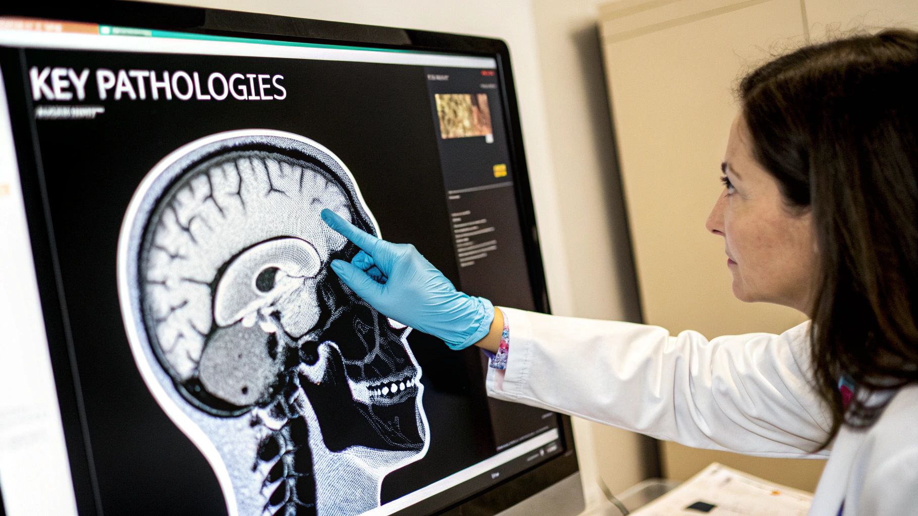
Identifying Key Pathologies with Confidence
This is where all the groundwork pays off. Once you’ve mastered the sequences and have a systematic search pattern down, you can start confidently connecting what you see on the scan to what’s happening with the patient. Learning how to read a head MRI isn't just about spotting an anomaly; it's about understanding the story that abnormality is telling.
You’re moving beyond just seeing a bright spot or a dark patch. You’re starting to think like a diagnostician, linking the visual evidence to the underlying pathology. It’s that critical leap from seeing the "what" (the imaging finding) to understanding the "why" (the disease process driving it). This transforms a grayscale image into a clear roadmap for patient care.
Classic Signs of Acute Ischemic Stroke
In neurology, we live by the motto "time is brain." When you suspect an acute ischemic stroke, Diffusion-Weighted Imaging (DWI) is your most powerful ally. During a stroke, brain cells starve for oxygen and begin to swell—a process called cytotoxic edema. This swelling traps water molecules, restricting their ability to move freely.
DWI is incredibly sensitive to this restricted movement. An acute stroke pops out as a bright, hyperintense signal on the DWI sequence. But don't stop there. You must cross-reference this with the Apparent Diffusion Coefficient (ADC) map. A true stroke will show a corresponding hypointense (dark) signal on the ADC map. This DWI-ADC mismatch is the definitive fingerprint of acute ischemia, and it can show up within minutes of the event.
The Telltale Footprints of Brain Tumors
Brain tumors are bullies. They often make their presence known through mass effect—a clinical term for how they push, squeeze, and displace healthy brain tissue. As you scroll through the slices, be on the lookout for these pressure signs.
- Ventricular Compression: Is a ventricle getting squashed? A growing tumor can make it look much smaller than its counterpart on the other side.
- Midline Shift: This is a big one. A large mass can literally shove the brain's hemispheres across the midline, a critical and often dangerous finding.
- Sulcal Effacement: Look at the grooves (sulci) on the brain's surface. If they look flattened or even wiped out, it’s a sign of swelling pushing the brain against the skull.
The tumor's behavior across different sequences gives you even more clues. Most tumors will show up as abnormal signals on T2 and FLAIR scans, thanks to the edema they cause. Then, after you give gadolinium contrast, many will "light up" on T1-weighted images. This enhancement tells you where the tumor has broken down the blood-brain barrier.
Spotting Demyelination in Multiple Sclerosis
When you're hunting for Multiple Sclerosis (MS), FLAIR is the star of the show. MS creates inflammatory plaques—scars of demyelination—in the white matter. These plaques are full of water, so they'd be bright on a standard T2 scan.
Here's the problem: classic MS plaques love to hang out right next to the ventricles (periventricular). On a T2 scan, the bright signal from the CSF in the ventricles could easily hide them. This is where FLAIR works its magic. By canceling out the CSF signal, FLAIR makes those bright periventricular plaques stand out in sharp relief. You'll see them as distinct, ovoid lesions, often called "Dawson's fingers."
The Subtle Scars of Traumatic Brain Injury
Sure, a large hemorrhage from a head trauma is easy to spot. But much of the real damage from a Traumatic Brain Injury (TBI) can be incredibly subtle. This is where you absolutely need Gradient Echo (GRE) or Susceptibility Weighted Imaging (SWI) sequences, as they are exceptionally good at detecting tiny traces of blood.
In a post-TBI scan, you might see tiny dark dots called microhemorrhages. These represent sheared, damaged small blood vessels, often right where the gray and white matter meet. Finding these is crucial for painting a complete picture of the patient's injury.
The ultimate goal is to build a strong differential diagnosis. Every finding you identify—a DWI restriction, a ring-enhancing mass, a periventricular plaque—is a piece of a larger puzzle. Your confidence grows when you can confidently assemble these pieces to tell a coherent clinical story.
It’s also critical to remember that brain anatomy changes dramatically over a person's life, which can complicate any diagnosis. A landmark study in Nature developed normative brain charts by analyzing MRI data from diverse populations to help benchmark these structural differences. The researchers emphasized that brain measurements are highly sensitive to scanner type, sequences, and processing methods—a strong argument for harmonizing our standards in the clinic.
Modern tools can certainly help manage this complexity. At PYCAD, we specialize in building custom web DICOM viewers and integrating them into medical imaging platforms, which streamline the analysis of complex scans. Furthermore, AI advancements like automated segmentation can help quantify these anatomical variations. If you're interested, you can check out our guide on https://pycad.co/brain-segmentation/ and its clinical applications.
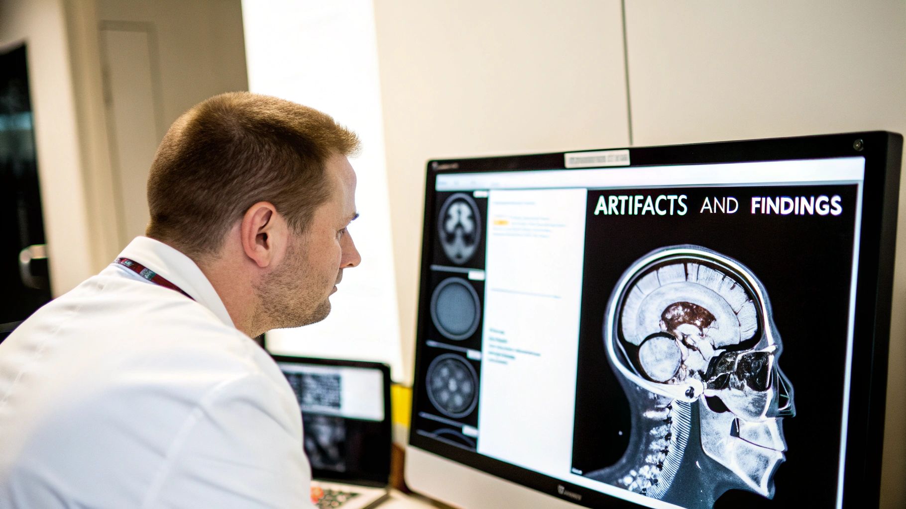
Navigating Artifacts and Incidental Findings
Not every bright or dark spot on a head MRI is a sign of trouble. The real artistry in reading these scans comes from honing your ability to tell the difference—to confidently separate a true clinical problem from the "noise" of imaging artifacts and the frequent surprise of an incidental finding. This is what takes you from just spotting abnormalities to making sharp, reliable clinical judgments.
Think of it as building a crucial layer of diagnostic confidence. It’s how we prevent both missed diagnoses and the unnecessary anxiety that comes from over-calling a benign quirk. You're learning to see the ghosts in the machine so you can focus on what really matters for your patient.
Recognizing Common Imaging Artifacts
Artifacts are essentially signal glitches that don't represent the patient's actual anatomy. They can be incredibly deceptive and mimic real pathology, so knowing what to look for is non-negotiable for anyone serious about reading a head MRI.
- Motion Artifacts: These are by far the most common offenders. Even a tiny patient movement during the scan can smear, ghost, or streak an image, hiding critical details. You'll often see them as repetitive, fuzzy lines that just don't look like anatomy.
- Susceptibility Artifacts: These pop up where tissues with different magnetic properties meet—think of the sinuses where bone and air sit side-by-side, or around any metal implants. They appear as dark, warped voids that can completely black out an area, especially on sequences like GRE or SWI.
Getting a feel for these patterns just takes practice. When you spot a potential lesion, stop and ask yourself: Could this be an artifact? Does it follow anatomical lines? Does it show up consistently across different sequences in a way that makes biological sense?
The World of Incidental Findings
Once you've ruled out artifacts, you'll still run into incidentalomas—unexpected findings that have nothing to do with why the patient was scanned in the first place. These are real anatomical variations or minor pathologies, but most of the time, they are completely asymptomatic and don't need any intervention.
And they are incredibly common. Global studies show that incidental brain abnormalities appear in roughly 9% to 54% of otherwise healthy adults. For instance, incidental tumors, usually benign meningiomas, have a prevalence of about 0.5% to 2.5% and become more common as we age. This is why we need to be so skilled at telling these apart from clinically urgent findings—it’s all about avoiding needless worry and workups. You can dig deeper into the data by checking out the full research on incidental findings to get a better sense of their prevalence.
Honestly, managing incidentalomas is one of the toughest parts of neuroimaging. It's a delicate balance between being cautious and avoiding the cascade of over-investigation that causes patients so much stress. Your clinical judgment is truly your most important tool.
A Practical Approach to Management
When you find something unexpected, the key is to have a clear strategy. Can you confidently dismiss it, or does it need a second look?
Here are a few you'll see all the time:
- Arachnoid Cysts: These are just harmless, CSF-filled sacs. They are extremely common and will match the CSF signal on every sequence. Unless one is massive and causing a significant mass effect, they almost never require follow-up.
- White Matter Hyperintensities (WMHs): Those little bright spots on T2/FLAIR images are everywhere, especially in older adults. While they can be linked to vascular risk factors, a few scattered, non-specific WMHs are usually of no clinical importance.
- Pineal Cysts: A small, simple cyst in the pineal gland is a normal variant for a huge portion of the population. They are almost universally benign.
Having the right software makes navigating these findings so much easier. At PYCAD, this is the exact challenge we focus on. We at PYCAD, build custom web DICOM viewers and integrate them into medical imaging web platforms. Our tools are designed to make it seamless to compare sequences, pull up prior studies, and take quick measurements. This lets you decide—quickly and with confidence—whether you're looking at an artifact, a benign incidentaloma, or something that needs attention.
To see how the right viewing technology can transform your workflow and give you more clarity on complex cases, have a look at our projects on the PYCAD portfolio page.
7. Using Technology to Your Advantage in Modern Reporting
https://www.youtube.com/embed/eawQB_IY0ZM
Your clinical expertise is, without a doubt, the most critical tool you have when interpreting a head MRI. But let's be honest—modern technology can be an incredible force multiplier, helping you work faster, become more consistent, and ultimately, deliver more powerful insights. Adopting these tools isn't about letting a machine take over; it’s about augmenting your own skills to raise the bar for patient care.
The real magic happens when you weave these intelligent tools into your daily rhythm. You can hand off the repetitive, time-consuming tasks and dig deeper into the images, freeing you to focus on the nuanced diagnostic thinking that only a human mind can do. It's the classic "work smarter, not harder" mantra brought to life in the reading room.
AI is Your New Diagnostic Partner
Artificial intelligence is no longer a sci-fi concept; it's a real, practical assistant in neuroimaging today. AI-driven software excels at jobs demanding incredible precision and stamina, like performing volumetric analysis or spotting tiny lesions.
Think of it as having a tireless second reader by your side. An AI algorithm can meticulously measure brain volumes or flag subtle white matter changes you might miss at the end of a long shift. It can scan thousands of images without a hint of fatigue, pointing out areas of interest for your expert review. This kind of collaboration boosts confidence and acts as a fantastic safety net.
The true value of AI in radiology isn't to replace you, but to manage the tedious, quantitative parts of the job. This frees up your mental energy for what truly matters: complex problem-solving and building a solid differential diagnosis. It's a partnership that elevates your own expertise.
These tools are getting more impressive by the day. If you want to get a feel for where this is all going, exploring the different applications of artificial intelligence for radiology is a great way to see what's on the horizon.
Writing Reports That Make a Difference
A brilliant read is worthless if it isn't communicated well. Your final report is the crucial link between your findings and the patient's treatment plan, so it has to be crystal clear, concise, and directly address the clinical question.
Here’s a simple framework for a modern, effective report:
- Conclusion First: Start with a brief "Impression" that gets right to the point. This gives the referring clinician the most critical information upfront.
- Logical Structure: Use straightforward subheadings like "Findings" and "Technique." When detailing your findings, follow the same systematic search pattern you used during your read.
- Speak with Confidence: Whenever the evidence supports it, ditch the wishy-washy language. Instead of "appears to be," go with stronger phrases like "is consistent with."
- Answer the Question: Always bring it back to why the scan was ordered. If the referral was to rule out an acute stroke, your report needs to explicitly confirm or deny the presence of ischemic changes.
Why a Modern DICOM Viewer is Non-Negotiable
Your entire workflow lives and dies by your viewing software. An old, clunky DICOM viewer is a constant source of friction, slowing you down and causing needless frustration. A modern, web-based viewer, on the other hand, can completely change the game for your efficiency and diagnostic power.
This is exactly what we live and breathe at PYCAD. We at PYCAD, build custom web DICOM viewers and integrate them into medical imaging web platforms, creating a seamless and intuitive workspace for clinicians. Just imagine—effortlessly pulling up prior studies for comparison, applying advanced measurements in a click, and securely accessing scans from any device.
Of course, when you're working with sensitive patient data from head MRIs, a solid grasp of essential data security concepts is absolutely critical for maintaining privacy and compliance. A powerful and secure platform is the bedrock of any modern practice.
You can see how the right tools can elevate a practice by checking out the projects we've delivered for our clients on our PYCAD portfolio page.
Common Questions on Reading Head MRIs
As you get deeper into reading head MRIs, you'll find certain questions pop up again and again. Don't see these as hurdles. They're actually signs that you're leveling up, moving from just knowing the theory to applying it with real clinical confidence.
Which MRI Sequence Should I Always Look at First?
Every experienced radiologist or neurologist has their own rhythm, but a solid starting point is always the FLAIR (Fluid-Attenuated Inversion Recovery) sequence. It's a fantastic first look because it blacks out the signal from cerebrospinal fluid (CSF), making anything abnormal right next to the ventricles or on the brain's surface just pop. It gives you a quick, high-yield overview.
Right after a quick scan of the FLAIR, my eyes jump to the DWI (Diffusion-Weighted Imaging). If a patient comes in with sudden neurological symptoms, DWI is your money shot. It's hands-down the most sensitive sequence for spotting a new stroke, sometimes within just a few minutes of it happening.
How Can You Tell a New Stroke from an Old One?
This is one of the most critical skills you'll develop. An acute stroke—something that just happened—has a very specific signature: it’s bright on DWI and dark on the corresponding ADC map. This classic "DWI/ADC mismatch" tells you that water molecules are trapped inside swollen, freshly injured cells.
An old, chronic stroke, on the other hand, is the brain's healed scar tissue. The acute DWI/ADC mismatch is long gone. Instead, you'll see a dark hole on T1 images and a bright, gliotic scar on T2/FLAIR, which is just encephalomalacia, or permanent tissue loss.
Getting this distinction down is a game-changer. It’s that moment when you go from just seeing a lesion to truly understanding its place in the patient's timeline. That understanding is everything when it comes to making the right call.
Is a Head MRI Without Contrast Enough?
Honestly, a lot of the time, yes. A non-contrast head MRI gives you a massive amount of information. It's often all you need to diagnose an acute stroke, look for brain atrophy, or size up chronic white matter changes.
But when you're hunting for something that breaks down the blood-brain barrier, that's when you need gadolinium contrast. Think tumors, infections like a brain abscess, or inflammatory diseases like multiple sclerosis. The way these things light up with contrast—the specific patterns of enhancement—gives you indispensable clues that point directly to the right diagnosis.
Here at PYCAD, we're all about creating tools that feel like an extension of your own clinical expertise. We at PYCAD, build custom web DICOM viewers and integrate them into medical imaging web platforms, so your workflow is as smooth and intuitive as possible. You can check out some of the solutions we've built for clinicians over on our project portfolio.


