
The Evolution of Lung Segmentation CT: From Manual to Modern
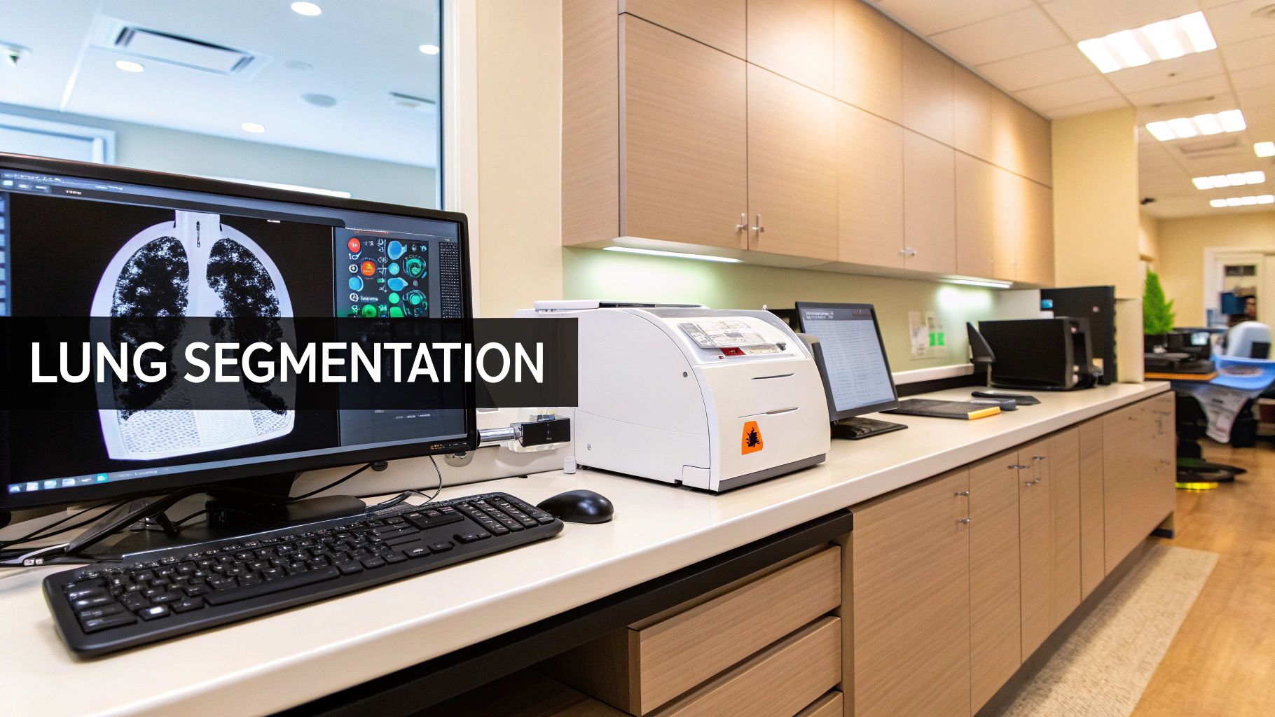
Lung segmentation CT has evolved significantly. Initially, radiologists manually traced lung contours on each CT image slice. This labor-intensive process was time-consuming and prone to inconsistencies, making reliable measurements a challenge.
The demand for faster, more accurate results led to the development of semi-automated techniques. These involve some user interaction, like placing seed points, followed by automated refinement. While faster and more consistent, these methods still relied on substantial user input which could affect the final result.
High-Resolution CT: A Key Advancement
The rise of high-resolution computed tomography (HRCT) was crucial. Gaining popularity in the late 1980s, HRCT provided significantly more detailed images of lung structures. By the early 1990s, it was a standard tool, thanks in part to publications like the 1985 study by Zerhouni et al. which presented a pattern-based approach for classifying diffuse pulmonary diseases. The field's growth is evident in the two issues the Journal of Thoracic Imaging dedicated to HRCT in 1993. You can explore this evolution further here.
The Rise of Automated Lung Segmentation
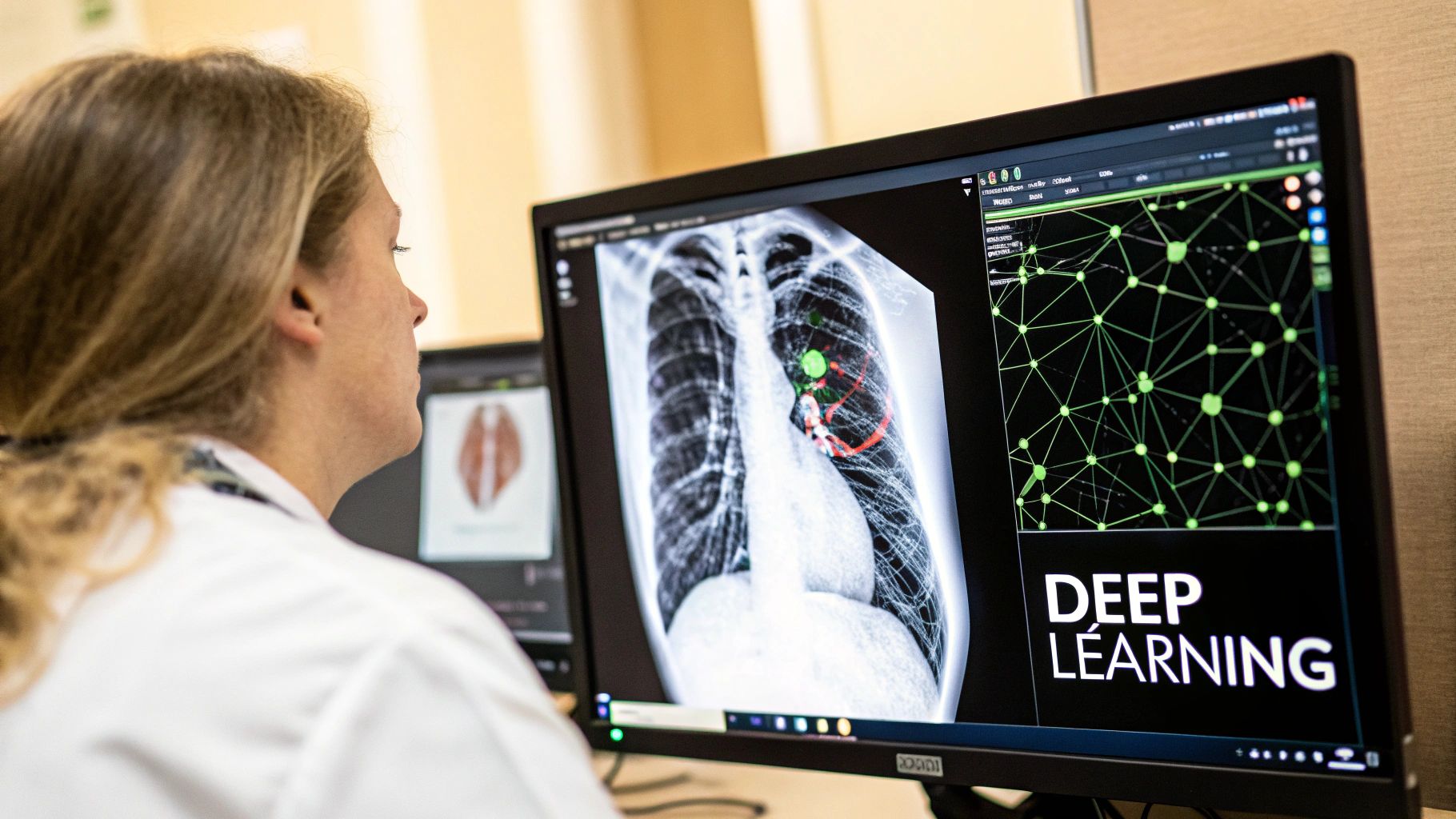
The biggest change in lung segmentation CT came with fully automated methods, especially those using artificial intelligence (AI). AI algorithms, based on deep learning models, analyze CT images and segment lungs with little to no human interaction, significantly reducing analysis time. This allows radiologists to focus on more complex diagnostic work.
AI-Powered Accuracy and Efficiency
AI-driven methods are faster, more accurate, and more consistent. They can detect subtle details and anatomical variations that a human might miss. Moreover, AI-powered segmentation enables the extraction of quantitative data like lung volume and density, crucial for monitoring disease progression and treatment effectiveness. This allows clinicians to make data-driven decisions.
The Future of Lung Segmentation CT
Further refinement of these AI algorithms is the future of lung segmentation CT. This includes creating methods that can handle difficult cases with severe lung disease or image artifacts. These advancements promise even greater precision and efficiency in lung disease diagnosis and management, ultimately improving patient care.
CT Technology Breakthroughs Transforming Lung Segmentation
Modern lung segmentation using CT scans has become remarkably precise. This is thanks to a number of key technological advancements. These improvements have not only made lung scans faster and more efficient, they have also significantly boosted the quality and detail of the images produced. This leads to more accurate diagnoses and improved treatment planning.
Increased Detector Rows and Faster Rotation Speeds
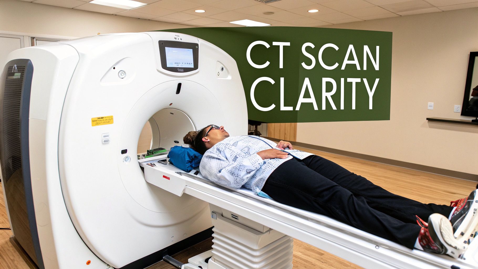
A major step forward has been the development of CT scanners with increased detector rows. Older scanners had just a few rows. This limited the amount of data gathered in a single rotation. Modern scanners have hundreds of rows. They capture a much larger lung volume in a fraction of the time.
Combined with faster rotation speeds, scans are quicker with fewer motion artifacts. This is particularly helpful for patients who struggle to hold their breath. Quicker scans also mean more patients can be seen.
Breakthrough Reconstruction Algorithms
The way CT data is processed after acquisition has also greatly improved. Advanced reconstruction algorithms are essential for lung segmentation. These algorithms process the raw data from the detectors. This data forms the cross-sectional images of the lungs.
Newer algorithms are more sophisticated. They compensate for patient movement and variations in tissue density. This results in clearer, more defined images, which is essential for accurate lung segmentation. Identifying subtle abnormalities is also easier with these improvements.
To illustrate the advancements, the introduction of spiral/helical CT in the early 1990s enabled faster scan times and the ability to image entire organs in a single breath-hold, reducing motion artifacts. The subsequent move to multi-slice CT further increased spatial resolution while reducing scan times. Modern CT scanners can image the entire chest in under 10 seconds, even for patients unable to hold their breath. More detailed information is available here.
Isotropic Resolution: The Gold Standard
A further vital development in lung segmentation CT is isotropic resolution. Isotropic resolution means having the same resolution in all three dimensions (x, y, and z). This is essential for 3D segmentation.
Isotropic resolution ensures consistent detail, regardless of the viewing angle or reconstruction method. It allows for the creation of precise 3D lung models. These models are essential for planning surgeries, radiation therapy, and other interventions. These advancements improve the visualization and measurement of lung structures, crucial for diagnosis and disease monitoring.
Reducing Radiation Exposure
Importantly, the evolution of CT technology has also focused on lower radiation doses. This makes frequent monitoring of chronic conditions like COPD and pulmonary fibrosis safer. It minimizes the risks associated with repeated imaging, particularly for patients requiring regular scans. These advancements are a considerable improvement for patient care and informed clinical decisions.
To understand the evolution of CT and its influence on lung segmentation, the following table offers a helpful overview:
Introduction: The following table summarizes the key milestones in CT technology, specifically focusing on their impact on lung segmentation capabilities.
| CT Technology | Time Period | Key Features | Impact on Lung Segmentation |
|---|---|---|---|
| Spiral/Helical CT | Early 1990s | Faster scan times, single breath-hold imaging | Reduced motion artifacts, improved image quality |
| Multi-slice CT | Late 1990s – Present | Increased spatial resolution, even faster scan times | Enhanced 3D imaging capabilities, more precise segmentation |
| Isotropic Resolution | Present | Equal resolution in all three dimensions | Highly accurate 3D models, improved visualization and measurement of lung structures |
| Low-dose CT | Present | Reduced radiation exposure | Safer frequent monitoring for chronic lung conditions |
Conclusion: The advancements in CT technology highlighted in this table have significantly improved the accuracy and efficiency of lung segmentation. These improvements have allowed for more precise diagnoses, better treatment planning, and safer monitoring of chronic lung conditions.
Lung Segmentation CT: Cutting-Edge Approaches That Deliver Results
Which lung segmentation CT techniques are truly impacting patient care? This section delves into the technologies that leading pulmonologists and radiologists are actively incorporating into their practices, exploring how these advancements are improving the precision and efficiency of lung disease diagnosis and treatment.
Photon-Counting CT: Transforming Lung Imaging
A significant advancement is photon-counting CT (PCCT). Unlike conventional CT, which measures the total energy of X-rays, PCCT counts individual X-ray photons and measures their energy levels. This allows for more refined differentiation between tissue types.
PCCT excels at detecting subtle ground-glass opacities, an important indicator of early interstitial lung disease. Traditional CT often misses these subtle signs. This increased sensitivity of PCCT is vital for early diagnosis and timely intervention. PCCT offers improvements in spatial resolution and contrast. This is especially useful for detecting interstitial lung disease and small airways diseases.
PCCT also reduces radiation doses while maintaining diagnostic accuracy, improving patient safety and outcomes. Learn more about the benefits of PCCT here.
Dual-Energy CT: Advancing Tissue Characterization
Dual-energy CT (DECT) utilizes two different X-ray energy levels during a single scan. This provides valuable information about tissue composition, enabling better characterization of lung nodules and other abnormalities.
DECT helps differentiate between benign and malignant lesions, often reducing the need for invasive biopsies. This non-invasive approach contributes to more accurate diagnoses and treatment plans. DECT is also helpful in characterizing various lung tissue types, essential for staging and evaluating the extent of lung diseases.
Ultra-High-Resolution CT: Visualizing Minute Details
Ultra-high-resolution CT (UHRCT) utilizes smaller detector sizes and advanced reconstruction algorithms to produce exceptional spatial resolution. This detailed imaging allows visualization of tiny airways and blood vessels, critical for diagnosing conditions like bronchiectasis and emphysema.
The ability to see these minute structures provides essential information for disease management and treatment. UHRCT plays a vital role in assessing the effectiveness of interventions, offering measurable results for clinicians. This precision enables more informed patient care decisions and facilitates personalized treatment strategies.
AI-Powered Lung Segmentation CT: Beyond the Buzzwords

The field of lung segmentation using CT scans is constantly evolving, with AI becoming increasingly important. But how much of this is real progress, and how much is just hype? This section cuts through the jargon to explain the practical impact of AI on lung segmentation, exploring real-world applications and key factors to consider.
Neural Networks: The Core of Automated Segmentation
AI-powered lung segmentation heavily relies on neural networks. These networks, modeled after the human brain, learn intricate patterns from image data. Trained on extensive datasets of annotated CT scans, they can automatically identify lung boundaries.
Several network architectures are particularly effective for lung segmentation in CT:
- U-Net: Shaped like a "U," this network excels at capturing both overall context and fine details, resulting in precise segmentations.
- V-Net: A 3D version of U-Net, V-Net is designed for volumetric medical images, making it ideal for lung CT scans.
- 3D Convolutional Networks: These networks directly process 3D image data, capturing spatial relationships within the lung for greater accuracy.
These networks drastically reduce segmentation time and offer improved consistency compared to manual methods, allowing radiologists to focus on more complex tasks.
Addressing Difficult Cases with AI
AI algorithms are remarkably effective in challenging scenarios where traditional lung segmentation CT methods often struggle. For instance, in cases of COVID-19 pneumonia, the diffuse nature of the infection makes accurate boundary delineation difficult. AI excels at identifying subtle boundaries in these hazy regions.
In cases of advanced tumors and large pleural effusions, anatomical landmarks can be obscured or distorted. AI's ability to learn from complex patterns allows it to segment these challenging cases with greater precision. This is vital for accurate staging, treatment planning, and monitoring.
Practical Implementation: More Than Just Algorithms
Implementing AI for lung segmentation in CT involves more than simply selecting the right algorithm. Computational resources are a critical factor. Powerful hardware is often required to train and deploy these complex models efficiently.
Integration with existing clinical workflows is also crucial. AI tools must seamlessly integrate into the radiologist's workflow to be truly effective.
Finally, striking the right balance between automation and human oversight is paramount. While AI can automate a significant portion of the segmentation process, expert review remains essential for quality control and handling especially difficult cases. Leading institutions are adopting strategies that combine the efficiency of AI with the expert judgment of radiologists. This collaborative approach ensures accurate and reliable results, ultimately benefiting patient care.
To better understand the strengths and weaknesses of different AI algorithms used in lung segmentation, let's examine the following comparison:
To further illustrate the differences between these algorithms, the following table provides a detailed comparison:
Comparison of AI Algorithms for Lung Segmentation
This table evaluates different AI methods used for lung segmentation on CT images.
| Algorithm Type | Accuracy | Processing Speed | Best Use Cases | Limitations |
|---|---|---|---|---|
| U-Net | High | Moderate | General lung segmentation, Nodule detection | Can be computationally intensive for 3D images |
| V-Net | High | Moderate to Slow | 3D lung segmentation, Complex anatomical structures | Requires significant computational resources |
| 3D Convolutional Networks | High | Slow | Detailed 3D analysis, Tumor segmentation | Very computationally intensive, Requires large datasets |
This table highlights the trade-offs between accuracy, speed, and resource requirements for each algorithm. Choosing the right algorithm depends on the specific clinical needs and available resources. While all three methods offer high accuracy, V-Net and 3D Convolutional Networks are better suited for complex 3D cases, albeit at the cost of increased processing time and computational demands. U-Net, on the other hand, offers a good balance for general lung segmentation tasks.
Real-World Clinical Applications of Lung Segmentation CT
Lung segmentation CT has evolved beyond simply visualizing lung structures. It's now a crucial tool with diverse applications, significantly improving diagnosis, treatment planning, and disease monitoring. Let's explore its impact on patient care.
Radiation Oncology: Precision Targeting
Radiation oncologists use precise lung segmentation to target tumors effectively while minimizing damage to healthy surrounding tissue. Accurate segmentation is essential for calculating radiation doses, allowing for higher doses to be delivered to the tumor while reducing the risk of side effects. This ultimately improves the patient's quality of life.
Pulmonology: Quantifying Disease Progression
Pulmonologists utilize lung segmentation CT to track the progression of lung diseases. Quantifiable measurements of emphysema, fibrosis, and air trapping provide critical information for treatment decisions. This data-driven approach empowers physicians to tailor treatment plans and offers objective measures of disease activity and response to therapy.
Predicting Treatment Responses and Personalized Interventions
Lung segmentation CT plays a vital role in predicting treatment responses and personalizing patient care. Biomarkers derived from segmentation data are used to identify patients most likely to benefit from specific therapies. This is especially important for diseases like interstitial lung disease, where treatment options can vary considerably.
For instance, segmentation can help determine the ideal time for a lung transplant or identify patients likely to respond well to new biologic therapies. This personalized approach maximizes treatment effectiveness and avoids unnecessary procedures.
Case Studies: Demonstrating the Impact
Numerous patient cases showcase the practical impact of lung segmentation CT. In COPD patients, it helps accurately measure lung volume changes, essential for assessing disease severity and monitoring treatment effectiveness. It also guides minimally invasive procedures, such as biopsies or targeted drug delivery. By precisely pinpointing lesions, these procedures become safer and less invasive. Lung segmentation CT is also invaluable in the fight against lung cancer, aiding in early tumor detection and surgical planning. These practical applications are leading to earlier interventions and more personalized treatment strategies.
Overcoming Lung Segmentation CT Challenges: Practical Solutions
Lung segmentation CT, significantly aided by AI, still encounters real-world obstacles. Addressing these is key for accurate and reliable results. This section explores common challenges and practical solutions used by experienced radiologists.
Handling Anatomical Variations
A major challenge is the natural anatomical variability of lungs. Accessory fissures, a common anatomical variant, can confuse segmentation algorithms. Post-surgical changes also create unique lung topographies that standard models may not recognize. The solution lies in utilizing AI models trained on diverse datasets that include these variations. Furthermore, incorporating expert oversight allows radiologists to correct inaccuracies and refine segmentations in complex cases.
Managing Pathological Conditions
Certain pathological conditions add further complexity to lung segmentation CT. Consolidation, the filling of air spaces with fluid, obscures normal lung boundaries. Large pleural effusions, fluid buildup around the lungs, present a similar issue. Advanced AI algorithms, like V-Net V-Net and 3D convolutional networks, are well-suited to handling these complexities because of their ability to process 3D spatial information. However, robust quality control protocols are still essential for verifying the results.
Minimizing Motion Artifacts and Noise
Motion artifacts, caused by patient movement during the scan, create blurring and distortions. Noise, inherent in CT image acquisition, reduces image clarity. Leading institutions use techniques like respiratory gating and iterative reconstruction algorithms to minimize these. Respiratory gating synchronizes image acquisition with the patient's breathing. Iterative reconstruction reduces noise while preserving important image detail. This improved image quality is fundamental for accurate lung segmentation.
Standardizing Results Across Different Scanners
Variations between CT scanner models create another obstacle. Image quality, resolution, and noise levels can differ substantially. To standardize results, many institutions use image pre-processing techniques like noise reduction and intensity normalization before segmentation. This harmonizes image characteristics, ensuring consistent segmentation performance regardless of the scanner used.
Realistic Quality Control Protocols: Balancing Efficiency with Accuracy
Maintaining accuracy without impacting workflow is critical. This requires practical quality control (QC) protocols. Automated QC tools can identify potential segmentation errors, while visual checks by trained personnel ensure quality in critical cases. This approach maximizes efficiency while preserving diagnostic accuracy. Overcoming these challenges requires a combination of advanced AI algorithms, robust QC protocols, and expert review to fully realize the power of lung segmentation CT.
Learn more about PYCAD's advanced solutions for enhanced diagnostics and operational efficiency.


