
Mastering the Fundamentals of Lung Segments CT Scan
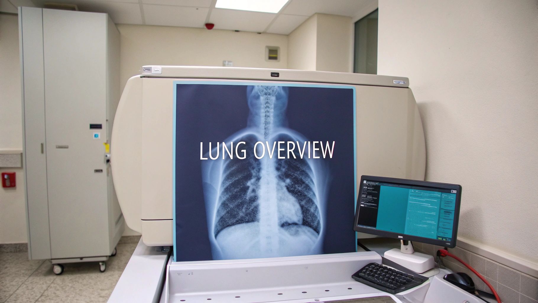
A lung segments CT scan offers a detailed visualization of the bronchopulmonary segments, the fundamental functional units of the lungs. Unlike conventional X-rays, this advanced imaging technique provides exceptional clarity in visualizing these segments, making it essential for modern pulmonary medicine. This enhanced clarity empowers doctors to diagnose and treat a broader spectrum of lung conditions with greater accuracy.
Understanding Bronchopulmonary Segments
Each lung is composed of lobes, which are further divided into bronchopulmonary segments. These segments function as independent units, each receiving its own air supply through a segmental bronchus and blood supply through a pulmonary artery branch. Understanding this unique anatomical structure is crucial as it influences disease progression and treatment strategies. For instance, a disease may initially be confined to a single segment, leaving the others unaffected.
Why Lung Segments CT Scans Matter
Lung segments CT scans enable doctors to precisely locate and determine the extent of abnormalities within the lung. This precision is particularly vital for conditions like lung cancer, where accurate localization is paramount for planning surgical interventions or other targeted therapies. Moreover, these scans allow doctors to distinguish between different lung diseases that may appear similar on conventional imaging methods. This increased diagnostic accuracy is a key advantage of this technology.
Visualizing the Complex Lung Anatomy
Lung segments CT scans illuminate the intricate network of bronchi, arteries, and veins within each segment. The visibility of segmental bronchi, for example, varies across different lung regions. Studies reveal that while almost all segmental bronchi in the right upper lobe are clearly visible on a CT scan, visibility diminishes in the middle and lower lobes. The lingular segmental bronchi, situated in the left upper lobe, pose even greater visualization challenges. For a more in-depth understanding of visualizing complex structures, you can explore various approaches to document analysis methodology. Accurate interpretation of these scans demands specialized training and a thorough understanding of lung anatomy.
The Impact on Patient Care
The detailed information provided by lung segments CT scans contributes to more precise diagnoses and tailored treatment plans. This precision results in improved patient outcomes through more effective interventions and a reduction in side effects. This technology is significantly impacting treatment approaches across pulmonary medicine, from surgical planning to radiation therapy. The ability to visualize and understand individual lung segments allows for highly targeted treatments, minimizing damage to healthy lung tissue.
Cutting-Edge Segmentation Techniques That Actually Work

Beyond visualizing lung segments lies the crucial process of segmentation: distinguishing individual segments and structures within a lung CT scan. This process is essential for accurate diagnosis and treatment planning. Several techniques exist for lung segmentation, each with its own advantages and disadvantages.
Five Major Segmentation Methodologies
Five primary categories of lung segmentation techniques are currently used. Let's explore each one.
-
Thresholding-Based: This method uses differences in pixel intensity to separate lung tissue from other structures. It’s computationally simple, making it relatively fast. However, variations in image quality can impact its accuracy.
-
Region-Based: This approach groups pixels with similar characteristics, forming regions that represent anatomical structures. It performs better with varying image quality than thresholding but can struggle with complex lung shapes.
-
Shape-Based: These techniques use predefined shapes to identify lung segments. They're useful for standardized analysis, but anatomical variations can limit their effectiveness. Not every lung conforms to a standard shape.
-
Neighboring Anatomy–Guided: This method uses the location and shape of surrounding structures, like the ribs and diaphragm, to guide segmentation. It’s helpful in complex cases but requires accurate identification of the neighboring anatomy first.
-
Machine Learning-Based: These techniques use algorithms to learn patterns from data and segment lung structures automatically. This is a rapidly evolving field with the potential for improved accuracy, especially in complex cases. Deep neural networks are a subset of machine learning becoming increasingly important for their high accuracy. Learn more about these varying lung segmentation techniques.
To better understand the differences between these methods, let's look at a comparison table.
To better understand the differences between these methods, the following table provides a comparison.
Comparison of Lung Segmentation Techniques
| Technique | Strengths | Limitations | Best Applications |
|---|---|---|---|
| Thresholding-Based | Computationally simple and fast | Accuracy affected by image quality variations | Cases with consistent image quality and simple lung structures |
| Region-Based | Handles varying image quality better than thresholding | Struggles with complex shapes | Cases with moderate image quality variations and relatively uniform lung structures |
| Shape-Based | Useful for standardized analysis | Limited by anatomical variations | Cases with standard anatomy and limited variations |
| Neighboring Anatomy–Guided | Helpful in complex cases | Requires accurate identification of neighboring anatomy | Cases with complex lung structures where surrounding anatomy is clearly discernible |
| Machine Learning-Based | High potential for improved accuracy, especially in complex cases | Requires large datasets for training | Complex cases with varied pathologies, including ILD, emphysema, and lung cancer |
This table highlights the specific strengths and weaknesses of each technique, assisting medical professionals in selecting the most appropriate method for their particular needs.
The Rise of Machine Learning in Lung Segmentation
Machine learning, particularly deep learning, is transforming lung segmentation. Deep learning networks, such as 2D and 3D U-Net models, have demonstrated impressive results in segmenting lung parenchyma on non-contrast chest CT images. These models are especially effective at segmenting diseases like interstitial lung disease (ILD), emphysema, and lung cancer, ultimately improving diagnostic accuracy.
Deep Learning and Its Impact on Accuracy
Deep learning algorithms excel at analyzing massive datasets to identify subtle patterns often missed by human observers. In lung CT scans, this translates to more accurate identification of segment boundaries, even in complex cases where traditional methods struggle. This improved accuracy has significant implications for patients with advanced pathologies, where precise segmentation is critical for effective treatment planning.
Choosing the Right Technique
The optimal segmentation technique depends on several factors, including the clinical scenario, available resources, and the pathology’s complexity. Leading institutions often combine techniques for the best results. While simpler cases might be adequately addressed with thresholding methods, complex pathologies, such as fibrosis or diffuse lung diseases, benefit from the higher accuracy of machine learning approaches. This informed selection process ultimately enhances diagnostic confidence and improves patient care. Companies like PYCAD, a leader in medical imaging AI, are constantly refining these techniques to enhance the accuracy and efficiency of lung segmentation.
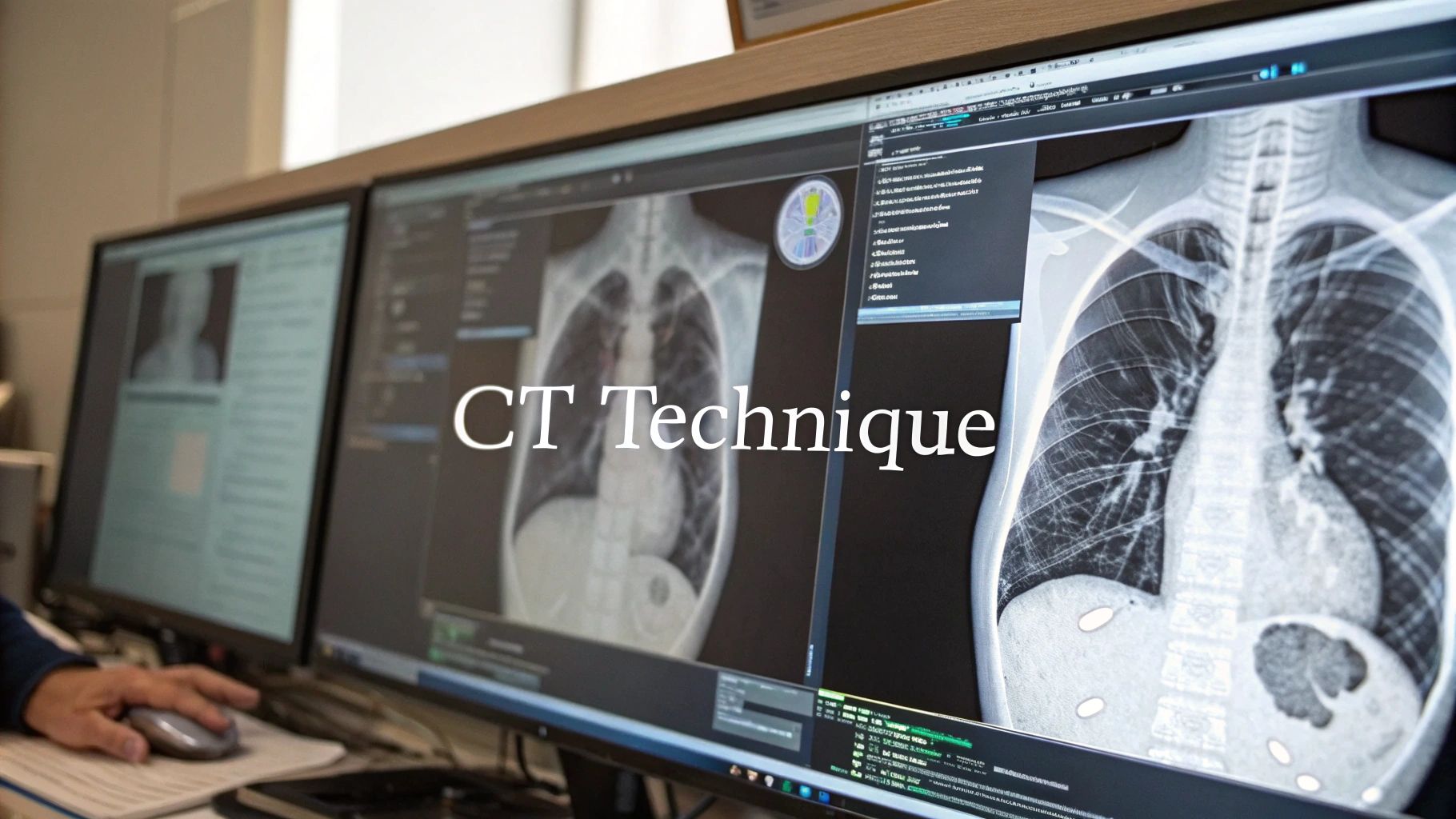
Visualizing Lung Anatomy: The CT Difference
Computed Tomography (CT) scans have become essential for visualizing the complex anatomy of the lungs, especially the bronchopulmonary segments. These segments are the smallest functional units of the lungs, each with its own dedicated air and blood supply. CT scans offer a level of detail not possible with traditional imaging, creating new opportunities for accurate diagnoses and treatments.
Visualizing the Invisible: Why CT Stands Out
CT scans provide superior visualization of lung segments due to their ability to create cross-sectional images. Imagine slicing a loaf of bread; each CT scan slice reveals the lung's internal structure at that specific level. This allows doctors to see individual segments with remarkable clarity, unlike a standard chest X-ray, which presents a flattened, two-dimensional view.
Variability in Visualization: From Upper to Lower Lobes
The clarity of lung segment visualization on a CT scan varies throughout the lungs. The right upper lobe segments are typically easier to see because of their anatomical position and the branching pattern of the bronchi. Visualizing segments in the middle and lower lobes, however, is more challenging because these areas are denser and the bronchi branch in a more complex manner.
This variability highlights the importance of expertise in interpreting CT scans of lung segments. Experienced radiologists use specific techniques to enhance visualization in these challenging areas. These techniques include adjusting the viewing plane, using contrast agents, and employing specialized reconstruction protocols. PYCAD, a company specializing in medical imaging AI, develops tools to refine these techniques further, assisting radiologists in navigating these complexities.
The Lingular Segment Challenge
The lingular segmental bronchi, situated in the left upper lobe, present a particularly difficult visualization challenge. These bronchi are smaller and less obvious than others, often hidden by surrounding lung tissue. This makes identifying abnormalities in this region complex, requiring highly skilled interpretation of CT scans. Understanding the normal anatomical variations within this region is crucial for accurate diagnosis.
Improving Diagnostic Confidence With CT
The enhanced visibility offered by CT scans of lung segments directly improves diagnostic confidence. By clearly defining individual segments, radiologists can pinpoint the precise location and extent of abnormalities. This allows for a more accurate evaluation of the disease process. This precise localization is essential for guiding treatment decisions, whether it involves surgical intervention, radiation therapy, or other targeted approaches.
Research Reinforces CT's Effectiveness
A study focusing on CT identification of bronchopulmonary segments highlighted the importance of this technology. Involving 50 patients, the study demonstrated CT's effectiveness in visualizing segmental bronchi and arteries for detailed segmentation. In the right lung, almost all segmental bronchi in the upper lobe were visible, while over half were identifiable in the middle and lower lobes. The lingular segmental bronchi, however, were more elusive, with less than half being visible. This detailed visualization improves the precise diagnosis and management of lung conditions, allowing healthcare providers to target specific segments. You can explore this topic further in this research publication.
Transforming Treatment Through Segment-Specific Analysis
Analyzing lung segments CT scans at the segmental level is significantly changing how doctors approach treatment for various pulmonary conditions. This detailed analysis provides more precise interventions, ultimately sparing healthy lung tissue and improving therapeutic effects. The implications are far-reaching across multiple medical disciplines, including thoracic surgery, interventional pulmonology, and radiation oncology.
Precisely Targeted Interventions: Maximizing Impact, Minimizing Harm
Pinpointing the exact location and extent of disease within individual lung segments allows for highly targeted interventions. This maximizes the therapeutic impact while minimizing damage to healthy tissue. Treatments are focused precisely where needed, reducing side effects and leading to better patient outcomes. For instance, if a tumor is limited to a single segment, surgeons can remove only the affected area, preserving more of the lung.
Thoracic Surgery: Planning Less Invasive Procedures
Thoracic surgeons are increasingly using segmental mapping from CT scans to plan video-assisted thoracoscopic surgery (VATS). This detailed roadmap enables less invasive procedures, resulting in smaller incisions, less pain, and faster recovery times. VATS also carries a lower risk of complications compared to traditional open-chest surgery. This focus on precision enhances patient comfort and speeds up healing. A segmentectomy, the removal of only the diseased segment, exemplifies this minimally invasive approach, preserving as much lung function as possible.
Interventional Pulmonology: Guiding Bronchoscopic Interventions
Interventional pulmonologists utilize segment-specific diagnoses from CT scans to guide bronchoscopic interventions with increased accuracy. They can navigate the complex bronchial tree and reach the target area with remarkable precision. This accuracy is essential for biopsies, stent placements, and other procedures, minimizing procedural risks and optimizing treatment effectiveness. This precise navigation is akin to using a GPS system within the lungs, ensuring the correct location is reached efficiently.
Radiation Oncology: Delivering Targeted Radiation With Precision
Radiation oncologists utilize segment-specific maps from CT scans to precisely deliver stereotactic body radiation therapy (SBRT). This technique delivers high doses of radiation directly to the tumor while sparing the surrounding healthy lung tissue. SBRT is a valuable tool in treating lung cancer and other localized lung diseases, offering the potential for tumor control with fewer side effects. Maintaining healthy lung function is paramount for overall patient well-being, and the precision of SBRT helps achieve this. Companies like PYCAD, specializing in medical imaging AI, are developing tools that further refine these techniques, enabling even greater precision in radiation treatment planning.
Improved Patient Outcomes Across Diverse Pulmonary Conditions
These real-world applications demonstrate that segment-level precision from CT scans leads to improved patient outcomes across a spectrum of pulmonary conditions. Whether the treatment involves surgical resection, a bronchoscopic intervention, or targeted radiation therapy, segment-specific analysis provides a more refined and effective approach. This translates to faster recoveries, fewer complications, and a better quality of life for patients. This focus on precision is shaping the future of pulmonary medicine and driving ongoing improvements in patient care.
Overcoming Complex Challenges in Lung Segmentation
Lung segmentation from CT scans offers powerful diagnostic capabilities, but it also presents unique challenges. Accurately defining the boundaries of lung segments becomes particularly difficult when certain pathologies are present. This impacts the precision of both diagnosis and treatment planning, especially for conditions like COVID-19 pneumonia and advanced fibrosis.
Why Some Pathologies Pose Segmentation Obstacles
Diseases that alter the normal architecture of the lungs, such as fibrosis, create blurry and indistinct boundaries between segments. Similarly, the patchy inflammation characteristic of COVID-19 pneumonia disrupts normal tissue density, making accurate segmentation a challenge. These situations require advanced techniques to overcome these inherent limitations.
Dual-Energy CT: Enhancing Tissue Characterization
Dual-energy CT (DECT) is emerging as a valuable tool in addressing these segmentation challenges. By acquiring images at two different energy levels, DECT provides more detailed information about tissue composition. This enhanced tissue characterization helps differentiate between healthy and diseased tissue, even when their densities appear similar on conventional CT scans. This is especially useful in cases of fibrosis where subtle density changes can indicate the extent of the disease.
Texture Analysis: Unveiling Parenchymal Changes
Texture analysis, a technique that quantifies subtle variations within the lung parenchyma (the functional tissue of the lung), offers another avenue for improved segmentation. This method analyzes the spatial arrangement and intensity of pixels in a CT image to identify patterns indicative of specific pathologies. This detailed analysis provides deeper insights into parenchymal changes, aiding in accurate segment boundary delineation, even with diffuse diseases like Interstitial Lung Disease (ILD).
Hybrid Segmentation Approaches: Combining Strengths for Accuracy
Combining traditional and machine-learning-based segmentation methods creates hybrid approaches that significantly improve accuracy in challenging cases. By incorporating anatomical knowledge from traditional methods with the pattern recognition capabilities of machine learning, these approaches offer more robust segmentation, even with distorted lung architectures. A hybrid approach might use shape-based segmentation for an initial estimate, then refine it using deep learning algorithms to account for anatomical variations and disease-related changes.
Handling Motion Artifacts and Anatomical Variations
Motion artifacts, caused by patient movement during the scan, can blur images and hinder accurate segmentation. Similarly, natural anatomical variations between individuals can complicate segment identification. Advanced reconstruction techniques and motion correction algorithms minimize the impact of motion artifacts, while statistical shape models account for anatomical variations, ultimately improving diagnostic quality.
Practical Strategies for Interpreting Challenging Cases
Practical strategies, like adjusting image windowing and leveling to optimize visualization, are essential for interpreting challenging cases. Furthermore, correlating CT findings with other clinical data, such as pulmonary function tests and lab results, enhances diagnostic accuracy and guides treatment decisions. These combined approaches ensure confident navigation of the complexities inherent in lung segment CT scan interpretation.
Lung segmentation plays a vital role in diagnosis and treatment planning. The Lung CT Segmentation Challenge (LCTSC) highlights this importance, where algorithms are compared for their accuracy in segmenting organs at risk during radiotherapy planning. This focus underscores the growing need for accurate automated segmentation in clinical settings. Explore this topic further by learning more about applications and competitions in lung segmentation.
From Scan to Diagnosis: A Practical Interpretation Guide
This guide provides a practical framework for interpreting lung segment CT scans. It incorporates insights from leading thoracic radiologists and offers a structured methodology for ensuring thorough and accurate evaluations, ultimately leading to better patient care. A systematic approach is crucial for avoiding missed critical findings, especially given the complexities of lung anatomy.
Anatomical Orientation: The First Step
Accurate interpretation begins with proper anatomical orientation. This involves correctly identifying the right and left lungs, their lobes, and the individual segments within each lobe. Think of the lungs like a map: understanding the landmarks is crucial for accurate navigation. This initial step is fundamental to all subsequent analyses and ensures accurate localization of any abnormalities.
Systematic Evaluation of Each Segment: A Step-by-Step Approach
Next, systematically evaluate each lung segment using a predefined sequence. This consistent approach minimizes the risk of overlooking subtle pathologies. For example, you might start with the right upper lobe, methodically examining each of its segments. Then, progress to the middle and lower lobes before repeating the process for the left lung.
Standardized Terminology: Enhancing Communication
Using precise anatomical terminology is vital for clear communication between radiologists and clinicians. Instead of a vague description, use specific terms like "apicoposterior segment of the left upper lobe". This precision eliminates ambiguity and facilitates collaborative discussions about patient care and treatment planning.
Measurement Standards: Ensuring Consistency
Consistent measurement standards are essential for tracking disease progression and response to therapy. Quantifying nodule size, for instance, requires adherence to established protocols, enabling accurate comparisons over time. This consistency is vital for informed treatment decisions and accurate prognosis assessment.
To assist in the systematic interpretation of lung segments on CT scans, the following checklist can be used:
Lung Segments CT Scan Interpretation Checklist
A systematic approach for radiologists and clinicians to evaluate lung segments on CT scans
| Anatomical Region | Normal Appearance | Common Variants | Key Pathologies |
|---|---|---|---|
| Right Upper Lobe | Clear lung fields, well-defined segmental boundaries | Accessory fissure | Pneumonia, Lung Cancer, Granuloma |
| Right Middle Lobe | Clear lung fields, distinct segmental bronchi | Absent middle lobe | Bronchiectasis, Atelectasis |
| Right Lower Lobe | Clear lung fields, basal segments clearly visible | Subsegmental atelectasis | Pulmonary Embolism, Pleural Effusion |
| Left Upper Lobe | Clear lung fields, lingular segment sometimes difficult to visualize | Incomplete fissure | Tuberculosis, Aspergilloma |
| Left Lower Lobe | Clear lung fields, similar appearance to right lower lobe | Basilar atelectasis | Pneumonia, Lung Cancer |
This table provides a quick reference for expected normal appearances, common anatomical variations, and potential pathologies within each lung segment. Using this checklist can help ensure a comprehensive evaluation and improve diagnostic accuracy.
Distinguishing Similar Pathologies: Mastering Subtle Differences
Many lung pathologies appear similar on CT scans. Mastering the subtle differences between conditions, such as a benign granuloma and a malignant nodule, significantly impacts treatment choices. This distinction often involves evaluating features like shape, margin characteristics, and density, and frequently requires correlation with clinical and laboratory data.
Real Case Examples: Bringing Theory to Practice
Real-world examples demonstrate these principles in clinical practice. Imagine a patient presenting with cough and shortness of breath. A lung segment CT scan reveals a ground-glass opacity confined to a single segment. Correlating these findings with the patient's symptoms and lab results can lead to a diagnosis of segmental pneumonia.
Correlation With Clinical Data: Enhancing Diagnostic Accuracy
Integrating CT scan information with clinical symptoms and laboratory findings is crucial for enhancing diagnostic accuracy. This comprehensive approach considers the patient’s overall health, not just the imaging data. This holistic view is essential for optimal patient management.
Ready to experience the power of AI in medical imaging? Discover PYCAD and learn how our solutions are transforming diagnostic accuracy and operational efficiency for medical device manufacturers and healthcare providers.


