Picture a future where diseases are spotted sooner, diagnoses are sharper, and treatments are perfectly matched to each patient. This isn't a scene from a futuristic movie; it's the reality taking shape right now, thanks to machine learning in medical imaging. This remarkable field is rapidly becoming an indispensable partner for radiologists and doctors around the globe.
The Dawn of a New Era in Medical Diagnostics
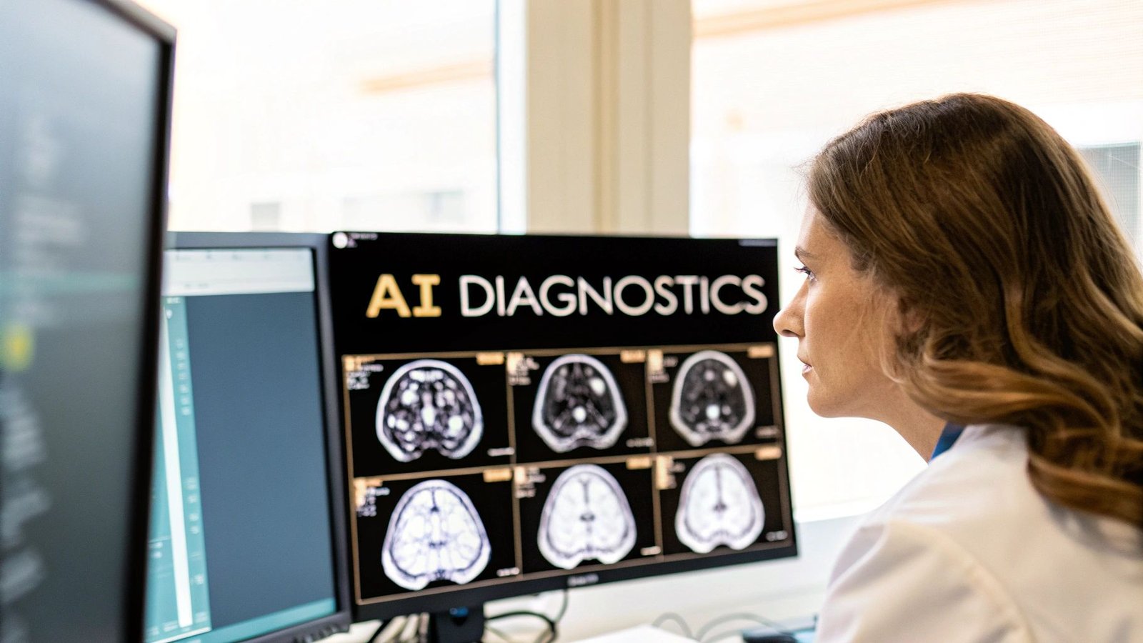
Think of machine learning in this context as an incredibly skilled co-pilot for healthcare professionals. It can sift through millions of medical images—from MRIs to CT scans—and pick up on subtle patterns that the human eye might miss. These algorithms are trained to spot anomalies and early signs of disease with incredible precision, giving specialists a powerful second opinion to solve the most complex medical puzzles.
This isn't about replacing the irreplaceable intuition and experience of doctors. It’s about supercharging their abilities. Machine learning takes on the heavy lifting of analyzing enormous amounts of data, freeing up clinicians to focus on what truly matters: critical thinking, patient connection, and crafting the best possible care plans.
Why This Matters for Healthcare
Weaving machine learning into our medical systems has a ripple effect that touches every corner of healthcare. It directly tackles some of our biggest hurdles, like the sheer volume of data and the need for faster, more accurate diagnoses.
The benefits are game-changing:
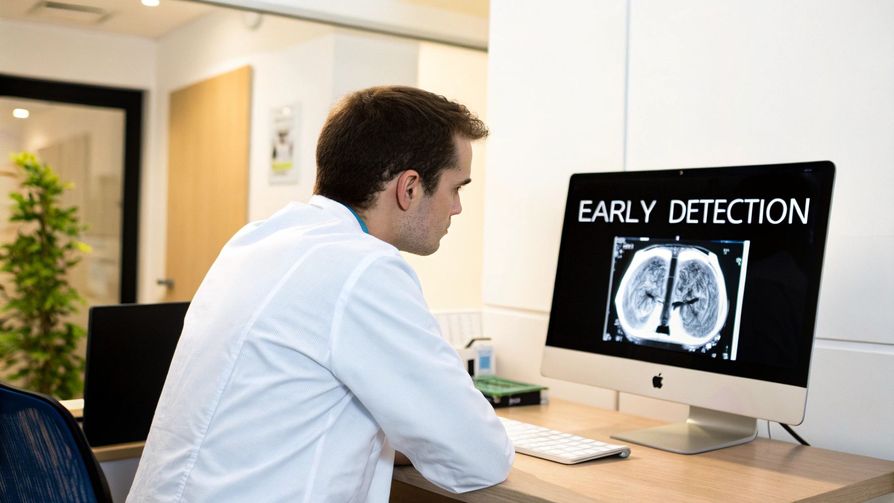
- Catching Diseases Earlier: Sophisticated algorithms can identify tiny cancerous nodules or the faintest signs of neurological conditions long before they might otherwise be seen.
- Bringing Precision to the Forefront: By measuring details like tumor volume or blood flow with objective accuracy, AI helps eliminate guesswork and leads to more definitive diagnoses.
- Crafting Personalized Treatments: AI can even help predict how a patient might respond to a specific therapy, guiding doctors toward truly customized care.
We're at the beginning of a fundamental shift. We’re moving from a healthcare model that reacts to disease to one that can predict it, changing patient outcomes for the better on a global scale.
The momentum here is undeniable. The global medical imaging market, valued at about USD 43.9 billion in 2024, is expected to soar to USD 75.8 billion by 2034. This incredible growth is driven by the urgent need for smarter diagnostic tools that can keep up with the ever-increasing amount of imaging data. You can explore more about these market trends and what they mean for the future of healthcare innovation.
To make this all work, machine learning medical imaging needs to fit seamlessly into the day-to-day clinical workflow. That’s where we come in. At PYCAD, we specialize in building custom web DICOM viewers and integrating them into medical imaging platforms. We create the bridge that allows clinicians to interact with these powerful AI insights effortlessly. Our mission is to ensure these incredible tools are practical, accessible, and ready to make a real difference in people's lives. Take a look at the advanced platforms we've built by visiting our portfolio.
How Machines Learn to See Inside the Human Body
How does a piece of software actually learn to read a complex CT scan or a detailed MRI? It’s not about programming a computer with a rigid set of rules. Think of it more like teaching a medical student. The whole journey begins with a core concept in machine learning medical imaging called supervised learning.
Picture a massive digital library—one filled with thousands, sometimes millions, of medical images. Each one has been carefully annotated by expert radiologists. A lung nodule is circled and tagged "cancerous," a brain lesion is outlined and identified as a sign of multiple sclerosis, and a healthy organ is simply marked "normal." The machine learning model studies this huge collection, learning to connect specific visual patterns with their labels. Just like a student pouring over textbooks and case studies, the algorithm trains itself to spot the telltale signs of disease.
This learning process is driven by some incredibly powerful tools, with the most important being Convolutional Neural Networks (CNNs).
The Digital Eyes of AI
You can think of a CNN as a set of digital eyes, built to mimic the way the human brain’s visual cortex processes information. Instead of taking in the entire image all at once, a CNN breaks it down into tiny, manageable pieces. It scans the image with a series of filters, and each filter is trained to look for something very specific.
- Simple Features: The first layers of the network might learn to spot basic elements like edges, corners, and simple textures.
- Complex Patterns: Deeper layers then start combining these simple features into more complex patterns—like the unique texture of a tumor or the distinct shape of an organ.
- Final Identification: Finally, the network pulls all this information together to make a prediction, like classifying an image as either benign or malignant.
This layered approach lets the model build a rich, sophisticated understanding of the visual data. It isn't just memorizing pictures; it's learning the fundamental visual building blocks that define different anatomical structures and diseases. This is a core idea you can explore further by understanding what computer vision is and how it works.
Through this layered analysis, a CNN can identify subtle patterns that might be nearly invisible to the human eye. It can pick up on minute changes in tissue density or faint structural abnormalities, offering a new level of perception that beautifully complements a radiologist's own expertise.
From Learning to Practical Application
Once a model is fully trained, it can perform specific, often life-saving, tasks with incredible speed and accuracy. Two of the most common jobs it's given in machine learning medical imaging are image classification and segmentation. These two tasks are absolutely essential for turning raw pixels into insights that can actually help patients.
1. Image Classification: Answering "What Is This?"
This is the most straightforward application. The algorithm looks at an entire medical image—say, a chest X-ray—and assigns it a single label. It might classify the X-ray as showing signs of pneumonia, tuberculosis, or being perfectly healthy. This gives clinicians a quick, data-backed assessment that can help them prioritize which cases need their attention first.
2. Image Segmentation: Drawing the Lines
Segmentation is a far more detailed task. Instead of just labeling the whole image, the algorithm outlines the precise boundaries of a specific object. It might trace the exact contour of a tumor on a brain MRI or measure the volume of a heart chamber from an echocardiogram. This pixel-level precision is vital for planning treatments, like guiding radiation therapy, or for tracking how a disease is progressing over time.
These powerful capabilities depend on sophisticated platforms that can handle and display complex medical data. At PYCAD, this is exactly what we specialize in. We build custom web DICOM viewers and integrate them into medical imaging web platforms, creating the seamless interface clinicians need to interact with these AI-driven insights. Our work ensures that the powerful outputs from these models are presented clearly and intuitively, making them truly useful in a real-world clinical setting. We invite you to explore our portfolio page to see how we bring these solutions to life.
Building the AI Medical Imaging Pipeline
Turning a raw medical scan into a life-saving insight isn't a single magic trick; it's a carefully choreographed journey. A truly effective machine learning medical imaging model comes to life through a solid, end-to-end pipeline. Think of it as a blueprint, where each step meticulously builds on the last to create a tool that’s not just powerful, but also worthy of a clinician's trust.
This journey starts long before an algorithm even sees its first pixel.
Data Acquisition and Preprocessing
The first and, frankly, most critical step is data acquisition and preprocessing. High-quality, diverse datasets are the absolute bedrock of any successful AI model. If you're curious about what goes into building these foundational resources, this guide on medical image datasets is a fantastic place to start.
This phase is all about gathering thousands of images and—this is key—making them consistent. Medical images, like DICOM files, often come from different scanners with their own unique settings. Preprocessing is the great equalizer. It normalizes things like brightness, contrast, and resolution, creating a clean, uniform dataset that's ready for the main event: training.
Training and Validating the Model
With our pristine dataset in hand, we move on to model training. This is where the machine truly learns to see. We feed the preprocessed images and their corresponding labels into powerful architectures like Convolutional Neural Networks. Through millions of tiny adjustments, the model starts to pick up on the subtle patterns and textures that separate a healthy organ from a diseased one.
But building a smart model is only half the battle. We have to prove it's reliable. That’s where rigorous validation comes in. We test the AI on a completely new set of images it has never seen before to gauge its real-world performance. By calculating metrics like accuracy, sensitivity, and specificity, we make sure the model can apply what it’s learned to new cases, not just "remember" the answers from its training data.
The infographic below beautifully captures this flow—from meticulously labeled data to a final, insightful diagnosis.
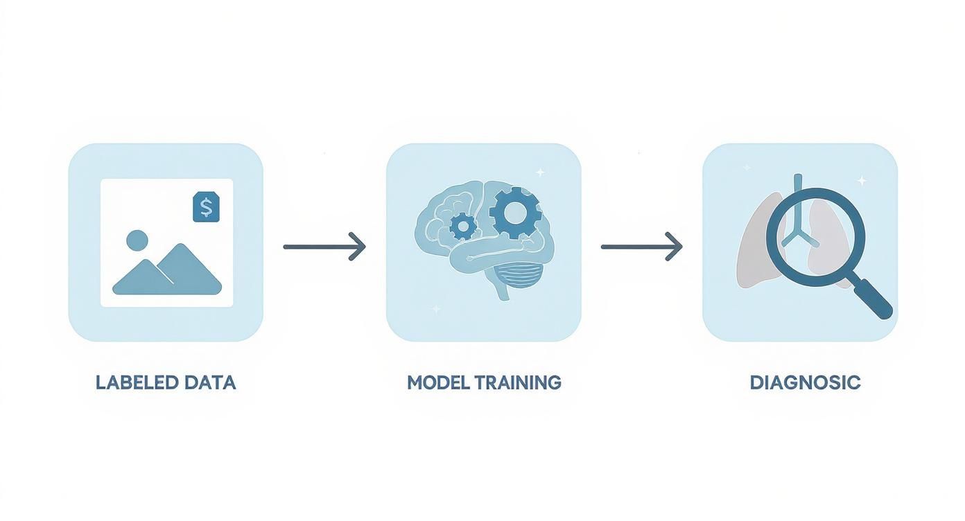
It’s a clear visual reminder: a successful diagnosis is born from a well-trained model, which, in turn, is completely dependent on expertly curated data.
Deploying Insights into Clinical Workflows
The final piece of the puzzle is deployment. An incredible AI model sitting on a server is useless. Its true value is only unlocked when its insights are woven directly into a real-world clinical workflow, transforming it from a brilliant algorithm into a practical tool for doctors.
This means plugging the AI into existing hospital systems, like the PACS (Picture Archiving and Communication System), so its analysis is right there when a radiologist opens a patient's scan.
The ultimate goal of deployment is to make advanced AI insights feel like a natural extension of a clinician's own diagnostic toolkit—intuitive, immediate, and actionable.
This is exactly where our expertise at PYCAD shines. We build custom web DICOM viewers and integrate them into medical imaging web platforms. These viewers are the bridge between the AI's powerful analysis and the medical professionals who rely on it. A great viewer doesn't just show a picture; it presents the AI's findings—highlighted tumors, precise segmentations—in a way that's interactive and instantly understandable.
Our solutions ensure clinicians can effortlessly engage with AI-generated data, compare it side-by-side with the original scans, and fold it into their diagnostic process. By building these intuitive platforms, we close the gap between complex code and better patient care. The final step is what turns a laboratory success into a clinical game-changer. Check out our portfolio page to see what we build.
Real-World Applications Transforming Patient Care
This is where the theory truly comes to life—where algorithms become trusted allies and data turns into saved lives. The potential of machine learning medical imaging is no longer just a concept; it’s making a real, tangible impact in clinics and hospitals everywhere. From busy oncology wards to specialized neurology centers, these tools aren’t just making diagnostics better; they're changing the entire patient experience for the better.
Think of each application as a story: a tough clinical challenge met with a brilliant, AI-powered solution. These aren't futuristic dreams. They are today's realities, giving clinicians new abilities and offering genuine hope to patients across the globe.
A Sharper Eye in Oncology
In the relentless fight against cancer, catching it early is often the most powerful weapon we have. Machine learning is giving us a remarkable new edge, particularly in breast cancer screening. AI models, trained on millions of mammograms, can now spot subtle, early-stage cancers with an accuracy that complements—and sometimes even surpasses—a human expert.
These systems work like a vigilant second pair of eyes, flagging tiny, suspicious areas that might otherwise be missed during a hectic day in the clinic. This means earlier diagnoses, which opens the door to more effective treatment options and, most importantly, better outcomes for patients. It’s a perfect example of technology amplifying human skill.
To bring these capabilities to the forefront, clinicians need the right tools. At PYCAD, we build custom web DICOM viewers and integrate them into medical imaging web platforms, making it effortless for radiologists to see and work with AI-generated insights. You can see examples of what we've built on our portfolio page.
Precision and Clarity in Neurology
For someone living with a condition like multiple sclerosis (MS), tracking how the disease is changing over time is absolutely crucial. This usually means a neurologist has to painstakingly analyze MRIs to measure the size and number of brain lesions—a process that can be both slow and subjective.
This is where machine learning algorithms shine. They can perform automated lesion segmentation, precisely outlining and measuring these areas on an MRI scan to provide objective, repeatable data.
This consistent tracking gives neurologists a much clearer picture of how a patient is responding to treatment. It replaces guesswork with a solid, data-driven foundation for making critical care decisions.
This level of detail gives patients and their families a clearer understanding of their own health journey, turning complex scan data into insights that help guide the path forward.
Empowering Cardiology with Quantitative Insights
The heart is an incredibly complex, dynamic organ. Assessing its function is key to diagnosing and managing cardiovascular disease. While cardiologists use tools like echocardiograms to see the heart in motion, quantifying its performance—like the ejection fraction—can still be a challenge.
AI is stepping in to automate this analysis. Machine learning models can instantly identify the heart's chambers on an echocardiogram and calculate key functional metrics with incredible precision.
This gives cardiologists objective data they can trust when planning treatments, from medication changes to surgical procedures. The speed and accuracy of these tools create more efficient workflows and foster more confident clinical decisions. By analyzing huge volumes of data from CT scans and X-rays to find subtle abnormalities, these tools are vital for early detection. You can discover more insights on the AI in medical imaging market to grasp the scale of this shift.
To see the bigger picture of how artificial intelligence is reshaping diagnostics and patient care, exploring an article on How AI BI Transforms Healthcare Analytics can provide a broader perspective. These applications are just the dawn of a new era in medicine, one where human expertise and intelligent technology collaborate to create a higher standard of care for everyone.
Navigating the Challenges on the Path to Adoption
The promise of weaving machine learning into the fabric of daily clinical practice is incredible, but the path forward isn't without its obstacles. While the potential of machine learning medical imaging is genuinely inspiring, getting there responsibly means tackling some serious hurdles head-on. Think of them less as roadblocks and more as crucial checkpoints that guarantee these powerful tools are safe, fair, and ultimately, trustworthy.
One of the first and most critical issues is data privacy and security. Medical data is intensely personal, guarded by stringent regulations like HIPAA. Since AI models need massive amounts of this sensitive information to learn, the demand for iron-clad security isn't just a feature—it's a necessity.
Tackling the challenges of adopting machine learning in medical imaging, particularly around data privacy and compliance, is non-negotiable. Strong data governance best practices are the bedrock for handling patient data ethically and securely. This ensures that even as we push the boundaries of innovation, we never compromise a patient’s fundamental right to privacy.
The Quest for Fair and Unbiased AI
Beyond just securing the data, we have to grapple with its quality and diversity. An AI model, at its core, is a reflection of the data it was trained on. If a dataset is skewed—say, it mostly contains images from one demographic—the final algorithm will likely struggle when it encounters patients from underrepresented groups.
This is how algorithmic bias creeps in, a critical flaw where a model can accidentally reinforce or even worsen existing health disparities. It’s a massive undertaking to build large, diverse, and perfectly labeled datasets, but it's the only way to create AI that works for everyone.
Unpacking the Black Box
Another major hurdle is the infamous "black box" problem. Many of today's deep learning models are phenomenal at reaching accurate conclusions, but their internal logic can be a complete mystery. For a clinician, just getting an answer isn't enough; they need to understand how the AI came to its conclusion.
For a doctor to trust an algorithm with a patient's health, they need confidence in its reasoning. Model interpretability isn't just a technical feature; it's the foundation of clinical trust.
This is precisely why there's a growing push for explainable AI (XAI). These techniques are designed to pull back the curtain on a model's thinking, showing exactly which pixels in an image led to its decision. A transparent model is a trustworthy model. This focus on rigorous testing is central to the entire development pipeline. You can dive deeper into the methods used to ensure AI reliability by exploring our guide on https://pycad.co/what-is-model-validation/.
Finally, we can't forget the regulatory landscape. Any AI software used in a medical context is considered a medical device and faces intense scrutiny from bodies like the FDA. Getting approval is a long and demanding journey, requiring extensive validation and meticulous documentation to prove an algorithm is both safe and effective.
At PYCAD, we live and breathe these complexities. We build custom web DICOM viewers and integrate them into medical imaging web platforms, with security and usability as our north star. Our solutions are built to deliver AI insights in a clear, understandable way, bridging the gap between sophisticated algorithms and the clinicians who depend on them. You can see our commitment to building practical, reliable systems by exploring our portfolio page. By facing these challenges with care and persistence, we can help build a future where AI stands as a powerful and trusted ally for healthcare professionals.
The Future Horizon of Intelligent Medical Imaging
The journey into machine learning for medical imaging has brought us to an incredible place. From here, we can see the dawn of a truly predictive and personalized era in medicine. What’s on the horizon isn't just a slightly better version of today’s tech; it’s a complete reimagining of what healthcare can be.
We’re moving toward a future where our tools don't just find diseases—they see them coming.
Imagine a world where an algorithm, trained on global data without ever seeing a single patient's private information, can predict your personal risk for a disease years before symptoms ever show up. This is the promise of concepts like federated learning, which allows models to learn collaboratively while keeping sensitive data completely secure.
Pushing the Boundaries of Data and Discovery
One of the most exciting frontiers is the use of generative AI to create synthetic medical data. Think about it: this gives researchers the ability to train algorithms on massive, diverse, and perfectly balanced datasets without ever touching real patient records.
It’s like having an infinite, privacy-safe library to build the next generation of diagnostic tools, speeding up breakthroughs in a way that’s both safe and ethical.
This kind of innovation is fueling explosive growth. The global market for AI in medical imaging is skyrocketing, with forecasts projecting a compound annual growth rate somewhere between 22.4% and 34.8% in the coming years. This surge, especially in regions with advanced healthcare systems, shows a deep, industry-wide commitment to making these intelligent tools a core part of clinical practice. You can read the full research about these market projections to grasp just how big this shift really is.
A New Partnership in Medicine
As the technology gets smarter, the role of the clinician will evolve right along with it. Routine analytical tasks will become more and more automated. This doesn't make radiologists obsolete; it frees them from the repetitive work so they can focus their incredible expertise on the most complex and nuanced cases.
They will shift from being interpreters of images to becoming strategic clinical guides, armed with powerful insights from their AI partners.
This future isn’t about replacing the physician. It's about augmenting their expertise, creating a powerful human-AI partnership that will define the next generation of medicine and bring unprecedented clarity to patient care.
The vision is crystal clear: a future where intelligent systems and human intuition work together seamlessly. This powerful synergy will unlock new possibilities, turning mountains of complex data into life-saving actions and paving the way for a healthier world for everyone.
At PYCAD, we're committed to building the very infrastructure for this future. We build custom web DICOM viewers and integrate them into medical imaging web platforms, creating the practical, intuitive interfaces needed to bring AI insights into the real world of clinical care. Explore the platforms we’ve brought to life on our portfolio page.


