In the world of modern radiology, PACS and RIS are the two indispensable pillars that hold everything together. Think of them as two sides of the same coin, each with a unique job, but working in perfect harmony to manage medical imaging. A Picture Archiving and Communication System (PACS) is the high-security digital vault for all medical images—MRIs, CT scans, X-rays, you name it. Meanwhile, a Radiology Information System (RIS) is the air traffic controller, managing the entire patient journey from start to finish.
When these two powerhouses work together, they create a fluid, highly efficient environment for diagnostics.
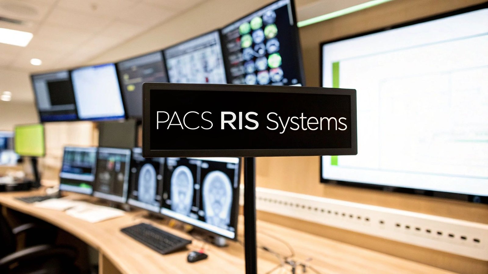
The Digital Heartbeat of Modern Radiology
Let's use an analogy. Imagine a busy hospital's imaging department is like a sophisticated, bustling library. Every single medical image is a detailed, irreplaceable book. In this scenario, PACS is the library's massive, state-of-the-art digital archive. It’s where every "book" is securely stored, meticulously cataloged, and made available in an instant to the right doctors. Its entire focus is on the images themselves.
The RIS, on the other hand, is the master librarian. This system doesn't hold the books; it orchestrates everything happening around them. The RIS is the brains behind the operation, managing the "who, what, when, and why" of the whole imaging process.
It’s the system responsible for:
- Patient Scheduling: Juggling appointments and keeping the calendars for all the imaging machines in check.
- Order Entry: Taking in and organizing scan requests from other physicians.
- Patient Tracking: Keeping a close eye on a patient's progress through the department, from the moment they walk in to when their exam is done.
- Reporting and Billing: Helping radiologists create their reports, sending those reports where they need to go, and handling the billing details.
To put it simply, PACS manages the image, while RIS manages the workflow. When these two are connected, they truly become the digital heartbeat of any modern imaging center.
PACS vs RIS Core Functional Roles
To really grasp how these systems create such a powerful duo, it helps to see their core responsibilities side-by-side. One is the guardian of the clinical data (the image), while the other is the conductor of the entire administrative and logistical orchestra.
| Function | PACS (The Digital Image Archive) | RIS (The Workflow Conductor) |
|---|---|---|
| Primary Focus | Securely storing, retrieving, and displaying medical images. | Managing the entire operational workflow of the radiology department. |
| Key Tasks | Image acquisition, long-term archiving, distribution to clinicians. | Patient scheduling, registration, exam ordering, and billing. |
| Core Data | DICOM image files (MRIs, CTs, X-rays, Ultrasounds). | Patient demographics, exam schedules, billing codes, and reports. |
| Main Users | Radiologists, specialists, and referring physicians who need to view images. | Radiologists, administrative staff, technologists, and billing departments. |
This clear separation of duties is what makes the whole process so organized and efficient. For a deeper look into the mechanics of this digital archive, check out our guide on what is PACS in healthcare.
The synergy between PACS and RIS does away with the old, clunky manual processes that used to slow down radiology departments, paving the way for a smarter workflow.
By digitizing both image storage and workflow management, integrated PACS RIS systems lead to faster diagnoses, better collaboration among medical teams, and ultimately, a higher standard of patient care. It’s a move from disconnected steps to a unified, data-driven operation.
Here at PYCAD, we take these systems to the next level. We specialize in building custom web DICOM viewers and integrating them directly into medical imaging web platforms, giving clinicians diagnostic tools that go far beyond the basics.
To see how we’re pushing the boundaries of what’s possible in medical imaging, feel free to explore our portfolio page. The integration of PACS and RIS isn't just a technical upgrade; it's about building the very foundation for a more intelligent and responsive future in healthcare.
How PACS and RIS Communicate Seamlessly
While PACS and RIS are powerful on their own, their real magic happens when they work together. This isn't just about plugging one system into another; it's a carefully choreographed dance of data, ensuring every bit of information gets where it needs to be, right when it's needed.
Think of it like a symphony orchestra. For everyone to play in harmony, they need a shared sheet of music. In the world of pacs ris systems, that music is written in two essential languages: DICOM and HL7. These standards are the universal translators that allow different systems, often from different manufacturers, to speak to one another without missing a beat.
The Role of DICOM and HL7
DICOM, which stands for Digital Imaging and Communications in Medicine, is the global gold standard for all things medical imaging. It’s the language that MRI machines, CT scanners, and X-ray units use to package and send images to the PACS. This is what guarantees that a scan taken on a GE machine can be perfectly viewed and analyzed on a workstation running Siemens software.
On the other side of the coin, we have HL7 (Health Level Seven). This is the language of healthcare administration and logistics. It handles all the non-image data—patient demographics, appointment schedules, exam orders, and the final diagnostic reports. HL7 is the thread that connects the RIS to the hospital's main Electronic Health Record (EHR), keeping everyone in sync with the latest patient information.
The real harmony comes from DICOM and HL7 working in concert. DICOM manages the "picture," while HL7 handles the "paperwork." Together, they create a seamless patient journey from check-in to diagnosis.
The flow is remarkably elegant. An imaging order created in the RIS kicks off a process that moves through the imaging modality and ultimately lands in the PACS for a radiologist to review.
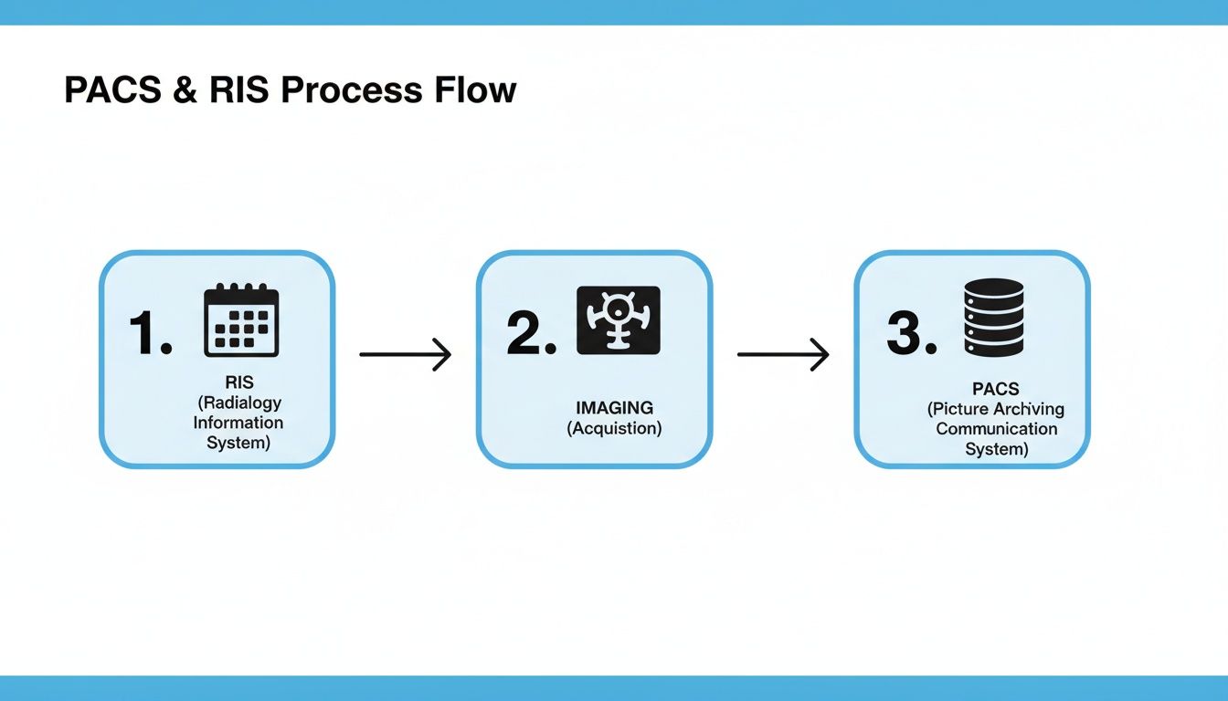
This diagram shows just how distinct yet interconnected each system's role is, all designed to drive efficiency and accuracy.
Tracing the Patient Data Journey
Let’s follow the data to see how this communication plays out in a real-world scenario. The whole workflow is a masterclass in automation, designed to slash manual data entry and minimize the risk of human error.
-
Order Placement in RIS: It all starts when a physician orders a chest X-ray. The order is placed in the RIS, which already holds the patient’s details and the specific instructions for the exam. This single action sets the entire chain in motion.
-
HL7 Transmits the Order: The RIS instantly sends an HL7 message to the X-ray machine. This message populates a worklist for the technologist, telling them exactly which patient is next and what scan to perform. No more typos or mix-ups.
-
Image Acquisition and DICOM Transmission: The technologist performs the X-ray. The machine captures the image and wraps it in a DICOM file—a smart container that holds not just the image but also critical metadata like the patient’s ID and scan parameters. The machine then sends this DICOM file directly to the PACS. If you want to get into the weeds of this crucial protocol, our guide on the DICOM communication protocol is a great place to start.
-
PACS Archives and Notifies: The PACS receives the DICOM file, securely archives it, and links it to the patient’s records. At the same time, it sends a message back to the RIS, confirming the images are ready for interpretation. This simple notification alerts the radiologist that a new study is in their queue, ready for their expert eye.
This beautifully synchronized process is the backbone of any modern radiology department. It protects data integrity, shaves down diagnostic turnaround times, and frees clinicians to focus on what they do best: taking care of patients.
Choosing Your System Architecture
Deciding where your PACS and RIS systems will live is one of the most important calls you'll make. It’s not just a technical choice; it’s a strategic one that will impact your budget, your daily workflow, and how you grow in the years to come. Think of it as deciding on the foundation for your entire imaging operation. Each option—on-premise, cloud, or a mix of both—comes with its own set of trade-offs in control, cost, and convenience.
The Fortress: On-Premise Systems
Going with an on-premise system is like building and owning your own house from the ground up. You control everything. The servers hum away in your own data center, guarded by your own security protocols, and managed by your own IT team. This gives you absolute authority over your data, which is a massive plus for large hospitals or research institutions with strict data sovereignty rules or legacy systems they can't part with.
But, just like owning a house, you're on the hook for all the upkeep. The initial cost for hardware is steep, and you're responsible for every single update, patch, and late-night troubleshooting session. It’s a model that offers maximum control but demands significant investment in both capital and people.
The Flexible Loft: Cloud-Based Systems
A cloud-based system, on the other hand, is like leasing a sleek, modern loft in a fully-managed building. You don't have to worry about the plumbing or fixing the roof—the building management (the cloud provider) handles all of that. You just get to enjoy the top-tier amenities and the freedom to get a bigger or smaller space whenever you need it.
This approach flips the financial model on its head, swapping a massive upfront investment for a predictable monthly operational cost. For healthcare, the benefits are compelling:
- Lighter IT Load: Your team is freed from the endless cycle of server maintenance, software updates, and security patches. The vendor takes care of it all.
- Work from Anywhere: Clinicians can securely pull up images and reports from their home office, another hospital, or anywhere with an internet connection. It’s a game-changer for teleradiology and cross-department collaboration.
- Scale at Will: As your patient load increases, you can instantly expand your storage and processing power without having to buy and install new hardware.
This move toward agility is fueling some serious growth. The global PACS and RIS market was valued at around USD 6.58 billion in 2024 and is on track to hit USD 13.15 billion by 2034, growing at a 7.2% clip each year. It's clear the industry is embracing a more flexible digital future. You can explore more market insights on this trend and see where things are headed.
The Best of Both Worlds: A Hybrid Approach
So what if you want the security of owning your own space but also love the convenience of managed amenities? That’s where the hybrid model comes in. It’s the condo of the architecture world. You own your private unit (your on-premise servers for critical, high-speed data) but share access to managed services (the cloud for things like long-term archiving or disaster recovery).
A hybrid setup offers a brilliant middle ground. It lets you keep your most sensitive data close while tapping into the cost savings and scalability of the cloud for everything else.
This is the perfect path for organizations that are easing their way into the cloud or have specific data rules that demand some information stay on-site. For example, a hospital might keep the last six months of patient scans on local servers for lightning-fast access in the ER, while older studies are automatically pushed to a cheaper cloud archive.
Here at PYCAD, we live and breathe these modern architectures. We build custom web DICOM viewers and integrate them seamlessly into medical imaging web platforms, no matter the setup—on-prem, cloud, or hybrid. Our goal is to make sure that no matter which foundation you choose, your team has powerful, intuitive, and secure tools to do their best work.
We've helped all kinds of organizations build platforms that are compliant, scalable, and ready for the future. To see what these advanced solutions look like in the real world, take a look at our portfolio page. In the end, the "right" architecture is the one that fits your budget, your team, and your vision for patient care.
The Next Frontier: Integrating AI and Advanced Viewers
The pacs ris systems we’ve come to rely on for managing images and workflows are undergoing a profound change. They're evolving from simple digital filing cabinets into intelligent, predictive platforms. This isn't some far-off concept; it's a shift happening right now, powered by incredible tools that are completely redefining what's possible in diagnostics. The static archive is becoming a living, breathing ecosystem where data drives real-time clinical insights.
At the very heart of this evolution is the custom web DICOM viewer. Standard, out-of-the-box viewers have their place, but they are quickly becoming the bottleneck in a modern radiology department. Today's diagnostic challenges demand tools that can keep up with the sheer complexity of medical imaging and the urgent need for remote, collaborative work. We at PYCAD, build custom web DICOM viewers and integrate them into medical imaging web platforms to address this exact challenge.
Beyond Viewing: Advanced DICOM Capabilities
Advanced web DICOM viewers go far beyond just displaying an image. They are sophisticated workstations you can access from any device with a web browser, completely shattering the physical walls of the traditional reading room. Just imagine a world where a specialist consults on a tough case from halfway across the globe, collaborating in real time with the on-site care team. It's already happening.
These next-generation viewers bring powerful features that turn flat images into interactive diagnostic models:
- 3D Rendering: Instantly build a detailed 3D model of an organ or anatomical structure from 2D slices, giving clinicians a much more intuitive grasp of spatial relationships.
- Multi-planar Reconstruction (MPR): Effortlessly slice through images from any angle—coronal, sagittal, or oblique—without ever needing to re-scan the patient.
- Collaborative Annotation: Let multiple users mark up, measure, and comment on the same study at the same time, creating a rich, shared diagnostic story.
This move toward more dynamic and accessible viewing tools is a massive step in modernizing the entire radiology workflow. To see how custom integrations elevate these capabilities, you can explore our portfolio page.
The Impact of Integrated Artificial Intelligence
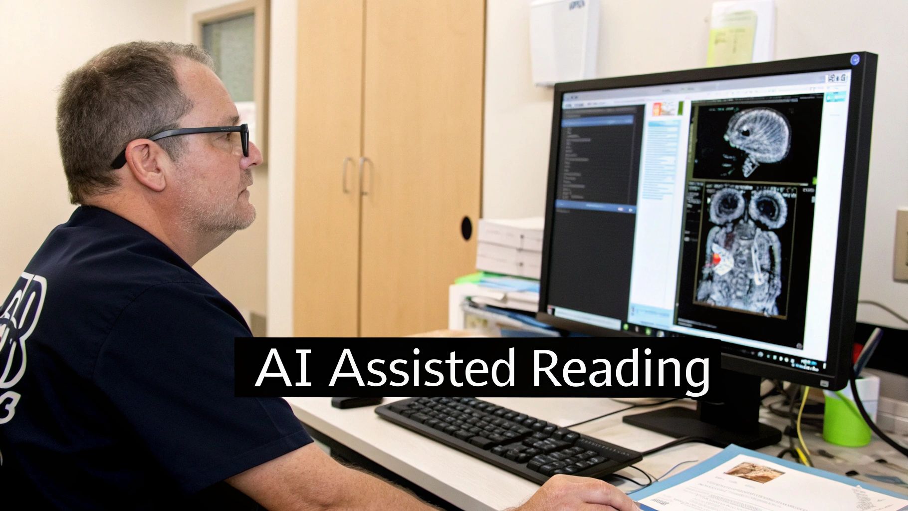
The real quantum leap happens when you pair these advanced viewers with artificial intelligence. AI is no longer just a buzzword; it’s a practical tool that acts as a second set of tireless eyes for the radiologist, scanning every single pixel to find patterns the human eye might otherwise miss.
Integrated AI modules can perform some truly incredible tasks right inside the PACS environment:
- Automated Anomaly Detection: Algorithms can sift through thousands of images and automatically flag potential trouble spots, like nodules or lesions, for immediate human review.
- Intelligent Triage: AI can analyze incoming studies and prioritize them based on urgency, pushing critical cases like potential strokes or internal bleeding right to the top of the worklist.
- Quantitative Analysis: AI delivers precise, repeatable measurements of tumors or other structures, providing objective data to track how a disease is progressing over time.
By taking over routine tasks and highlighting critical findings, AI gives radiologists the freedom to work at the absolute top of their game. It frees them from repetitive analysis and lets them focus their deep expertise on the most complex diagnostic puzzles, which ultimately improves both speed and accuracy.
The future of radiology is undoubtedly built on these kinds of intelligent systems. Getting a handle on AI for medical imaging and diagnostics is essential for clinicians and IT professionals who want to stay ahead of the curve.
Where the Rubber Meets the Road: PACS and RIS in Action
Theory and technical specs are one thing, but the real magic of integrated PACS and RIS systems happens when they touch a patient's life. These aren't just IT projects; they're the digital backbone of modern medicine, directly impacting how quickly a patient gets a diagnosis, how new treatments are discovered, and whether someone gets access to expert care, no matter where they live.
Let's walk through a few real-world scenarios to see just how profound this impact can be.
The Clock is Ticking: Accelerating Care in the ER
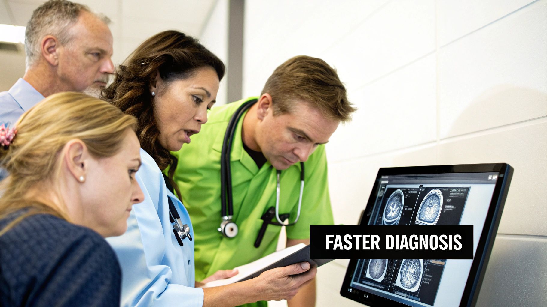
Picture this: a patient rushes into the emergency room, clutching their side in severe pain. The ER doctor needs a CT scan, and they need it now. This is where an integrated system becomes a lifesaver.
The doctor places the order in the RIS. Instantly, it appears on the CT tech’s worklist, complete with all the patient's info. No phone calls, no lost paperwork. As soon as the scan is done, the images fly into the PACS and are automatically tied to the patient's record. The radiologist gets an immediate ping: a high-priority case is ready.
Within minutes—not hours—the radiologist has reviewed the images, dictated their findings, and the report is back in the hands of the ER physician and the waiting surgical team. That seamless flow, from order to diagnosis to action, shaves off precious time that can make all the difference in a critical situation.
Connecting Minds: Powering Global Clinical Trials
Now, think bigger. Imagine a groundbreaking clinical trial for a new cancer drug, with research teams spread across North America, Europe, and Asia. How do you keep everyone on the same page? A cloud-based PACS/RIS platform is the answer. It becomes the central nervous system for the entire operation.
- A Single Source of Truth: Researchers anywhere can securely upload, view, and analyze imaging data as if they were in the same room.
- Ironclad Consistency: The RIS enforces a standard protocol, ensuring every scan is performed and documented the exact same way, no matter the location.
- Teamwork from Anywhere: Multiple experts can annotate the same image at the same time, sharing insights and speeding up the discovery process.
This kind of global teamwork simply wouldn't be possible with disconnected, old-school systems. The integrated platform ensures the data is solid and empowers researchers to function as one cohesive unit, pushing medical science forward faster.
In these high-stakes environments, a unified system is the difference between siloed data and collective intelligence. It connects experts across continents, ensuring that every piece of information contributes to a larger goal, whether it's saving a single life or advancing medical science for millions.
Bridging the Distance with Teleradiology
What about a small, rural clinic that can't afford a full-time radiologist? With a web-based PACS and RIS system, they can offer the same level of diagnostic expertise as a major city hospital.
When a local patient gets an X-ray, the images are securely uploaded to a cloud PACS. Miles away, a board-certified radiologist sees the study pop up in their queue. They can review the images on a high-resolution viewer, write up a detailed report, and send it back. The report is instantly available to the physician at the rural clinic.
This model completely changes the game for healthcare access. It brings world-class specialists to underserved communities, ensuring every patient gets a timely, expert diagnosis without having to travel for hours.
Here at PYCAD, we live and breathe this technology. We're passionate about building the tools that make these stories a reality. We at PYCAD, build custom web DICOM viewers and integrate them into medical imaging web platforms. Our goal is to create systems that foster collaboration, speed up diagnoses, and extend the reach of medical expertise. To see how we engineer these powerful solutions, take a look at our work on our portfolio page.
Your Implementation and Selection Checklist
Choosing the right PACS and RIS system is a defining moment for any healthcare organization. This isn't just another IT upgrade; it’s a foundational investment in the future of your patient care, the efficiency of your operations, and your clinical excellence. Taking a thoughtful, methodical approach will help you find a true partner, not just a product—one that genuinely aligns with your vision and can grow with your ambition.
The journey starts with a deep dive into your own operational DNA. Before you even sit through a single vendor demo, you need to map out your current workflows, identifying every bottleneck and frustration point. What are the biggest headaches for your radiologists, technologists, and administrative staff? Where do communications fall apart? Getting a crystal-clear picture of your present challenges is the only way to define what your ideal future looks like.
Assembling Your Evaluation Framework
Once you have a solid handle on your needs, it's time to build a robust evaluation framework. Think of this as your compass, guiding you through the complex world of available pacs ris systems and making sure your choice is objective and data-driven. Your criteria should be thorough, covering everything from core technical features to the quality of the long-term vendor partnership.
Your evaluation must include these non-negotiable elements:
- Interoperability and Integration: How smoothly will the new system talk to your existing Electronic Health Record (EHR) and other vital hospital systems? Insist on clear answers about their experience integrating with your specific EHR vendor.
- Security and Compliance: The system must provide uncompromising, multi-layered security. Verify that it is fully HIPAA compliant and ask for specifics on their data encryption, access controls, and how often they conduct security audits.
- Scalability and Future-Readiness: Can the architecture actually support your growth plans? Ask if it can easily handle a surge in imaging volume or expand to new clinics without needing a complete and costly overhaul.
The right system isn’t just about fixing today’s problems. It's about laying a flexible foundation that can adapt to the technological leaps and clinical demands of tomorrow. It should be a platform that grows with you, not one you’ll quickly outgrow.
Critical Questions for Potential Vendors
With your framework ready, you can start talking to vendors with real purpose. Treat these conversations less like sales calls and more like interviews for a long-term strategic partnership. Push past the glossy presentations and ask the tough questions that reveal the true character and capability of a potential partner.
Here are a few essential questions to guide your vendor discussions:
- Data Migration and Support: What is your detailed plan for migrating our legacy data? Walk us through the process, the timeline, and the level of support your team will provide to ensure a seamless transition with minimal disruption.
- Training and Onboarding: How do you approach training for our different user groups—from radiologists to front-desk staff? Is training an ongoing process, and how do you support new hires who join after the initial rollout?
- Customization and Flexibility: How much can we tailor the system to fit our unique workflows? Can we adapt reporting templates, user interfaces, and workflow rules to match how our team actually works?
As you navigate this process, remember that ensuring business continuity is paramount. It’s always wise to have a plan for the unexpected, and that includes building a robust recovery plan to safeguard your operations.
At PYCAD, we believe in creating these exact kinds of forward-thinking partnerships. We at PYCAD, build custom web DICOM viewers and integrate them into medical imaging web platforms that are secure, scalable, and built around your unique needs. To see how we help organizations achieve their clinical and operational goals, we invite you to explore our portfolio page.
Your Questions Answered: PACS and RIS Explained
Diving into the world of medical imaging technology can feel a bit overwhelming, and it naturally brings up a lot of questions. As you look to build a smarter, more connected system, getting to grips with the real-world ins and outs of PACS and RIS is key. Here are some straightforward answers to the questions we hear most often.
Are PACS and RIS Only for Big Hospitals?
Not anymore. That idea is a holdover from a different era. Today, modern cloud-based solutions have completely changed the game, making these powerful systems both affordable and accessible for clinics of any size.
For smaller practices, adopting a scalable system means gaining the kind of efficiency that was once only possible for large institutions. It smooths out your workflow, sharpens diagnostic quality, and—most importantly—makes it simple to collaborate with specialists anywhere. This ensures your patients get the best care possible, no matter how big your clinic is.
What's the Real Difference Between PACS and a VNA?
Here’s a helpful way to think about it. A PACS is like a specialized library within the radiology department, often built to work with one specific vendor's equipment. A Vendor Neutral Archive (VNA), on the other hand, is like the main, central library for the entire hospital.
A VNA is designed to be the single source of truth for all medical images—from cardiology, pathology, dermatology, you name it. It stores everything in a standard format, creating a complete imaging history for each patient. This gives you the freedom to switch out viewers or other software down the road without facing a massive, expensive data migration project.
How Does a Custom DICOM Viewer Make a PACS Better?
Most PACS come with a basic, built-in viewer that gets the job done. But if you want to tap into the true power hidden in your imaging data, a custom viewer is a total game-changer. At PYCAD, we build custom web DICOM viewers and integrate them into medical imaging web platforms, unlocking a whole new level of capability.
Our custom solutions bring advanced tools to the table—things like 3D reconstruction, AI-driven analysis, and real-time collaboration features that you just won't find in a standard package. This transforms a PACS from a simple storage archive into a vibrant, intelligent hub for diagnostics. You can see what this looks like in practice by exploring our advanced medical imaging projects.
At PYCAD, we’re not just developing software; we're building the future of medical diagnostics. We craft secure, scalable medical imaging web platforms that come supercharged with powerful custom DICOM viewers and integrated AI modules. To see how we can bring your vision to life, take a look at our work at https://pycad.co/portfolio.


