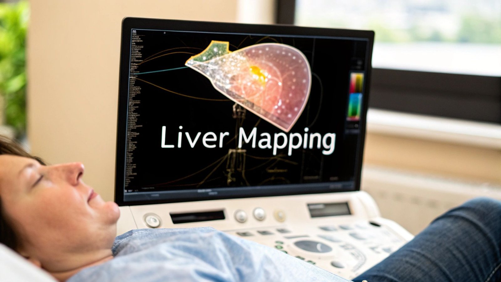
When it comes to the liver, precision isn't just a goal—it's everything. Think of understanding ultrasound liver segments as having a highly detailed, internal GPS for one of the body's most complex organs. This mastery allows clinicians to navigate the liver's intricate geography, pinpointing issues with an accuracy that was once unimaginable.
This skill has fundamentally changed the game in oncology, transplant surgery, and managing chronic liver disease, turning what could be a high-stakes guessing game into a precise, planned procedure.
Why Accurate Liver Mapping Is a Modern Medical Marvel
Picture a surgeon viewing the liver not as one large, uniform organ, but as a territory composed of eight distinct, functional districts. That's the power that a deep understanding of ultrasound liver segments unlocks. It transforms this vital organ into a clearly charted map, where each segment functions almost independently with its own dedicated blood supply and drainage.
This isn't just a fascinating academic exercise; it's the very foundation of modern liver surgery and diagnostics. It's what separates a good outcome from a great one.
This segmental approach makes incredibly precise interventions possible. For example, if a tumor is confined to a single segment, a surgeon can perform a segmentectomy—removing only that specific "district" while leaving the surrounding healthy tissue completely intact. This preserves vital liver function, dramatically speeds up patient recovery, and lowers surgical risk. It's a world away from the older methods that often required removing much larger portions of the organ.
The Fusion of Anatomy and Technology
Seeing these segments in real-time is where classic anatomical knowledge meets modern technology. Ultrasound gives us a safe, non-invasive window into the body, but it takes a trained eye and immense skill to interpret those grayscale images and accurately map out the segmental boundaries.
This powerful combination has had a massive impact on patient care across several key areas:
- Surgical Planning: It allows surgeons to plan resections for tumors or cysts with millimeter-level precision.
- Transplant Viability: Clinicians can assess the health and volume of individual segments, which is critical for living donor transplants.
- Disease Monitoring: It helps track the progression of diseases like cirrhosis, which can affect different segments in unique ways.
- Targeted Biopsies: It ensures that a biopsy needle hits the exact location of a suspected lesion, avoiding inconclusive results.
The real breakthrough is treating the liver not as a single entity, but as a collection of eight functional units. This granular view empowers clinicians to make more informed, life-saving decisions that were once thought impossible.
This field continues to push forward, driven by powerful new visualization tools. Here at PYCAD, we specialize in building custom web DICOM viewers and integrating them into medical imaging web platforms. We create the intuitive interfaces that clinicians need to interact with these complex anatomical maps effectively. To see what this technology looks like in action, feel free to explore the advanced imaging solutions in our portfolio.
Decoding the Couinaud Map: Your Guide to Liver Anatomy
To truly get a handle on ultrasound liver segments, we first need to get familiar with the brilliant map that defines them: the Couinaud classification. Forget those old, dense anatomical charts for a moment. Instead, let's picture the liver as a bustling city, neatly organized into eight self-sufficient districts.
Each of these districts, or segments, is a little marvel of biological engineering. It has its own dedicated supply lines—a branch of the portal vein and the hepatic artery—and its own unique drainage system, a bile duct. This incredibly clever design means a surgeon can address a problem in one district, like removing a tumor, without shutting down the vital functions of its neighbors.
This functional independence is the secret sauce of the Couinaud system. The major hepatic veins act like the grand highways dividing the city into larger territories, while other vessels create the smaller, local streets that define each segment’s borders.
The Blueprint of Liver Function
The Couinaud classification, which became the gold standard back in the mid-20th century, gives us a framework for understanding the liver. It divides the organ into eight functionally independent segments, each with its own vascular inflow, outflow, and biliary drainage. Think of them as individual, resectable units. As a key landmark, the gallbladder is tucked neatly between segments IVb and V.
The system follows a logical clockwise numbering pattern. It starts with Segment I (the caudate lobe) at the very back and spirals outwards. While ultrasound is fantastic for visualizing these structures, it's not the only tool in the box. To see how these same segments look through a different lens, you can explore our guide on CT liver segmentation.
This infographic really brings home the relationship between understanding the anatomy, using ultrasound technology, and performing precise surgical procedures.
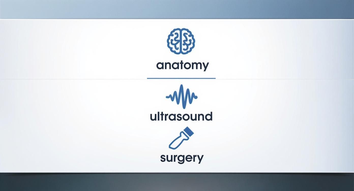
It perfectly illustrates how a solid foundation in anatomy is the bedrock upon which we build advanced imaging techniques and achieve surgical success. At PYCAD, we build custom web DICOM viewers and integrate them into medical imaging web platforms to help clinicians visualize this complex data effectively. Our work can be seen in our portfolio at https://pycad.co/portfolio.
Visualizing the City Plan
Mastering this map is about more than just memorizing numbers. It’s about building a three-dimensional model of the liver’s geography in your mind. This spatial awareness is absolutely critical for any clinician looking at an ultrasound scan. For example, knowing that Segment VII sits superiorly and posteriorly in the right lobe helps a sonographer orient themselves instantly during an exam.
The genius of the Couinaud map is that it's a functional blueprint, not just a static one. It tells us how the liver works, district by district, which is infinitely more powerful for diagnosis and treatment.
This table provides a quick reference to the eight segments and the key landmarks you'll be looking for on an ultrasound.
The 8 Couinaud Liver Segments at a Glance
| Segment | Anatomical Lobe | General Location | Key Vascular Landmarks |
|---|---|---|---|
| I | Caudate Lobe | Most posterior | Posterior to portal vein |
| II | Left Lobe | Superior-lateral | Left hepatic vein |
| III | Left Lobe | Inferior-lateral | Left portal vein branches |
| IVa | Left Lobe | Superior-medial | Middle hepatic vein (left side) |
| IVb | Left Lobe | Inferior-medial | Near gallbladder fossa |
| V | Right Lobe | Inferior-anterior | Right portal vein (anterior branch) |
| VI | Right Lobe | Inferior-posterior | Right portal vein (posterior branch) |
| VII | Right Lobe | Superior-posterior | Right hepatic vein |
| VIII | Right Lobe | Superior-anterior | Middle hepatic vein (right side) |
Having this mental map allows you to navigate the liver’s complex internal structure with confidence and precision.
This deep anatomical knowledge is what transforms a grayscale ultrasound image into a confident, accurate clinical decision. It’s this very precision that makes modern medical procedures possible, allowing surgeons to save liver tissue and dramatically improve patient outcomes.
Making the Invisible Visible with Ultrasound
Having the Couinaud map is like holding a detailed city blueprint, but the real magic begins when you learn to read that map in real-time. This is where the true artistry of sonography comes alive, translating abstract anatomical knowledge into clear, actionable images on a screen. With an ultrasound transducer, clinicians can literally peer inside the body, making the invisible structures of the liver visible.
This process is a dynamic dance between technical skill and deep anatomical understanding. Sonographers don't just place a probe on the skin; they meticulously angle and maneuver it. They use specific scanning planes—like transverse and longitudinal views—to slice through the liver's architecture and reveal its hidden landmarks. It's a bit like being a detective, piecing together clues from vascular pathways to build a complete picture of the liver's geography.
Navigating with Vascular Landmarks
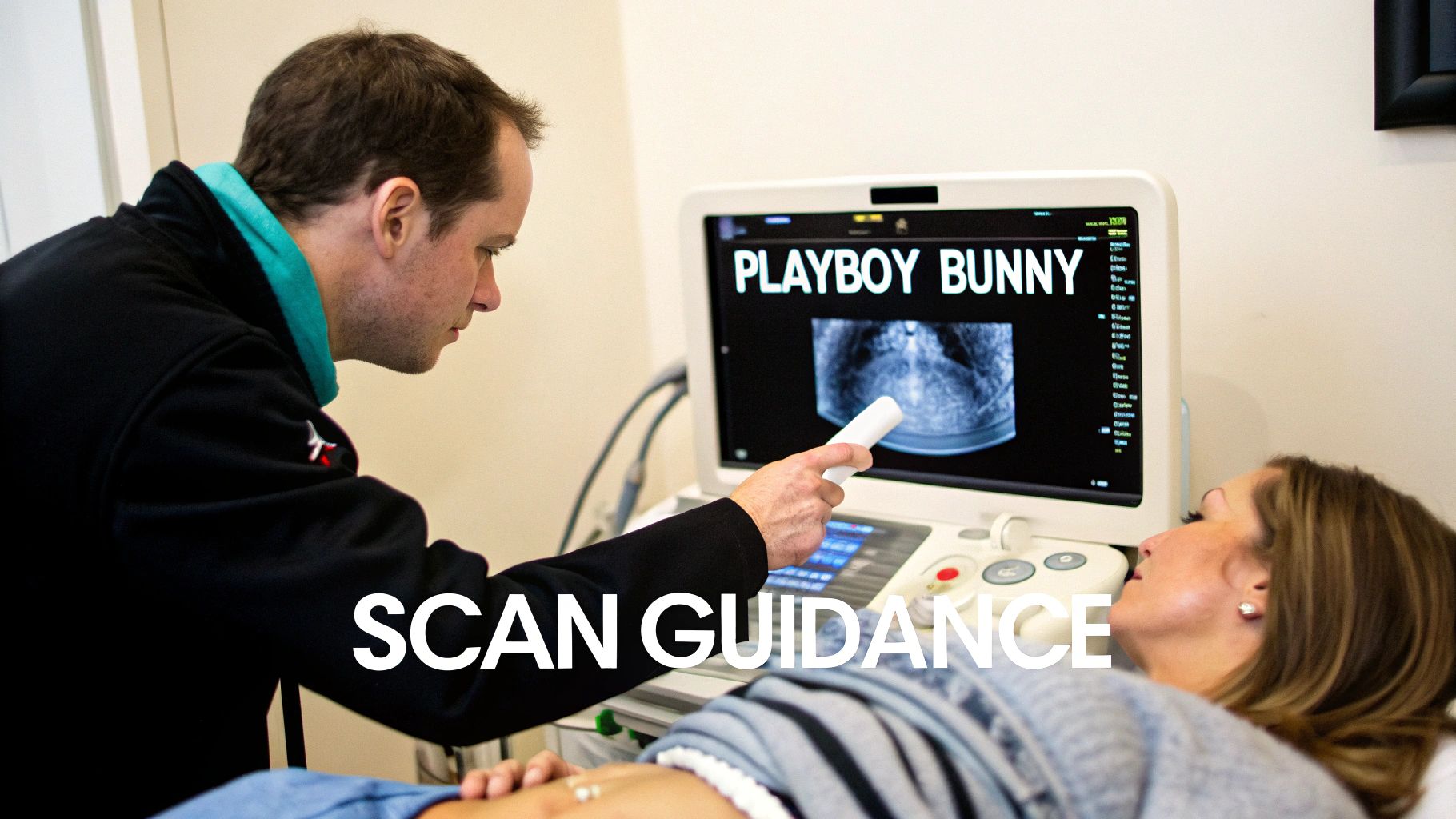
The secret to identifying ultrasound liver segments is learning to recognize the major blood vessels, which act as the primary road signs. The hepatic veins and portal veins crisscross the liver at different angles, creating a natural grid that defines the boundaries of each segment. A skilled sonographer learns to follow these vessels to get their bearings.
For instance, the three main hepatic veins—right, middle, and left—are crucial dividers. When you see them together in a specific transverse view, they form a distinctive shape often called the 'Playboy Bunny' sign. This isn't just a quirky name; it's a foundational landmark that instantly helps a sonographer confirm their position and identify the superior segments (like VII, VIII, IVa, and II). This knack for recognizing patterns within a grayscale image is what separates a novice from an expert.
Visualizing liver segments with ultrasound is a cornerstone of clinical practice worldwide, thanks to the organ's sheer size and complex anatomy. As the body's largest internal organ, its right lobe is typically five to six times larger than the left. Recognizable sonographic landmarks are absolutely vital for demarcation, and ultrasound's accessibility and real-time feedback make it an indispensable tool. You can discover more insights on liver sonography on sonographictendencies.com.
From 2D Slices to a 3D Mental Model
Each press and tilt of the ultrasound probe creates a 2D slice, a single snapshot of the liver's interior. The real genius happens in the sonographer's mind, where they stitch these individual slices together into a cohesive, three-dimensional mental model of the patient’s unique anatomy. It's a skill honed over thousands of scans, requiring an intuitive feel for how a tiny adjustment of the transducer will reveal the next piece of the puzzle. You can learn more about how modern imaging builds these complex views in our article on ultrasound in 3D.
The ultimate goal is to move beyond seeing just shapes and shadows. It’s about interpreting those images to tell a story about the liver’s health, segment by segment, and providing the clear, confident answers that clinicians and patients depend on.
This interpretive skill is what truly matters. At PYCAD, we understand that clear visualization is everything, which is why we build custom web DICOM viewers and integrate them into medical imaging web platforms. Our solutions provide the clarity needed to navigate complex anatomies with absolute confidence. You can see examples of our work in our portfolio of innovative medical imaging solutions.
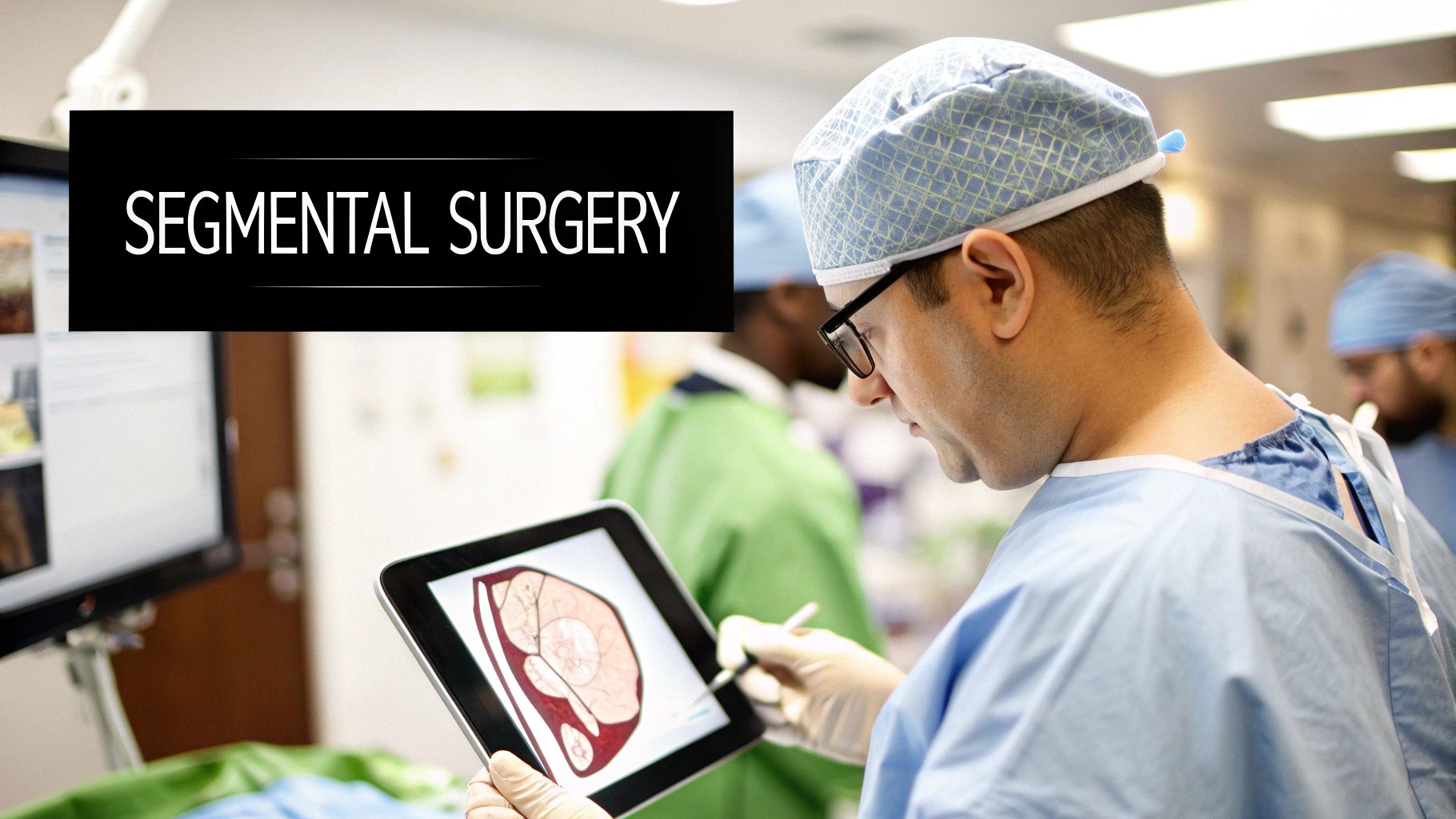
How Segmental Knowledge Saves Lives in the Clinic
Anatomical maps are fascinating on their own, but their true power is unleashed when they guide life-altering decisions in the clinic. Understanding ultrasound liver segments isn't just an academic exercise; it’s the crucial link between a grayscale image and a life-saving intervention. It transforms the liver from a complex, intimidating organ into a navigable map, empowering clinicians to act with incredible precision and confidence.
This knowledge translates directly into better outcomes for patients. Imagine a surgeon sees that a tumor is confined neatly within Segment VI. With this information, they can plan a precise segmentectomy, removing only the affected "district" while preserving the maximum amount of healthy, functional liver tissue. This means a faster recovery, lower surgical risk, and a much better quality of life for the patient. It's a world away from older, more aggressive approaches that required removing huge portions of the organ.
Precision in Diagnosis and Treatment
The real-world impact of segmental anatomy goes far beyond the operating room. This detailed mapping is the backbone for a whole range of diagnostic and therapeutic procedures, each one made safer and more effective by the clarity it provides.
- Guided Biopsies: When a radiologist knows a suspicious lesion is tucked away in the hard-to-reach Segment VII, they can plot the safest path for the needle, steering clear of major blood vessels to get a clean tissue sample. One nationwide study on biopsies found that the specific liver segment being sampled was a direct risk factor for complications, with Segments VII and VIII being the trickiest. This just underscores how critical this anatomical knowledge is for keeping patients safe.
- Metastatic Cancer Treatment: For patients whose cancer has spread to the liver, treatments like radiofrequency ablation or chemoembolization can be aimed at specific segments. This allows doctors to destroy tumor cells with pinpoint accuracy while sparing the surrounding healthy liver.
- Cirrhosis Assessment: In diseases like cirrhosis, the liver doesn't shrink evenly. Some segments might wither while others swell in compensation. Tracking these volume changes with ultrasound gives doctors vital clues about how the disease is progressing and helps them stage the severity of liver failure.
Every precise biopsy, every targeted therapy, and every successful resection is built on the foundation of segmental anatomy. It’s the invisible framework that makes modern liver care possible, turning complex challenges into manageable, targeted actions.
Of course, this level of detail demands powerful visualization tools. At PYCAD, we build custom web DICOM viewers and integrate them into medical imaging web platforms, creating the clear, interactive interfaces clinicians need to apply this life-saving knowledge. Our tools are designed to make complex anatomical data feel intuitive and accessible. You can see what we're building on our PYCAD portfolio page and get a glimpse of how we’re helping clinicians visualize the future of medicine.
The Next Frontier: AI-Powered Liver Segmentation
Welcome to the future of liver imaging—a place where a sonographer's hard-earned skill is powerfully amplified by artificial intelligence. Manually identifying ultrasound liver segments is a true art form, but it's also incredibly time-consuming and relies heavily on years of hands-on experience. A new horizon is opening up, one where AI and deep learning are bringing in a fresh wave of speed, consistency, and precision.
Think of these advanced algorithms as a seasoned assistant. They've been trained on thousands upon thousands of ultrasound scans, learning to automatically recognize and outline liver segments with a level of accuracy that’s truly remarkable. They can spot subtle anatomical differences and faint vascular landmarks that are sometimes tough for the human eye to catch, helping to clear up diagnostic uncertainty and boost clinical confidence. This is where technology and medicine come together to create something special.
Automating Precision and Bringing Images to Life
At its heart, the magic of AI-powered segmentation is its ability to take a flat, 2D ultrasound image and transform it into a living, breathing 3D model in just seconds. This automated process gives clinicians an instant, comprehensive map of a patient's liver, with every segmental boundary clearly defined.
The impact this has on day-to-day clinical work is massive:
- Surgical Planning: Imagine a surgeon getting an exact 3D model of a patient's liver. They can meticulously plan resections, understanding the precise spatial relationship between a tumor and the vital structures surrounding it.
- Training and Education: New sonographers and radiologists can get up to speed much faster. They can learn liver anatomy by interacting with these AI-generated models and seeing how they connect directly to live scan images.
- Rock-Solid Consistency: AI smooths out the natural human variability in manual segmentation. This means measurements and reports are standardized and reliable from one scan to the next, no matter who performs them.
The real goal of AI isn't to replace experts but to empower them. By taking on the demanding work of segmentation, AI frees up clinicians to concentrate on what they do best: interpreting results, diagnosing conditions, and planning life-saving treatments.
Bringing this kind of AI into a clinical setting requires specialized talent, and many healthcare organizations find themselves needing to bring in outside experts. If you're exploring that path, this guide on choosing the right staff augmentation company for AI talent is an excellent starting point.
Here at PYCAD, we live at this very intersection of AI and medical imaging. We specialize in building custom web DICOM viewers and integrating them smoothly into medical imaging platforms. These viewers are the windows that allow clinicians to see, manipulate, and truly understand these incredible AI-generated 3D models. For a deeper look into the technology that makes this all possible, our guide on deep learning segmentation is a great resource. You can also view our completed projects at https://pycad.co/portfolio.
Common Questions About Ultrasound Liver Segments
As we journey through the intricate world of liver anatomy and imaging, a few questions always seem to pop up. Getting clear on these points is what turns head knowledge into hands-on confidence, giving you the power to master ultrasound liver segments and use that skill to make a real difference for your patients. Let's tackle some of the most common ones.
Why Is the Couinaud System the Gold Standard?
The Couinaud classification reigns supreme because it’s based on function, not just pure anatomy. It brilliantly divides the liver into eight segments, where each one has its own independent blood supply and drainage. Think of them as eight self-contained, functional units.
This design is a game-changer for surgeons. It means they can remove a diseased segment while leaving the maximum amount of healthy, working liver tissue behind. It’s this direct impact on smarter surgical planning and better patient outcomes that cements its place as the undisputed standard.
What Makes Segment Identification So Challenging?
The biggest hurdles in real-time ultrasound come down to two things: patient variability and image quality. Factors like obesity or bowel gas can easily obscure your view, hiding the crucial vascular landmarks that act as your map. Plus, every patient's internal anatomy is slightly different, which means you have to be ready to adapt on the fly.
The real art is in navigating these variables. On top of that, diseases like cirrhosis or large tumors can completely distort the liver's normal architecture, turning accurate segmentation into a complex puzzle that truly relies on deep experience and intuition.
How Does Color Doppler Elevate the Exam?
Color Doppler is the tool that brings a static, grayscale image to life. By showing you the direction and velocity of blood flow, it lets you definitively identify the portal and hepatic veins—the primary "road signs" for segment boundaries.
This isn't just a helpful feature; it's essential. It removes the guesswork and provides a layer of certainty, ensuring that every diagnosis and surgical plan is built on a rock-solid understanding of the patient's unique vascular map.
At PYCAD, we're passionate about advancing what’s possible in medical imaging. We build custom web DICOM viewers and integrate them into medical imaging web platforms, creating the tools clinicians need to see complex data with absolute clarity. To see how we're helping shape the future of imaging, explore our portfolio of innovative imaging solutions.


