
The Evolution of Volume Rendering CT: From Pixels to Precision
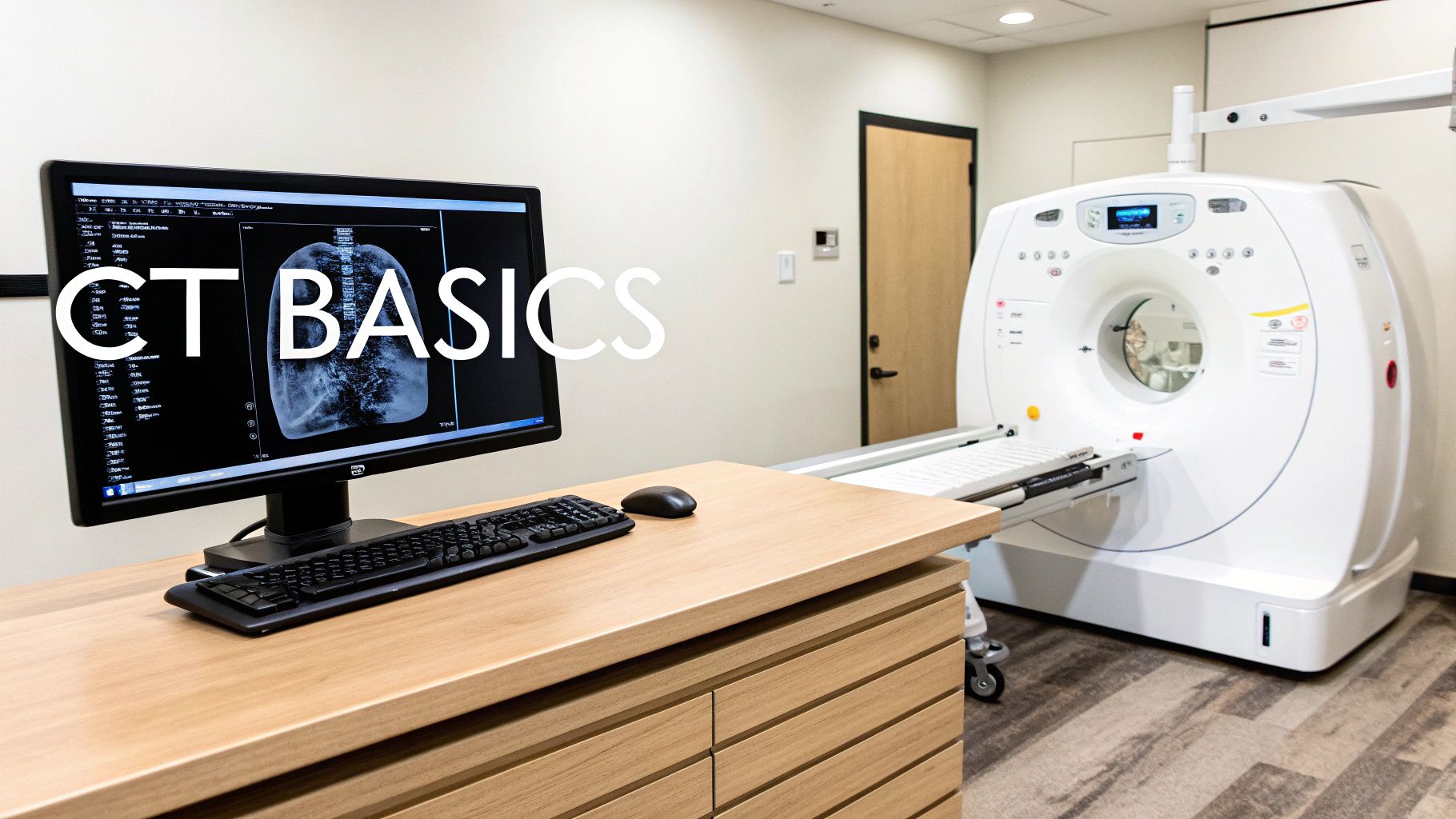
Visualizing the human body's intricate inner workings in three dimensions has long been a goal of medical imaging. Early computed tomography (CT) scans offered valuable cross-sectional images, yet they lacked the ability to fully showcase the complex interplay of anatomical structures. This spurred the development of volume rendering CT, a technique that transforms 2D image slices into dynamic 3D visualizations, fundamentally changing how clinicians view and understand the body.
The Impact of Multi-Detector CT (MDCT)
The arrival of multi-detector CT (MDCT) in the early 2000s marked a major turning point for volume rendering CT. Acquiring the necessary data for volume rendering was previously a lengthy process. MDCT, however, enabled the rapid acquisition of volumetric datasets, drastically improving both spatial and temporal resolution. This resulted in clearer images captured in significantly shorter timeframes, making volume rendering more practical and effective. MDCT wasn't merely a technical upgrade; it ushered in a new era in diagnostic imaging.
This advancement facilitated the creation of isotropic voxels, cube-shaped units of image data essential for volume rendering. These voxels possess the same dimensions in all three spatial directions, allowing clinicians to reconstruct images in any plane – axial, sagittal, or coronal – without losing detail. The ability to explore anatomy from any angle revolutionized diagnostic imaging. Volume rendering in CT powerfully transforms 2D slices into 3D visualizations, particularly after the introduction of MDCT, enhancing the comprehension of complex structures.
Real-World Applications and Improved Accuracy
The evolution of volume rendering CT has brought tangible improvements to patient care. Studies reveal that volume rendering CT can increase diagnostic accuracy by up to 30% in certain cases compared to traditional 2D imaging. This improvement stems from the enhanced clarity and the ability to visualize complex anatomical relationships in 3D. This is especially crucial in fields like cardiology, where understanding the precise pathways of coronary arteries is vital for accurate diagnoses and effective treatment plans. Learn more about volume rendering CT
The impact extends beyond diagnosis. Volume rendering CT has become indispensable for surgical planning. By providing surgeons with a detailed 3D roadmap of a patient's anatomy, it allows for more precise planning, anticipation of potential challenges, and ultimately, improved patient outcomes. This technology has made images not only visually compelling but also clinically valuable, leading to more informed decisions and better patient care.
Behind the Magic: How Volume Rendering CT Actually Works
The journey from a CT scan to a striking 3D image of the body involves some impressive technology. Volume rendering CT takes data from multiple 2D image slices and transforms them into a dynamic, three-dimensional visualization. This process allows doctors to see the interplay of different anatomical structures with greater clarity. But how does it actually work? This section unveils the process behind volume rendering CT.
From Data Points to 3D Structures
The process begins with the raw data acquired by the CT scanner. This data represents varying tissue densities within the body. Volume rendering algorithms then use these data points, called voxels, to construct the 3D representation. Think of it like connecting the dots to create a picture, but on a vastly more complex scale.
This resulting 3D model can then be manipulated and viewed from any angle, providing a far richer understanding of the body's internal structures compared to traditional 2D images. This enhanced view allows medical professionals to better diagnose and treat a variety of conditions.
Understanding Different Rendering Techniques
Several techniques exist within volume rendering CT, each possessing its own advantages. Maximum Intensity Projection (MIP), for instance, highlights the brightest structures along a line of sight. This proves particularly useful for visualizing blood vessels and bony structures, aiding in the identification of potential blockages or fractures.
Another frequently used technique is Direct Volume Rendering (DVR). DVR assigns colors and opacities to the different tissue densities, creating a more comprehensive and realistic representation of the anatomy. This technique allows for a more nuanced visualization of soft tissues and organs.
To help illustrate the differences between these approaches, let's examine a comparison table:
To help understand the nuances between different volume rendering techniques, the following table summarizes key aspects:
Volume Rendering Techniques Comparison: Comparison of different volume rendering approaches used in CT imaging.
| Technique | Best Applications | Limitations | Processing Requirements |
|---|---|---|---|
| Maximum Intensity Projection (MIP) | Visualizing high-contrast structures like blood vessels and bones | Can obscure underlying structures | Relatively low |
| Direct Volume Rendering (DVR) | Creating realistic representations of complex anatomies | Requires careful adjustment of transfer functions | Higher than MIP |
This table summarizes the best applications, limitations and processing needs of MIP and DVR. By understanding these differences, medical professionals can select the most appropriate technique for a given diagnostic task.
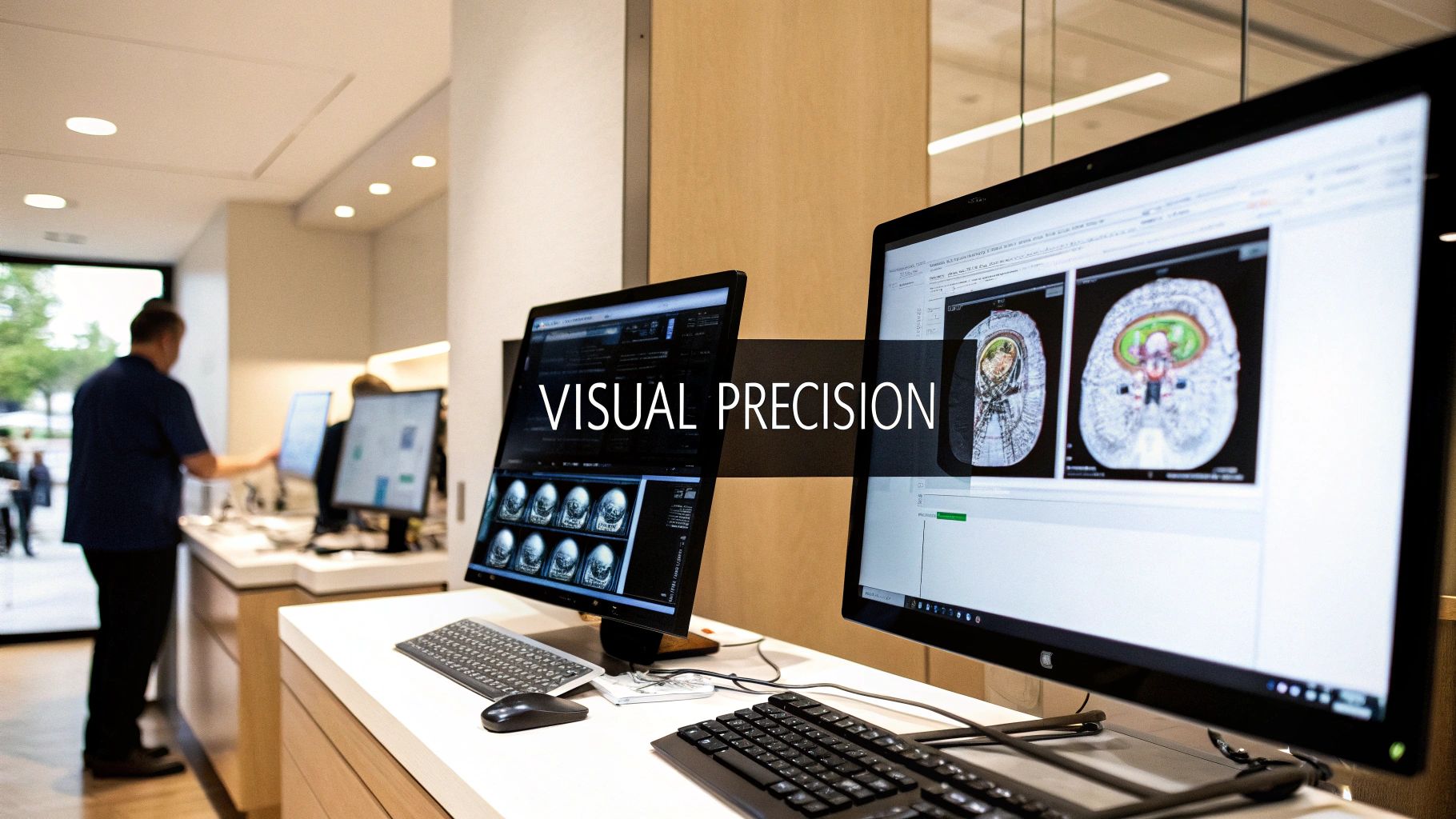
The Power of Transfer Functions
A crucial component of volume rendering CT is the transfer function. This function maps tissue density values to color and opacity. For example, dense bone might be assigned a bright white color and high opacity, making it clearly visible. Conversely, less dense tissues like muscle might be assigned a darker color and lower opacity.
By adjusting the transfer function, radiologists can highlight specific anatomical structures of interest. This effectively makes other structures transparent, allowing for unobstructed viewing of the target area. This fine-tuning capability is invaluable for various diagnostic tasks, ranging from assessing vascular structures to characterizing tumors. This level of control allows for precise visualizations tailored to the specific diagnostic needs.
Volume rendering CT offers remarkable insight into the human body. Its ability to translate complex data into understandable visualizations continues to advance medical diagnostics and treatment planning. Understanding the mechanics of this technology offers valuable insight into the capabilities of modern medical imaging.
Cinematic Rendering: Hollywood Meets Hospital Imaging
While previous discussions explored the mechanics of volume rendering CT scans, this section unveils a groundbreaking advancement: cinematic rendering (CR). This technique elevates volume rendering to a new level, producing strikingly realistic images that appear more at home in a Hollywood film than a hospital setting. This leap forward significantly impacts how medical professionals visualize and interpret CT scan data.
The Power of Light Simulation
Cinematic rendering goes beyond traditional volume rendering by incorporating advanced light simulation algorithms. Instead of simply assigning colors and opacities to tissue densities, CR simulates the complex interaction of light with these tissues. This process mirrors techniques used in computer-generated imagery (CGI), creating images with unprecedented depth, texture, and anatomical clarity. This represents a significant departure from standard methods, which can sometimes lack realism.
This enhanced realism isn't merely aesthetic; it has practical diagnostic implications. Cinematic rendering simulates light propagation through volumetric data, creating lifelike image quality. A recent survey indicated that 85% of medical professionals found CR images easier to interpret, particularly in complex cases. Unlike standard volume rendering, which can sometimes produce artifacts due to its reliance on scalar data, CR utilizes multiple light-simulating algorithms for more visually compelling results. Researchers have observed that CR can improve anatomical structure perception by as much as 50%, boosting diagnostic confidence and potentially leading to improved surgical outcomes. Explore this topic further.
Transforming Perception and Treatment
The improved clarity and depth offered by cinematic rendering fundamentally change how clinicians perceive anatomical relationships. It's comparable to upgrading from standard definition to high-definition television—the increased detail unlocks a new level of understanding. This enhanced visualization is especially valuable in cases involving intricate anatomical structures, where even subtle details can be critical for accurate diagnosis and treatment planning.
Real-World Impact of Cinematic Rendering
Cinematic rendering is already making a tangible impact in real-world medical practice. For example, it enhances surgical planning by providing surgeons with exceptionally detailed, realistic representations of the surgical field. This allows for more effective pre-operative planning, potentially minimizing surgical risks and improving patient outcomes.
Cinematic rendering is also enhancing communication between doctors and patients. The realistic images generated by CR make it easier for doctors to explain complex medical conditions to patients, leading to improved patient understanding and engagement in their own care. As computational power continues to advance, cinematic rendering is poised to become even more prevalent in medical imaging, setting a new standard for visualization and interpretation. This advancement will undoubtedly contribute to more precise and personalized treatment strategies.
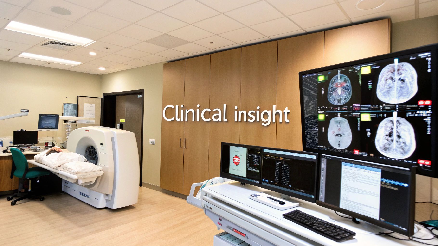
Volume Rendering CT in the Clinical Trenches
Volume rendering CT is transforming patient care. More than just visually impressive images, this technology offers clinicians a powerful tool for 3D visualization of the human body, fundamentally changing diagnosis and treatment planning across various medical disciplines. Let's explore its impact.
Cardiology: Visualizing the Coronary Network
Cardiologists use volume rendering CT to navigate the complex web of coronary arteries. This allows them to visualize intricate anatomies with remarkable clarity, providing a deeper understanding of vessel structure and potential blockages.
For instance, the precise location and severity of a stenosis can be clearly visualized, crucial for deciding the best treatment. This enhanced visualization leads to more informed decisions about interventions like angioplasty or bypass surgery.
Oncology: Precise Tumor Characterization
Volume rendering CT plays a vital role in oncology, aiding in characterizing tumors. Oncologists can visualize the size, shape, and location of tumors, allowing for accurate staging and assessment of tumor spread.
This detailed information is essential for planning precise radiation therapy, ensuring treatment targets the tumor while minimizing harm to healthy tissue. Furthermore, it's useful in monitoring treatment response and making necessary adjustments.
Orthopedics: Understanding Complex Fractures
Orthopedic surgeons benefit from volume rendering CT to understand intricate fracture patterns. Traditional 2D images can sometimes mask a fracture's full extent, making assessment and surgical planning challenging.
Volume rendering provides a 3D representation of the fractured bones, allowing surgeons to visualize the fracture from all perspectives. This leads to more effective surgical planning and improved patient outcomes.
To better understand the applications of volume rendering CT, let's look at the table below. It summarizes key clinical applications across various medical specialties, highlighting the primary uses, benefits, and real-world case examples.
Volume Rendering CT Applications by Specialty
Summary of key clinical applications across medical specialties
| Medical Specialty | Primary Applications | Key Benefits | Case Examples |
|---|---|---|---|
| Cardiology | Coronary artery visualization, stenosis assessment | Improved visualization of vessel structure, informed decision-making for interventions | Pre-operative planning for bypass surgery, guiding angioplasty procedures |
| Oncology | Tumor characterization, treatment planning, monitoring response | Accurate staging and assessment of tumor spread, precise radiation therapy targeting | Determining tumor volume for chemotherapy dosage, evaluating treatment efficacy |
| Orthopedics | Complex fracture visualization, surgical planning | 3D representation of fractures, improved surgical approach and reconstruction | Planning complex fracture repairs, assessing bone fragment displacement |
This table clearly demonstrates the versatility and power of volume rendering CT across different medical fields. Its ability to provide detailed 3D visualizations significantly improves diagnosis, treatment planning, and ultimately, patient care.
Improvements in Clinical Decision-Making
The advantages of volume rendering CT extend beyond these specific examples. Across various specialties, this technology drives improvements in clinical decision-making. The 3D visualization of anatomical structures provides a comprehensive understanding of a patient's condition, leading to more informed and effective treatment.
This results in:
-
Improved surgical planning: Providing surgeons with a 3D anatomical roadmap facilitates precise surgical interventions.
-
Better patient outcomes: Improved diagnosis, treatment planning, and surgical interventions contribute to better overall patient outcomes.
-
Enhanced communication: Clear 3D visualizations are valuable for communicating complex medical information to patients, improving understanding and engagement in their care.
Volume rendering CT is more than just enhanced imaging; it empowers clinicians to make the best decisions for their patients. As technology advances, volume rendering will continue to shape the future of medical imaging and patient care.
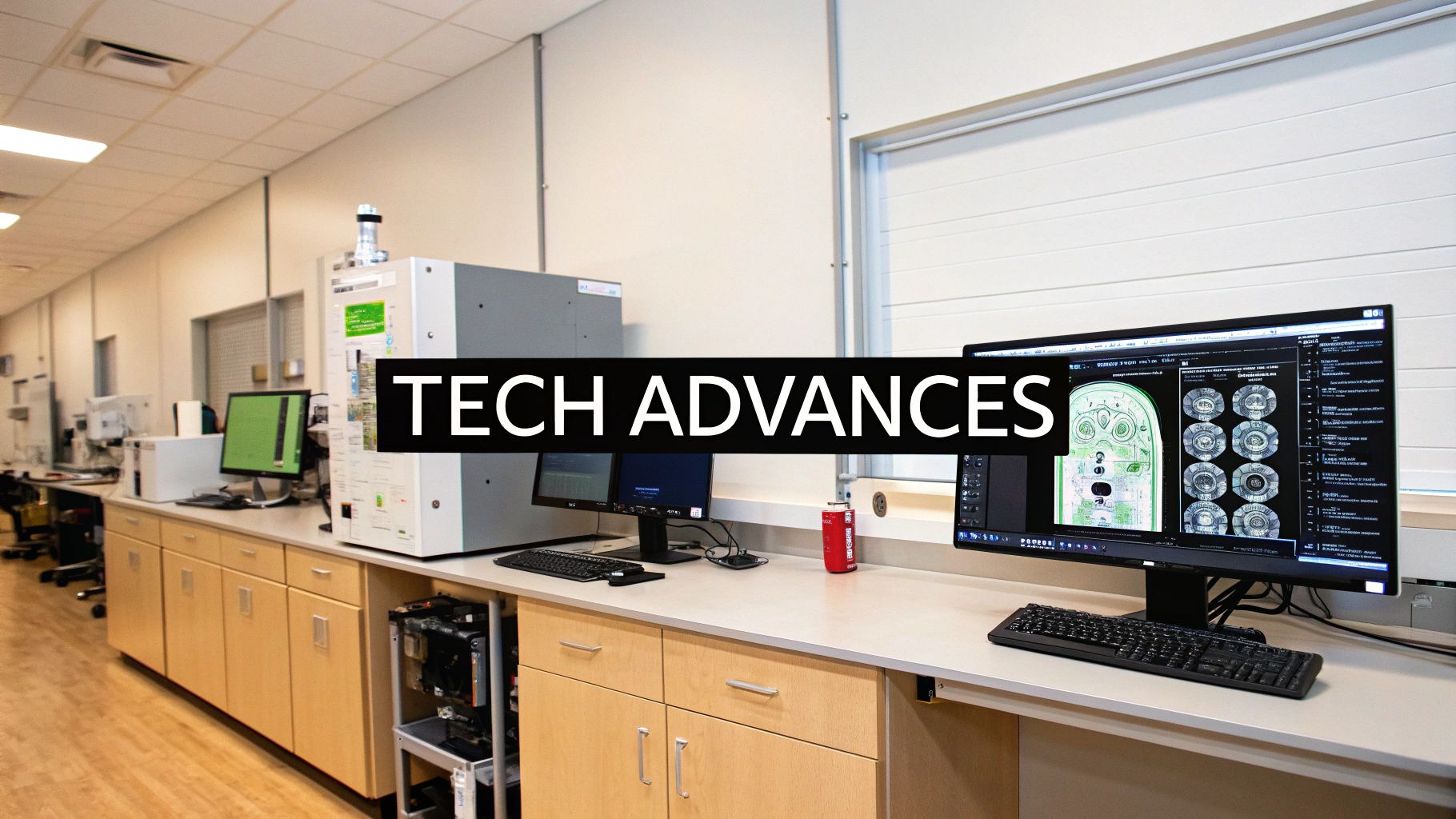
The Technological Engine Driving Volume Rendering CT
The impressive 3D visualizations created by volume rendering CT scans are a direct result of ongoing technological advancements. These improvements haven't just enhanced the clarity and detail of the images; they've also significantly sped up the rendering process. Let's explore the core technologies driving this evolution.
The Role of GPUs
Graphics Processing Units (GPUs) are now crucial for volume rendering CT scans. These specialized processors are designed to handle the massive parallel processing required to render complex 3D images. Generating these visualizations used to take hours, making real-time interaction and analysis difficult.
GPUs have dramatically reduced processing times, often down to mere seconds. This allows for real-time manipulation and interaction with large datasets. Clinicians can now quickly explore anatomy from various angles, which is vital for interactive exploration, diagnosis, and surgical planning.
Specialized Rendering Software
Advances in volume rendering CT wouldn't be possible without dedicated software. These programs offer a range of features designed to optimize the entire rendering workflow.
- Automated Segmentation: Software can now automatically identify and isolate specific anatomical structures, saving radiologists significant time and effort.
- Advanced Visualization Tools: Tools like Maximum Intensity Projection (MIP) and Direct Volume Rendering (DVR) allow clinicians to fine-tune visualizations and highlight specific tissues or structures for improved clarity.
- Integration with PACS: Many rendering software solutions integrate seamlessly with Picture Archiving and Communication Systems (PACS), streamlining workflow and access to patient data.
These software advances have been accompanied by sophisticated hardware improvements. The development of powerful GPUs capable of processing large datasets has transformed the speed and quality of volume rendering. The ability to process 3D datasets in real time allows medical professionals to interact directly with volumetric data, leading to more effective collaborative decision-making. Studies show that 70% of practitioners reported improved workflow efficiency due to these advancements. Some facilities have seen a 30% increase in daily case throughput. More detailed statistics can be found here.
Democratizing Access to Advanced Visualization
Technological progress is making advanced visualization tools more accessible. These tools were once primarily available only at large academic medical centers.
Smaller hospitals and clinics can now leverage the power of volume rendering CT. This wider access leads to earlier and more accurate diagnoses, ultimately improving patient care across various healthcare settings. It also simplifies collaborative decision-making during patient consultations and multidisciplinary meetings.
Value Beyond Radiology
Volume rendering CT offers value beyond radiology. These visualizations are increasingly used in other areas:
- Surgical Planning: Surgeons can use detailed 3D models of patient anatomy to improve pre-operative planning and increase surgical precision.
- Patient Education: Realistic 3D images help patients better understand their conditions and treatment plans.
- Medical Education: Volume rendering has become a valuable training tool for medical students and residents, offering a more intuitive and interactive way to learn anatomy.
As technology continues to evolve, volume rendering CT will become even more essential to medical imaging, driving improvements in patient care and medical knowledge.
Future Horizons: Where Volume Rendering CT Is Heading
Volume rendering CT has significantly impacted medical imaging, providing detailed 3D visualizations that have transformed how clinicians view the inside of the human body. But what’s next for this ever-evolving technology?
The Rise of Artificial Intelligence
One of the most promising advancements is the integration of artificial intelligence (AI). AI algorithms are already automating time-consuming tasks like image segmentation, the process of identifying and isolating specific anatomical structures. This automation allows radiologists to dedicate more time to image interpretation and diagnosis.
AI is also being used to improve image quality. By reducing noise and artifacts, the resulting visualizations are clearer and more diagnostically useful, further assisting clinicians in their data interpretation.
Cloud-Based Rendering: Expanding Access
Cloud-based rendering solutions are making advanced visualization tools more accessible. The high computational demands of volume rendering CT previously limited its use to large academic centers with powerful hardware.
Now, cloud platforms allow hospitals and clinics of any size to access these capabilities on demand, without a large initial investment. This wider availability can enhance patient care in diverse settings. Cloud computing also enables greater scalability and broader access to processing power.
Market Growth and Economic Considerations
The increasing adoption of volume rendering CT is evident in market trends. The global market for 3D medical imaging, which includes volume rendering technologies, was valued at approximately $1.2 billion in 2020.
Projections indicate it will reach $2.3 billion by 2025, growing at a 15.2% CAGR. This significant growth demonstrates the increasing recognition of volume rendering's clinical value. More detailed statistics can be found here.
Hospitals and imaging centers are carefully evaluating their budgets to prioritize these advanced imaging techniques. They see the return on investment (ROI) not just financially, but, more importantly, in improved patient outcomes. By reducing surgical complications and streamlining clinical workflow, volume rendering contributes to more effective and efficient patient care.
Measuring the Real Impact
Forward-thinking organizations are concentrating on measuring the tangible impact of volume rendering CT. This goes beyond assessing image quality and focuses on concrete benefits:
- Reduced Surgical Complications: Pre-operative planning with volume rendering CT gives surgeons a more thorough anatomical understanding, potentially leading to fewer complications during procedures.
- Improved Patient Outcomes: More accurate diagnoses and better surgical planning contribute to faster recovery times and better overall health outcomes.
- Enhanced Clinical Workflow: Automating segmentation and other tasks through AI integration makes the entire diagnostic process more efficient.
By focusing on these measurable outcomes, healthcare institutions can demonstrate the true value of investing in volume rendering CT technology. This data-driven approach will further drive adoption and continued development within the field.


