
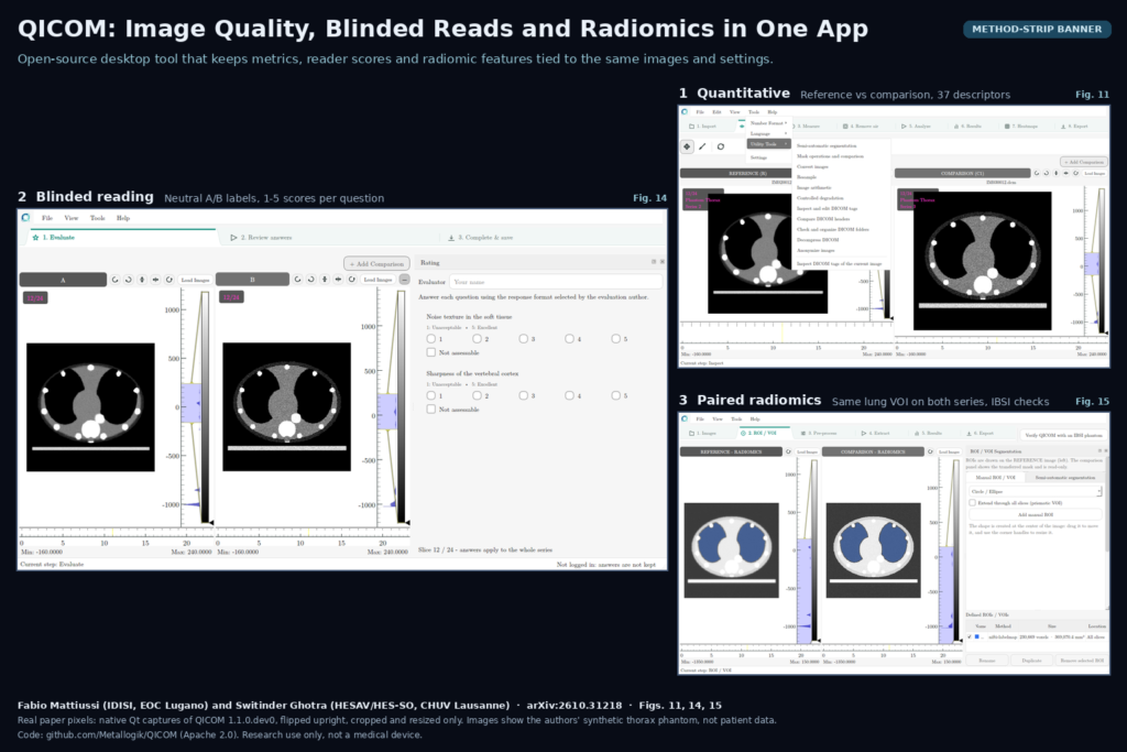
Testing a new CT denoiser or MRI reconstruction usually goes like this. SSIM and PSNR come from one script, radiomic features from PyRadiomics in a notebook, and the reader study from a spreadsheet and a folder of renamed PNGs. Three months later nobody can say which window, which mask or which file version produced the numbers in the slide deck. Fabio Mattiussi (Integrated Diagnostics Institute of Southern Switzerland, EOC Lugano) and Switinder Ghotra (HESAV and CHUV, Lausanne) posted arXiv:2610.31218 on 22 October 2026 describing QICOM, the Quality Image COMparator. It is an open-source desktop application that puts quantitative image comparison, blinded reader assessment and paired radiomics in one place, and saves enough about each analysis that it can be reopened and recalculated later.
Why a viewer or platform team should care
Most of the AI work we see in hospitals stops at the question “is the processed image better?”, and the answer usually depends on how it was measured. QICOM adds no new metric or model. It handles the bookkeeping around that question by keeping the images, masks, settings, measurements and reader responses linked to each other. For a team shipping a denoising, super-resolution or reconstruction feature inside a DICOM viewer, that is the evidence a radiologist or a procurement committee will ask for. The paper also shows why one score is not enough. In its synthetic filtering example, stronger Gaussian smoothing raised SSIM from 0.8724 to 0.9119, while MSE and the edge-based HFEN error got worse and GLCM contrast moved 55.9% below the clean reference. Picking the filter by SSIM alone would have chosen the image with more intensity error and flattened texture, which matters if those images later feed a radiomics model.
What goes in and what comes out
Inputs are DICOM series, NIfTI volumes or ordinary bitmaps, plus masks and, for the qualitative side, a list of questions. There are three workbenches. The quantitative workbench takes a reference image, one or more comparison images and optional ROIs, and computes 37 descriptors covering intensity error, structure, distributions, perceptual backends such as LPIPS and ROI statistics like SNR and CNR, along with noise power spectrum and task-based observer tools. The qualitative workbench turns a set of images and questions into a blinded package for readers, with randomised panel order and hidden repeats, and later aggregates the returned answers into agreement statistics. The radiomics workbench compares paired volumes under one mask and one preprocessing setup, using PyRadiomics or the optional MIRP backend, and reports feature differences. Outputs are tables (CSV, Excel, JSON), maps and a reproducibility receipt with hashes of the inputs, masks, configuration and results. A command-line entry point, qicom-cli confronta reference.dcm comparison.dcm, runs the full metric set without the interface.
How it works
QICOM is written in Python with a Qt interface, but the numerical, input/output, session, radiomics and reporting packages run headless, so the GUI and scripted experiments call the same functions. Every analysis records its data range, ROI, geometry, discretisation and slice aggregation, and it logs calculations that were unavailable, inapplicable or failed instead of dropping them silently. Analysis runs locally with no network access, and the optional learned metrics only use checkpoints already installed and identified by hash.
The blinded side is where spreadsheets usually fail. Each delivery uses presentation identifiers, and a separate author-side map holds the randomisation seed and the location of hidden repeats, so the reader package does not reveal which panel is the reference. Images are de-identified again on the way into a delivery. Returned scores feed weighted and unweighted kappa, Fleiss’ kappa, ICC, Krippendorff’s ordinal alpha, Gwet’s AC2 and a crossed reader-by-case bootstrap. Later versions add eleven utility tools that most viewer teams end up writing themselves: semi-automatic threshold segmentation, mask comparison with Dice and Hausdorff distance, resampling, image arithmetic, controlled degradation, DICOM tag editing and header comparison, folder checks for missing or repeated slices, decompression of lossless JPEG and multi-frame objects, and de-identification using the DICOM PS3.15 Basic Profile.
What the evaluation shows
The validation is about whether the software computes what it says it computes. Against analytic references, the largest discrepancies were 1.67 x 10⁻⁸ for MSE and 4.63 x 10⁻⁷ for SSIM. For radiomics, the QICOM and MIRP route met the Image Biomarker Standardisation Initiative (IBSI 1) tolerances for all 482 numeric digital-phantom entries and all 1,622 entries across the five preprocessing configurations of the public CT reference case. The PyRadiomics route covers a narrower set, 104 of the 482 phantom rows, and passed all of them. Reopened radiomics projects reproduced all 43 feature pairs exactly, and scripted reader responses were mapped back to their source conditions without error. In simulated reader-by-case data the bootstrap intervals covered the true value in 95 of 100 datasets, both with complete responses and with 15% missing.
What it does not show
No human readers took part in this study. The images are synthetic, the CT is a single public case analysed five ways, and the reader responses are scripted. The authors state that perceptual validity, clinical usefulness, cross-site behaviour and usability are untested, and that QICOM is not a medical device. Some of the no-reference scores are calibrated on photographs or on QICOM’s own reference distribution, so their scales mean little for CT or MRI without further work. Blinding strips text metadata and EXIF fields but does not detect burned-in labels. The later version blocks clinical DICOM that does not declare BurnedInAnnotation as NO, and runs no OCR, which matters for ultrasound and secondary captures. DICOM SEG and RTSTRUCT are not supported yet, so masks travel as NIfTI label maps, and the README lists manual stack concatenation and a polygon-only freehand ROI as known limits. The utility tools shown in the supplement belong to a later version than the one evaluated.
Code, data and license
The source is on GitHub at Metallogik/QICOM under Apache 2.0. The paper says the repository would go public before the arXiv posting, and it was public when we checked on 23 October 2026. Installation is pip-based with extras for the GUI, radiomics, MIRP and learned metrics, and bundles target Windows x64, macOS on Apple Silicon and Linux x64. MIRP is an optional dependency under EUPL 1.2. The IBSI CT reference case is CC BY-NC 3.0 from its original source, and the RIQE CT quality model used in later versions is archived on Zenodo under CC BY 4.0. There are no trained weights to worry about beyond those optional learned-metric checkpoints, which have to be fetched and verified separately. The experiment bundle used for the paper is held locally and is not part of the public repository.
How we would use it
We build DICOM viewers and imaging platforms, and the place we would use QICOM is the evaluation step before a processing feature ships. A typical case is a denoising or reconstruction option a client wants to switch on in their viewer. We would export the original and processed series, run the quantitative workbench with ROIs on the structures that matter for the task, check radiomic stability if downstream models depend on texture, and send a blinded package to two or three radiologists with a question tied to the clinical task, for example low-contrast lesion visibility. The receipt and hashes give a record that can be attached to a validation report. We would not wire QICOM into a production PACS; it is a research desktop tool. Its de-identification and folder-check utilities are useful on their own when preparing data exports, and its design, saving settings and hashes next to every number, is a pattern worth copying into any in-house evaluation pipeline. For related reading, see our posts on RIQE, the no-reference CT quality score from the same author, on DICOM anonymizer software, and on what a DICOM viewer does.
Sources
- Mattiussi, F., Ghotra, S. QICOM: Open-Source Software for Reproducible Image Quality Evaluation, Blinded Assessment, and Radiomic Comparison. arXiv:2610.31218, 2026. https://arxiv.org/abs/2610.31218. PDF: https://arxiv.org/pdf/2610.31218.
- Code: https://github.com/Metallogik/QICOM (Apache 2.0).
- Featured image: Figs. 11, 14 and 15 of arXiv:2610.31218 (quantitative, blinded qualitative and radiomics workbenches on the authors’ synthetic thorax phantom), real paper pixels, cropped and resized only.