
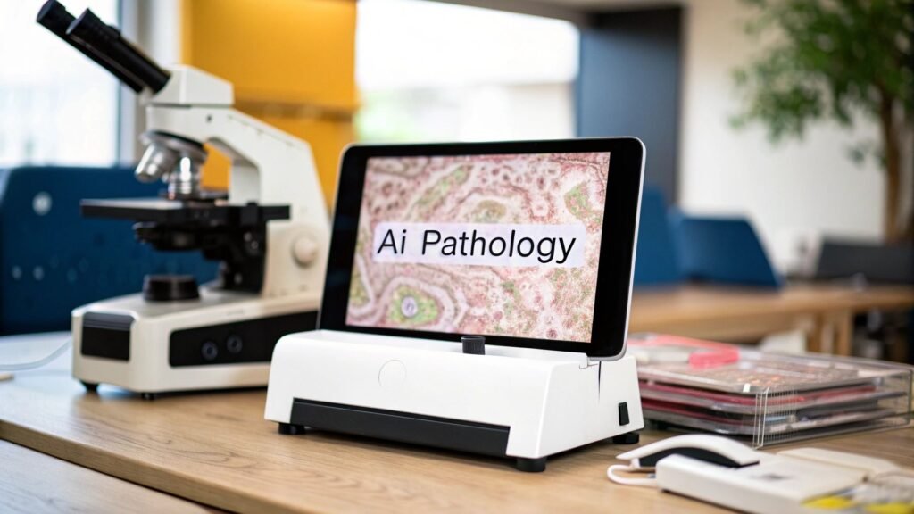
Artificial intelligence in pathology is software that reads a whole-slide image (WSI) and returns a triage flag, a count, or a heat map a pathologist then accepts or rejects. The pathologist signs. It is not radiology CAD in a lab coat. It is not a PYCAD product.
If you meant generic diagnosis / CAD → AI for medical diagnosis. If you meant the scanner / LIS / viewer stack → digital pathology software (a leftover software page; different job). If you meant what AI in healthcare is → what is artificial intelligence in healthcare. If you meant FDA class / 510(k) → FDA medical device approval process.
PYCAD builds custom web DICOM viewers and imaging models. A pathology WSI is a different file and a different viewer. We do not ship a digital-pathology LIS, a slide scanner, or a HER2 product.
Pixels first: the slide has to be digital
Glass under a microscope is not a training set. A WSI is a gigapixel pyramid from a slide scanner — often a .svs / .tiff, not a DICOM CT. Until the lab scans, there is nothing for a model to read. That conversion (scanner, storage, LIS hook) is the software page, not this one. This page starts after the file exists.
The model is the same second-reader pattern as radiology CAD, on a different pixel type. 5927 already says so in one table row. The rest of that article is radiology-shaped. This one is the lab.
Three jobs on a slide
| Job | What the model returns | What the pathologist still does |
|---|---|---|
| Triage | A worklist bump: this slide is more likely to hold tumor / this one is more likely normal | Opens the case. The model does not sign “benign.” |
| Quant | A number: mitotic figures, tumor cellularity, a biomarker percentage (ER / PR / Ki-67 / HER2 membrane) | Accepts or edits the number. Inter-observer spread is why people buy this job. |
| Second reader | A heat map or outline on regions the model thinks are tumor, Gleason pattern, mets | Confirms or dismisses each region and writes the report. |
Prostate biopsies are the textbook second-reader: the model highlights glands, the pathologist jumps to those fields instead of walking every millimeter. Mitotic count and IHC percentage are the textbook quant: a tired Friday afternoon number vs a whole-slide count. Neither is a diagnosis. Detection is not the signed report — same split as 5927.
What is actually hard (and what was invented)
Inter-observer variability on grade and on HER2-low / ultralow is a real problem. Vendors publish agreement lifts when pathologists use their overlay. Those lifts are study-specific. This page does not reprint an unsourced “+12.9 points” table. If you need a number, read the paper that produced it and check the stain, the cutoff, and who was in the panel.
A 2024 Proscia-commissioned survey of 360 lab leaders put workforce shortage at 38% as the top operational pressure and declining reimbursement at 31% as the top financial one. That is a vendor survey, not a registry. It is why labs buy automation. It is not a prevalence of disease. Source: Proscia, 2024.
What you have to buy before the model
- Scanners. Throughput and a file the model can open. A model trained on one scanner’s stain and compression will drift on another.
- Storage and a viewer. A WSI is large. The viewer has to tile. A CT DICOM viewer is the wrong tool.
- LIS integration. If the flag does not land on the case, it is a demo.
- A validated intended use. Clinical use in the US or EU needs a cleared / CE-IVD tool for that claim. An unvalidated research overlay is a research overlay. The pathway is 7568.
Alert fatigue is the failure mode: too many false positives and the overlay gets ignored. A tool that cannot show why it marked a region is the XAI problem — explainable AI — not a second pathology article.
What this page is not
- Not radiology diagnosis. Slides ≠ CT. Who signs is a pathologist.
- Not a digital-pathology software buy-list. Scanners, LIS, viewers → leftover 7619.
- Not a PYCAD pathology platform or a HER2 SKU.
- Not a predictive-biomarker future essay, a YouTube embed, or a kyve.network / vizule.io outbound. Dropped.
- Not “will AI replace pathologists.” No. The model counts and flags. The report is a person.
If the work is a viewer or a model on medical images — annotation through deployment — that is the imaging piece. Pathology WSI is a different file. Case studies.