
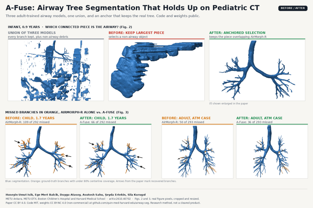
Airway segmentation on chest CT looks like a solved problem until you run an adult model on a child. The trachea and main bronchi come out fine, the small peripheral branches go missing, and every so often the post-processing step keeps the wrong object entirely and hands you a blob of soft tissue labelled “airway”. In arXiv:2610.40702, Huseyin Umut Isik, Ege Mert Balcik, Duygu Atasoy, Asutosh Sahu, Şeyda Ertekin and Sila Kurugol (METU in Ankara, and Boston Children’s Hospital with Harvard Medical School) describe A-Fuse, a way to combine three existing airway models that recovers more branches and does not lose the tree, on both adult and pediatric scans, without any pediatric training data.
The main idea is a post-processing rule, and that is why I picked this paper. Anyone who ships segmentation has written “keep the largest connected component” at the end of a pipeline. This paper shows when that line breaks and gives a simple replacement.
What the method does
A-Fuse runs three airway models on the same CT and merges their masks.
- AirMorph-R. The authors’ PyTorch reimplementation of the AirMorph ensemble, since its training code and checkpoints were not available. Five networks (three 3D U-Nets and two WingsNets) trained on 239 volumes from the ATM’26 challenge, combined by a three-of-five vote and a largest-component step. Its vote removes most false positives, and its remaining largest piece was the airway in every test scan.
- MEDPSeg-R. MEDPSeg retrained on the same 239 volumes, because the released weights were trained on ATM data that could overlap the test set.
- TfeNet. Used as released, with its checkpoint trained on the BAS dataset.
The fusion takes the voxel-wise union of the AirMorph-R mask and the raw outputs of the other two, before their own clean-up. The union keeps every branch that any model found, but it also keeps every false positive. So the union is split into connected pieces and the method keeps the single piece that overlaps the AirMorph-R mask the most. The AirMorph-R mask is only used to pick the piece; it does not cut the result. A branch that only TfeNet found stays in, as long as it is attached to the tree.
The reason for this rule is in the infant case in the figure above. On a 0.9-year-old, the union of the three models is full of non-airway debris, and the largest piece in it is not the airway at all. Both MEDPSeg-R and TfeNet, with their own largest-component clean-up, also returned a non-airway object on that scan. Choosing by overlap with the conservative model instead of by size keeps the real tree.
The fused mask also gets a voxel confidence map. Each voxel’s pattern of votes across the three models is mapped to how often that pattern was correct on 30 adult validation scans. Low-confidence voxels are where a reviewer should look first.
What comes out of it
The output is a binary airway mask on the CT grid, with a confidence value per voxel. The authors wrapped the full pipeline in a web application: upload a de-identified CT volume and it returns the airway mask, its centerline and per-branch measurements (length, diameter, branching angle and generation) in an interactive 3D viewer, downloadable in the orientation of the input volume. The paper reports about ten minutes for a 512 x 512 x 800 volume on one NVIDIA A10G GPU.
They also release a modified ITK-SNAP with 3D editing tools for tubular structures: paint and erase in 3D, delete a connected component, bridge a gap between two branch ends with a tube, and local morphological closing. The pediatric radiologists used it to build the ground truth for the paper. For a team that has to correct airway masks before measuring them, this editor may be the most useful piece in the release.
For a viewer or clinical platform, I see two uses. Airway trees with branch generations feed bronchoscopy planning and branch-level airway measurements, which is what pediatric airway growth and disease studies need. And the confidence colouring is a ready-made review layer: show the tree, colour the doubtful voxels red, and let the reader fix those first. If you already deal with broken tubular masks, our post on repairing broken vessel masks into connected centerlines covers the vessel version of the same problem.
Results that matter
Testing used 30 adult CTs held out from the ATM’26 training set (a local split, not the official challenge test set) and nine non-contrast chest CTs from eight children aged 0.9 to 16 years at Boston Children’s Hospital, all read as normal.
The number I would quote is branch detection, the share of ground-truth branches with at least 80% of their centerline covered by the mask. In the nine pediatric scans, A-Fuse detected 68.8% of branches against 59.8% for AirMorph-R alone, and it was best on all six reported metrics in children. In adults the gain over AirMorph-R is about one percentage point. The bottom row of the featured image shows the difference on one child and one adult, with the missed branches in orange.
The second result is about reliability. Anchored selection and largest-component selection gave different answers in four of the 39 test scans, three adults and the infant, and in all four the largest component contained no airway. A-Fuse kept the airway tree in every scan.
Plain majority voting had the best adult Dice of every strategy compared, yet it detected far fewer branches than A-Fuse. Voxel overlap is dominated by the trachea and main bronchi, so a method can look better on Dice while losing the small branches that branch-level measurements depend on.
Where it falls short
- Small pediatric test set. Nine scans from eight children at one hospital, all with normal lungs. Children with cystic fibrosis, bronchiectasis or other airway disease were not tested. The authors call for larger multicentre pediatric cohorts.
- It cannot remove false positives that touch the airway. The rule throws away disconnected pieces only. Leakage attached to the tree stays in, and at the tenth generation A-Fuse’s Dice was slightly below AirMorph-R’s because of extra voxels along thin branches.
- Everything hangs on the anchor. If AirMorph-R fails to find the airway component, the selection picks the wrong piece. It found the airway in all 39 test scans here, but that is a small sample.
- Branch measurements are not validated. The web app reports diameters, lengths and angles, but the paper does not compare them with an independent reference. Treat them as research output.
- Three models to run. You need the AirMorph repository, the MEDPSeg package and the TfeNet repository side by side, and about ten minutes of GPU time per large volume. The repository scripts work on NIfTI or MHA masks, so a DICOM pipeline needs its own conversion step.
- Web app access. When I checked on 2 November 2026, the web application link in the README asked for a username and password, so I could not test it.
Code, weights and licence
The code is public at github.com/quin-med-harvard-edu/airway-seg under the MIT licence, including the fusion rule, AirMorph-R and MEDPSeg-R training code, the evaluation scripts and the data split. Trained weights are in the repository’s models-v1 release. The AirMorph-R and MEDPSeg-R weights were trained on ATM challenge data and are released under CC BY-NC 4.0, so non-commercial research use only. The TfeNet checkpoint is the original authors’ file under their MIT licence. The ITK-SNAP extension is a separate repository under GPL-3.0. The pediatric scans are not public, and the ATM data come from the challenge under its own agreement. The preprint is under CC BY 4.0 and has been submitted to an IEEE venue (the repository names ISBI 2027). The authors report no funding and no conflicts of interest. This is research software, not a cleared medical device.
For another take on combining several models and using their disagreement as a review signal, see our post on hippocampal segmentation ensembles with uncertainty review.
My take
The non-commercial weights mean a product team cannot ship this as released. You would retrain AirMorph-R and MEDPSeg-R on data you are allowed to use commercially, which the MIT code makes possible but not quick. What I would take from the paper today costs nothing: go through your own pipelines, find every “keep the largest component” step, and ask what happens on a scan where the largest object is not the organ. For airways, vessels or any thin tree, choosing the piece that overlaps your most conservative model is a few lines of code, and on four of 39 scans here it was the difference between an airway and a blob. For pediatric airway research groups, the open code, weights and the 3D editing tool in ITK-SNAP together make a usable starting point.