
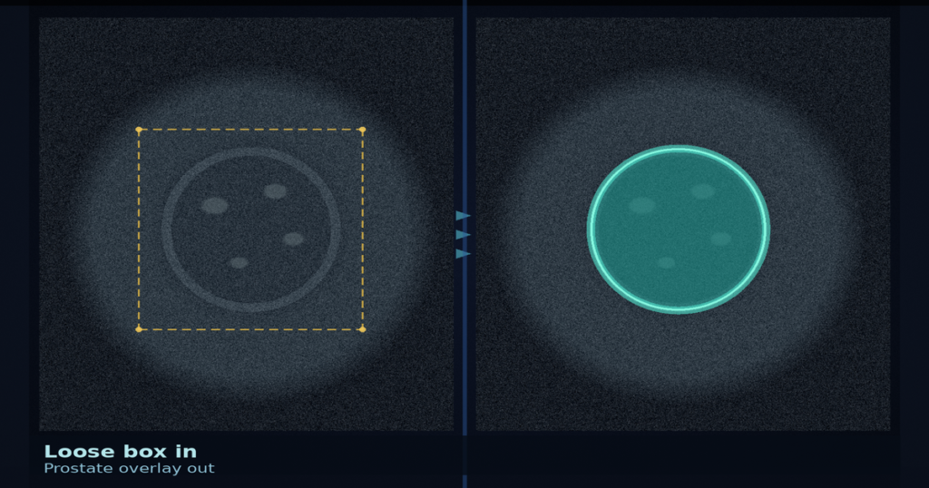
Drop an approximate box on a 29 MHz micro-ultrasound prostate slice and you want a gland overlay you can hang for PSAD or biopsy registration. Point clicks drown in dense speckle. Ayusha Abbas, Saram Abbas, and Kabita Adhikari (Newcastle) posted arXiv:2608.14796 around 14 August 2026. The paper is Zero-Shot Adaptation of Medical Vision Foundation Models for High-Frequency Micro-Ultrasound Prostate Segmentation. Code is planned at github.com/anonymous/medsam-microus upon acceptance. Public data sit on Zenodo 10475293.
ExactVu-style 29 MHz micro-US resolves about 3 to 4× finer than 6 to 12 MHz TRUS, and that gain comes with heavy acoustic noise. Two clinicians on the same image can differ by more than 10% in outlined area. The OPTIMUM trial (2025) already put micro-US biopsy in the same clinical conversation as mpMRI-guided targeting. Both PSAD and targeted biopsy still need a gland boundary, so a training-free overlay that tolerates a loose box is the viewer-facing ask.
Frozen MedSAM, then CLAHE, dilation, Fourier smooth
The pipeline keeps MedSAM frozen. An oracle-style bounding box (ground-truth box expanded by 20 px) prompts the model. The raw mask is then cleaned without any fine-tuning: CLAHE on the grayscale slice (clip limit 0.03, 8×8 tiles), binary dilation for three iterations with a 4-connectivity cross, then Fourier contour smoothing that keeps M=4 low-frequency modes and scales the contour by s=1.05 to recover wall pixels lost to shadowing. Inference is about 0.8 s per slice on a laptop CPU or MPS path.
Point prompts fail on this modality. Centre point, multi-point grids, and hybrid point-plus-loose-box setups top out around Dice 0.350. Speckle gives no stable local contrast for a click. A bounding box is necessary if you want a usable gland mask.
Where the overlays landed on the public cohort
Evaluation uses the public Micro-Ultrasound Prostate Segmentation dataset (Zenodo 10475293): 75 patients, 2,621 axial slices with prostate. A 20-patient held-out set carries four annotators (expert urologist as GT, plus a master’s student, a medical student, and a clinician with limited US experience).
On that n=20 oracle-box test, raw MedSAM scores Dice 0.749±0.043 and HD95 217.2±36.9 px. After CLAHE, dilation, and Fourier smoothing the same prompts reach 0.865±0.029 / 120.1±26.1, about a 45% cut in boundary distance error. Across all 75 patients the pipeline lands at Dice 0.859±0.037 and HD95 127.0±36.9; 74 of 75 improved over raw MedSAM. Mean Dice is not significantly different from the three non-expert rater groups (padj>0.19). Consistency is tighter: model inter-patient SD 0.029 versus 0.047 to 0.061 for the human groups (about 38 to 52% lower). Supervised MicroSegNet reports Dice 0.939 on this data, but that number needs labels and is not a zero-shot comparison.
Automatic localisers still trail the oracle box. A statistical prior box reaches Dice 0.691±0.056; a two-pass MedSAM cascade reaches 0.698±0.058. Closing that gap without a human or better detector remains open work.
How this lands in a viewer
If you already hang micro-US or TRUS in a DICOM viewer, treat this as a box-prompt gland overlay rail. Load the axial micro-US slice, accept a loose box (or seed one from a prior), run frozen MedSAM plus the CLAHE / dilation / Fourier cleanup, hang the smoothed prostate contour for PSAD area or biopsy registration, and fail closed when only point clicks are available, when the box misses the gland, or when you need a supervised MicroSegNet-level mask without labels. Do not hang the auto-localiser Dice as if it matched the oracle-box ceiling.
ExiL turns a rough bone brush into a refined US surface overlay. This paper is approximate box on speckled 29 MHz micro-US in, training-free prostate gland overlay out.
Rebuild from arXiv:2608.14796. As of 7 September 2026 the abstract and PDF respond (HTTP 200). Data: Zenodo 10475293. Code planned at github.com/anonymous/medsam-microus upon acceptance. The preprint is the source of record until a camera-ready version exists.
Sources
- Abbas, A., Abbas, S., Adhikari, K. Zero-Shot Adaptation of Medical Vision Foundation Models for High-Frequency Micro-Ultrasound Prostate Segmentation. arXiv:2608.14796, posted ~14 August 2026. https://arxiv.org/abs/2608.14796 (HTTP 200 on 7 September 2026). PDF: https://arxiv.org/pdf/2608.14796 (HTTP 200 on 7 September 2026). Code planned at github.com/anonymous/medsam-microus upon acceptance.
- Micro-Ultrasound Prostate Segmentation dataset. Zenodo 10475293. https://zenodo.org/records/10475293
- Ma, J., et al. Segment Anything in Medical Images. Nat. Commun. 2024. (MedSAM.)
- Jiang, H., et al. MicroSegNet: A deep learning approach for prostate segmentation on micro-ultrasound images. (Supervised reference cited in the paper.)
- OPTIMUM randomized clinical trial on micro-ultrasound for prostate cancer diagnosis, 2025. (Clinical context cited in the paper.)