Skip to content
  • Home
  • Services
    • All Services
    • Custom DICOM Viewer
    • Case Studies
  • Products
    • All Products
    • PYCAD DICOM Viewer
    • PYCAD Dental Viewer
    • PYCAD SSR Engine
  • Case Studies
  • Blog
  • About us
  • Home
  • Services
    • All Services
    • Custom DICOM Viewer
    • Case Studies
  • Products
    • All Products
    • PYCAD DICOM Viewer
    • PYCAD Dental Viewer
    • PYCAD SSR Engine
  • Case Studies
  • Blog
  • About us
Get in Touch

Category: Computer Vision

Saliency Maps Do Not Flag Chest X-Ray AI Errors at New Hospitals
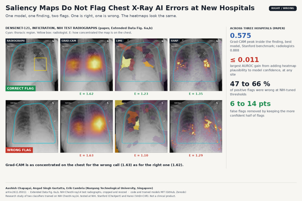
Comparison built from Extended Data Fig. 6a,b of arXiv:2611.05011 by Aashish Chapagai, Angad Singh Gurtatta and Erik Cambria: two NIH ChestX-ray14 test radiographs where DenseNet-121 flagged infiltration, one correct and one wrong, each shown with Grad-CAM, LIME and SHAP heatmaps of nearly equal concentration on the chest.

A three-site study finds Grad-CAM, LIME and SHAP look the same on right and wrong calls. Model confidence is the better alarm.

Dx-Rx: An MRI Agent That Measures Artifacts Before Fixing Them
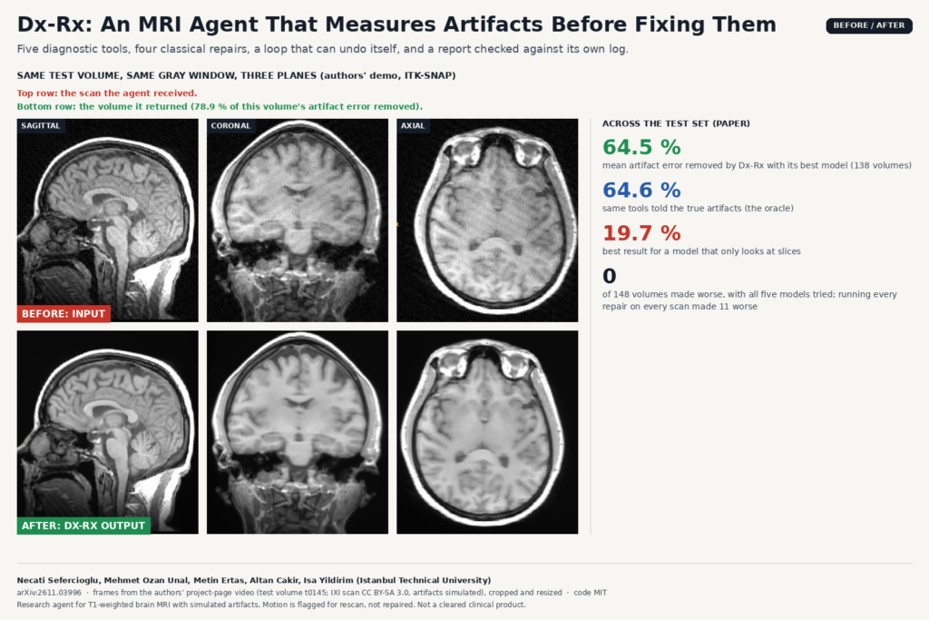
Before and after frames from the project-page video of arXiv:2611.03996 by Necati Sefercioglu, Mehmet Ozan Unal, Metin Ertas, Altan Cakir and Isa Yildirim: sagittal, coronal and axial slices of IXI test volume t0145 with simulated bias field, spike and noise, and the volume returned by the Dx-Rx agent.

Dx-Rx gives a language model five MRI artifact detectors and four classical repairs, lets it undo its own steps, and checks its report against the log.

EventMC: Training-Free Motion Correction for 3D Brain MRI
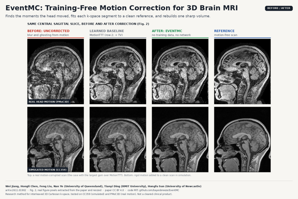
Before and after figure built from Figure 2 of arXiv:2611.01902 by Wei Jiang, Hongli Chen, Tianyi Ding, Feng Liu, Nan Ye and Hongfu Sun: central sagittal brain MRI slices from a real motion-corrupted scan and a simulated case, shown uncorrected, after the MotionTTT baseline, after EventMC, and the motion-free reference.

EventMC finds when the head moved, fits each k-space segment to a clean reference and rebuilds one sharp brain volume.

LAPIS: Chest X-Ray Anonymization Beyond the DICOM Header
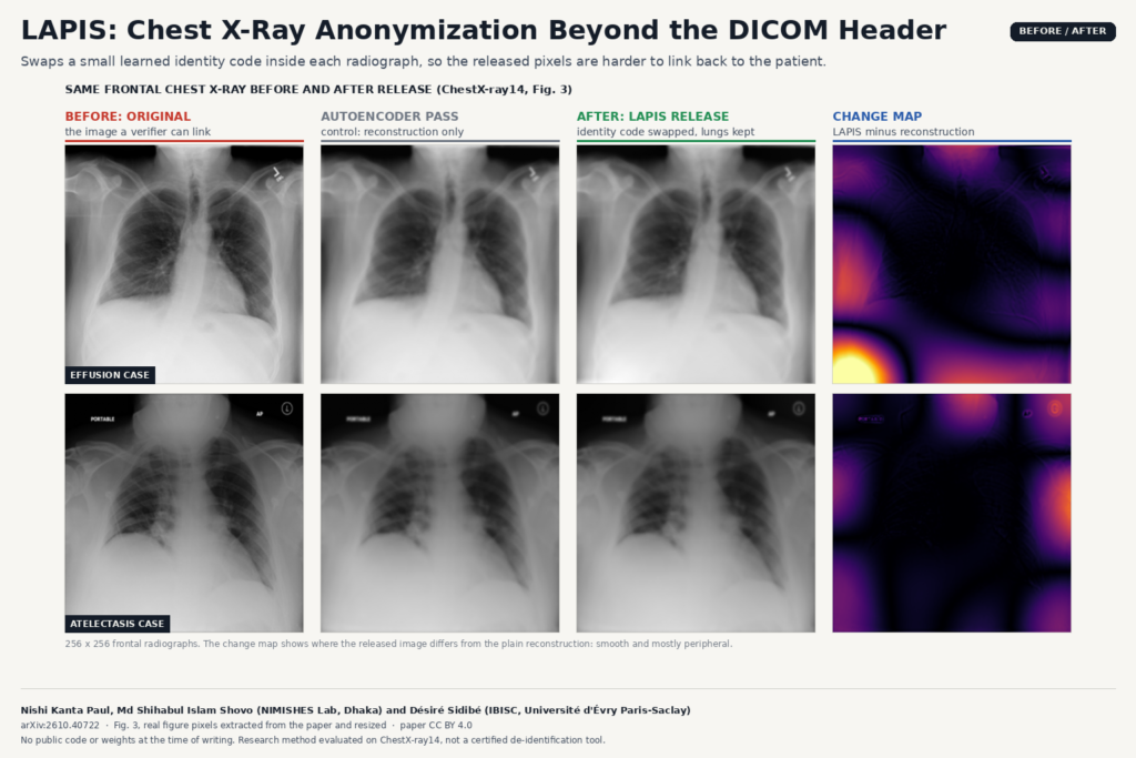
Before and after figure built from Figure 3 of arXiv:2610.40722 by Nishi Kanta Paul, Md Shihabul Islam Shovo and Désiré Sidibé: two frontal chest radiographs (effusion and atelectasis cases) shown as original, autoencoder reconstruction, LAPIS anonymized release and a change map that is smooth and mostly peripheral.

LAPIS swaps a small learned identity code inside each chest X-ray so released images are harder to link back to the patient.

EUReCA: One Network for Sparse-View Linac CBCT
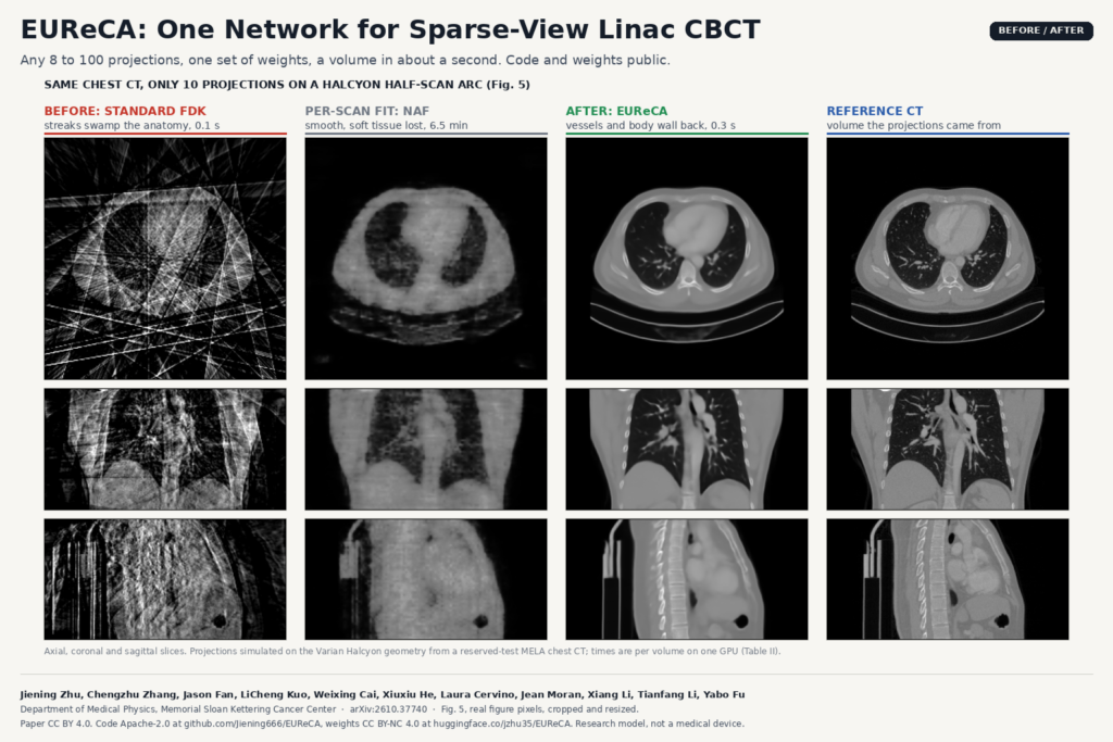
Before and after figure built from Figure 5 of arXiv:2610.37740 by Jiening Zhu, Yabo Fu and colleagues (Memorial Sloan Kettering Cancer Center): axial, coronal and sagittal chest slices reconstructed from 10 projections with standard FDK (streaks), NAF per-scan fitting (smooth, soft tissue lost), EUReCA (vessels and body wall recovered) and the reference CT.

Any 8 to 100 projections on a Halcyon arc, one set of weights, and a CBCT volume in about a second instead of minutes.

Spine MRI-to-CT Synthesis With Vertebra-Matched Pseudo-Pairs
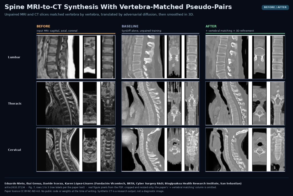
Figure 7 of arXiv:2610.37134 by Eduardo Nieto, Ibai Genua, Davide Scorza and Karen Lopez-Linares (Vicomtech, Cyber Surgery, Biogipuzkoa): lumbar, thoracic and cervical spine MRI in sagittal, axial and coronal views, the synthetic CT from the SynDiff baseline, and the synthetic CT from the full pipeline with vertebral matching and 3D refinement.

Unpaired MRI and CT slices are matched vertebra by vertebra, translated with adversarial diffusion, then cleaned up in 3D.

Pix2Point: Spine X-ray Landmarks From Dense Pixel Embeddings
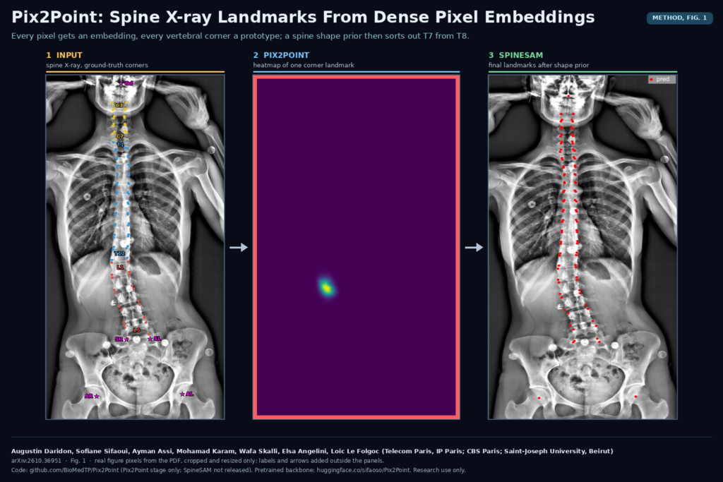
Figure 1 of arXiv:2610.36951 by Augustin Daridon, Sofiane Sifaoui, Ayman Assi, Mohamad Karam, Wafa Skalli, Elsa Angelini and Loic Le Folgoc (Telecom Paris, CBS Paris, Saint-Joseph University Beirut): a coronal full-spine X-ray with ground-truth vertebral corners, the Pix2Point heatmap of one corner landmark, and the final landmarks after the SpineSAM shape prior.

Every pixel gets an embedding and every vertebral corner a prototype; a spine shape prior then decides which corner is which.

SlimFormer++: Lean 3D Segmentation for CT, MRI and Ultrasound
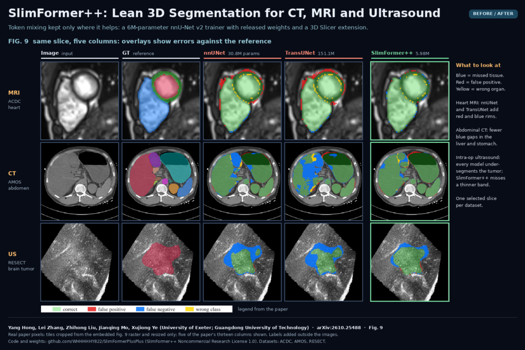
Figure 9 of arXiv:2610.25488 by Yang Hong, Lei Zhang, Zhihong Liu, Jianqing Mo and Xujiong Ye: cardiac MRI, abdominal CT and intraoperative brain ultrasound slices with ground truth and nnUNet, TransUNet and SlimFormer++ predictions, errors colored as false positive, false negative and wrong class.

An nnU-Net v2 trainer that keeps token mixing only where it helps, with released weights and a 3D Slicer extension that runs on CPU.

MedVTok: One Open Tokenizer for 2D Images and 3D Scans
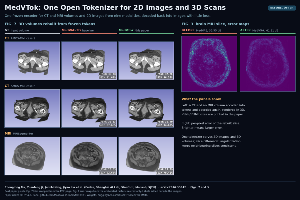
Figures 7 and 3 of arXiv:2610.35842 by Chenglong Ma, Yuanfeng Ji, Junzhi Ning, Jiyao Liu et al.: 3D renderings of two CT volumes and one MRI volume as ground truth, MedVAE-3D reconstruction and MedVTok reconstruction, next to brain MRI error maps for MedVAE and MedVTok.

One encoder turns CT and MRI volumes and 2D images from nine modalities into shared tokens, with MIT code and public weights.

XGLASS: Radiologist Gaze Tells AI Which Chest CT Slices to Read
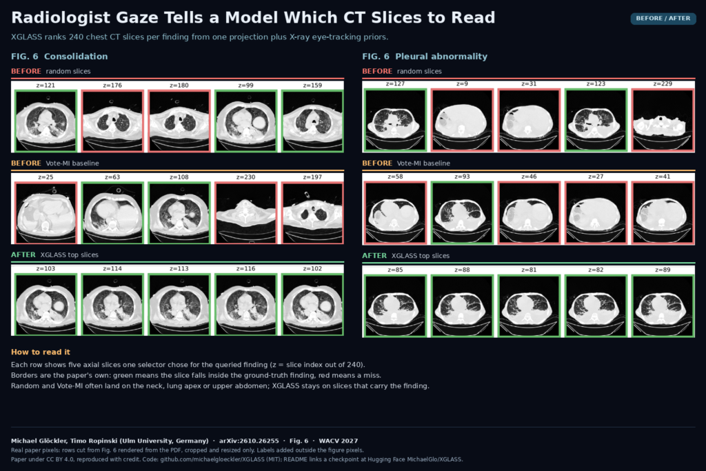
Figure 6 of arXiv:2610.26255 by Michael Glöckler and Timo Ropinski (Ulm University): five axial chest CT slices chosen by random selection, by Vote-MI and by XGLASS for consolidation and for pleural abnormality, with green borders on slices inside the ground-truth finding and red borders on misses.

A 2D ranker built on chest X-ray eye-tracking picks the CT slices a 2D VLM should check, at a fraction of 3D cost.

← Previous
Next →