Skip to content
  • Home
  • Services
    • All Services
    • Custom DICOM Viewer
    • Case Studies
  • Products
    • All Products
    • PYCAD DICOM Viewer
    • PYCAD Dental Viewer
    • PYCAD SSR Engine
  • Case Studies
  • Blog
  • About us
  • Home
  • Services
    • All Services
    • Custom DICOM Viewer
    • Case Studies
  • Products
    • All Products
    • PYCAD DICOM Viewer
    • PYCAD Dental Viewer
    • PYCAD SSR Engine
  • Case Studies
  • Blog
  • About us
Get in Touch

Category: Computer Vision

BasisSimulator.jl: One Spectral CT Decomposition for Every Scanner
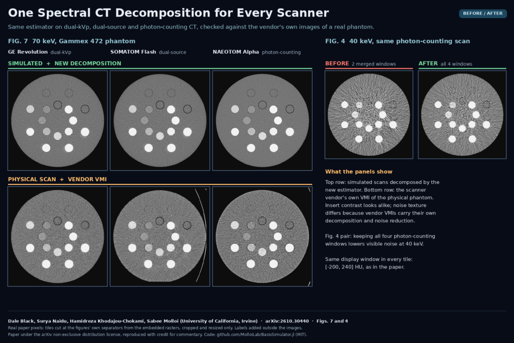
Figures 7 and 4 of arXiv:2610.30440 by Black, Naidu, Khodajou-Chokami and Molloi (University of California, Irvine): 70 keV virtual monoenergetic images of the Gammex 472 phantom on GE Revolution, SOMATOM Flash and NAEOTOM Alpha, simulated and decomposed with the new method above the vendor images of the physical scans, plus 40 keV photon-counting tiles with two merged windows and with all four.

A likelihood estimator in an open Julia CT simulator handles dual-kVp, dual-source and photon-counting data and keeps every energy window.

Plausible but Unimplantable: Foundation Models vs Cranial Implants
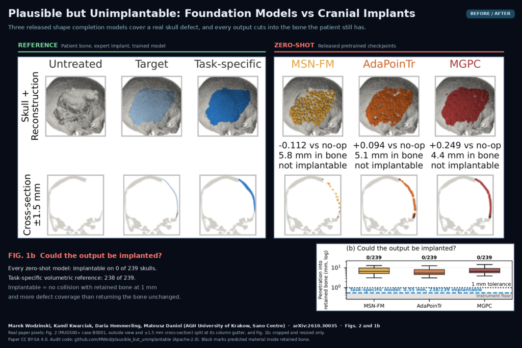
Figure 2 of arXiv:2610.30035 by Wodzinski, Kwarciak, Hemmerling and Daniol (AGH University of Krakow): a real craniectomy skull from MUG500+ with the expert target implant and a task-specific model beside the outputs of MSN-FM, AdaPoinTr and MGPC, each marked not implantable, plus Fig. 1b showing 0 of 239 implantable outputs per model.

An audit of three released shape completion models on 239 skull defects found no output that could be placed in a patient.

MedBOE: Rebuilding the Full Body Surface From Truncated PET/CT
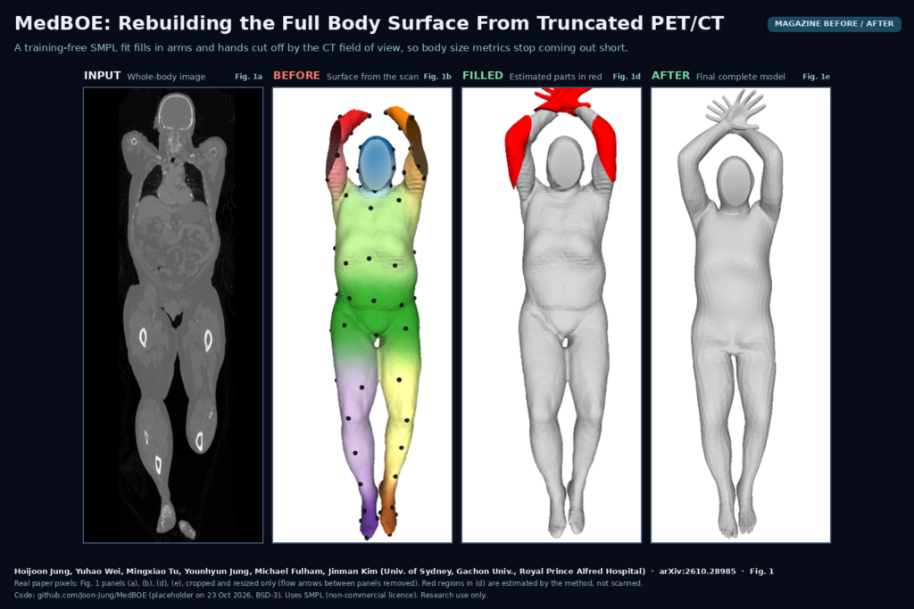
Four panels from Fig. 1 of the MedBOE paper: a coronal whole-body image, the incomplete body surface extracted from the scan with virtual-marker colours, the initial complete model with estimated arms and hands in red, and the final complete grey body model. Hoijoon Jung, Yuhao Wei and colleagues, University of Sydney, arXiv:2610.28985

A training-free SMPL fit fills in arms and hands cut off by the CT field of view, so body size metrics stop coming out short.

PCXR-Copilot: Pediatric ICU Chest X-Rays Without the Device Clutter
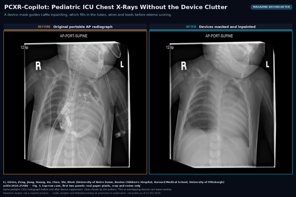
Magazine before/after from Fig. 3: the same portable AP chest radiograph of a child in a cardiac ICU, on the left with sternal wires, tubes, lines and ECG leads, on the right after PCXR-Copilot masked the devices and filled them in with LaMa inpainting. Li, Girten, Zeng, Jiang, Huang, Xu, Chen, Shi, Kheir, arXiv:2610.25486

Masks tubes and wires, paints them out, then scores lung water and body-wall edema. Live at Boston Children’s. Code not out yet.

TB-PET-DDS: A Diffusion Prior for Low-Count Total-Body PET
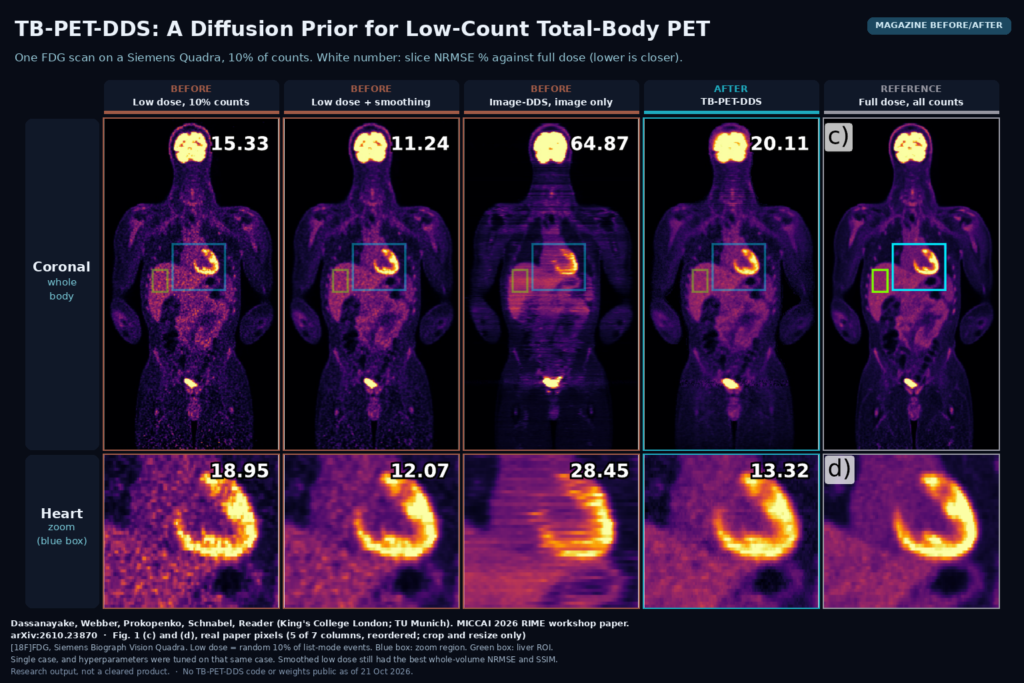
Magazine before/after from Fig. 1: one total-body FDG PET scan reconstructed from 10% of counts by the vendor, with smoothing, with Image-DDS and with TB-PET-DDS next to the full-dose reference, coronal view and heart zoom with slice NRMSE numbers. Dassanayake, Webber, Prokopenko, Schnabel, Reader, arXiv:2610.23870

A CT-conditioned diffusion prior plus a list-mode PET data step, tested on one total-body FDG scan at 10% counts. No code yet.

NV-Generate-Body: Synthetic Whole-Body CT Masks You Can Dial In
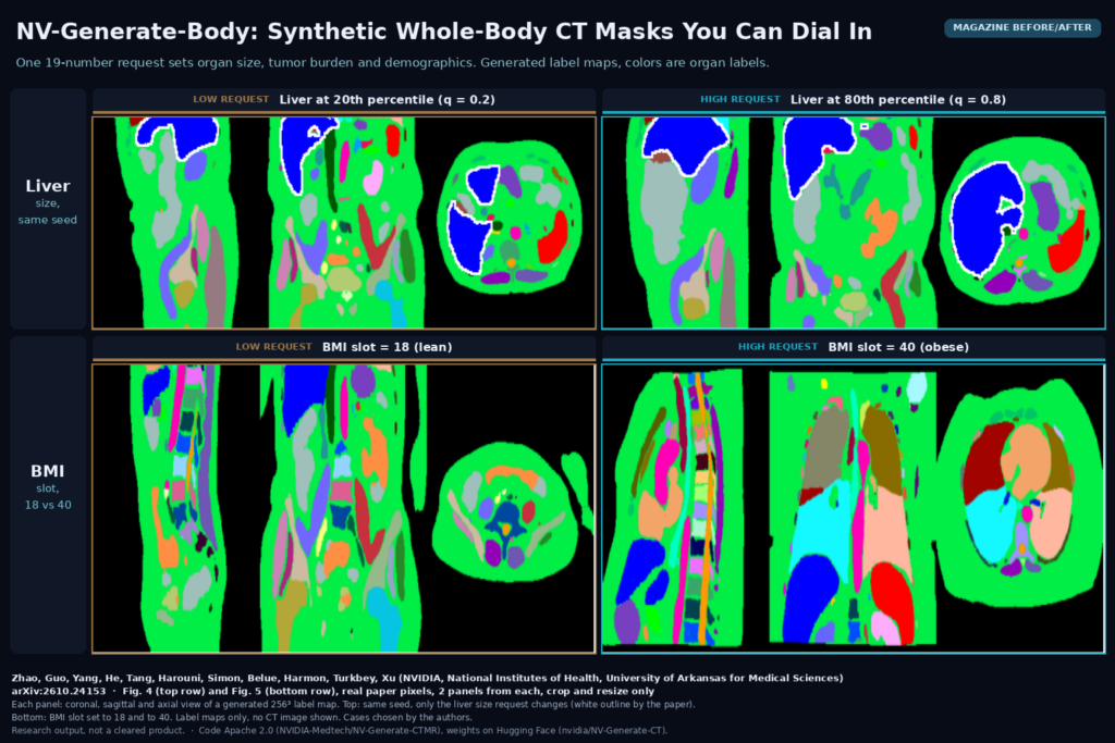
Magazine before/after from Figs. 4 and 5 of NV-Generate-Body: coronal, sagittal and axial views of generated 256-cube CT label maps, top row liver requested at the 20th and 80th percentile with the same seed, bottom row BMI slot set to 18 and 40. Zhao, Guo, ..., Turkbey, Xu, NVIDIA, NIH, UAMS, arXiv:2610.24153

NVIDIA’s NV-Generate-Body draws whole-body CT label maps with the organ sizes, tumors, sex and BMI you ask for. Code Apache 2.0, weights public.

GSMR3D: One Model Turns Thick-Slice Brain MRI Into 1 mm Volumes
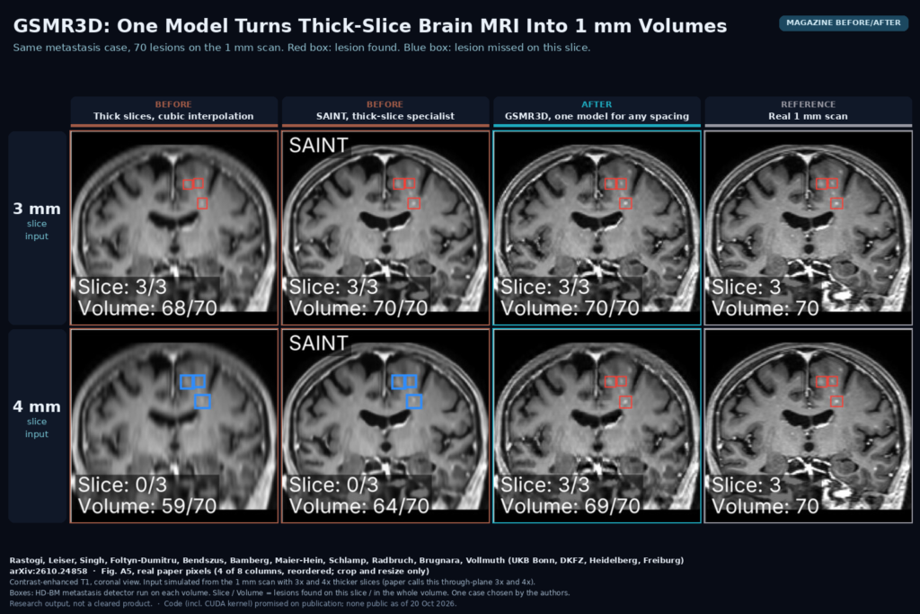
Magazine before/after from Fig. A5: coronal contrast-enhanced T1 of one brain-metastasis case rebuilt from 3 mm and 4 mm slices by cubic interpolation, SAINT and GSMR3D next to the real 1 mm scan, with detected (red) and missed (blue) lesion boxes. Rastogi, Leiser, ..., Brugnara, Vollmuth, arXiv:2610.24858

GSMR3D rebuilds thick-slice brain MRI on a 1 mm grid at any spacing, one model per contrast, and kept small metastases detectable.

LGGS: Faster Follow-Up Brain MRI Using the Patient’s Last Scan
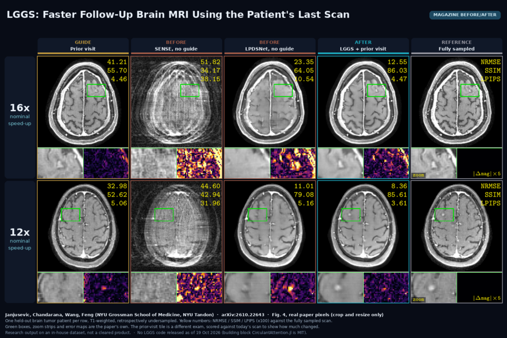
Magazine before/after from Fig. 4: two held-out brain tumor patients at 16x and 12x acceleration, each shown as the prior visit used as guide, SENSE, unguided LPDSNet, LGGS and the fully sampled scan, with zoom and error strips. Janjusevic, Chandarana, Wang, Feng, arXiv:2610.22643

LGGS rebuilds heavily undersampled follow-up brain MRI using the patient’s prior exam as a guide, without copying it into the new image. arXiv:2610.22643.

UniField++: Making Low-Field Brain MRI Look Like 3T and 7T
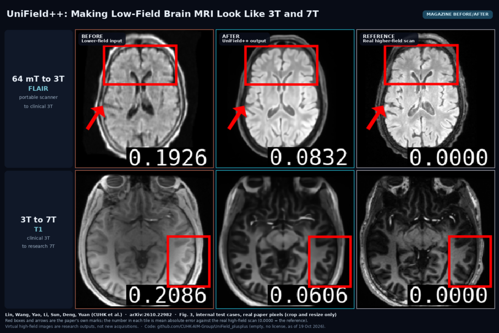
Magazine before/after from Fig. 3: two brain MRI test cases, a 64 mT FLAIR and a 3T T1, each shown as the lower-field input, the UniField++ virtual higher-field output and the real 3T or 7T scan. Lin, Wang, Yao, Li, Sun, Deng, Yuan, arXiv:2610.22982

UniField++ turns 64 mT brain MRI into virtual 3T and 3T into virtual 7T, with a vision-language planner that flags regions likely to go wrong. arXiv:2610.22982.

NoduleFlow: Automatic Lung Nodule Masks Across Follow-Up CT
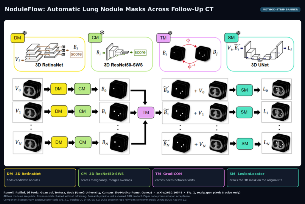
Method-strip banner from Fig. 1: chest CT volumes at several time points pass through a 3D RetinaNet detector, a ResNet50-SWS malignancy classifier, GradICON box transfer between visits and LesionLocator segmentation. Romoli et al., arXiv:2610.16548

NoduleFlow chains four public, frozen models to detect, score and segment lung nodules across a patient’s follow-up CTs with no clicks. arXiv:2610.16548.

← Previous
Next →