Skip to content
  • Home
  • Services
    • All Services
    • Custom DICOM Viewer
    • Case Studies
  • Products
    • All Products
    • PYCAD DICOM Viewer
    • PYCAD Dental Viewer
    • PYCAD SSR Engine
  • Case Studies
  • Blog
  • About us
  • Home
  • Services
    • All Services
    • Custom DICOM Viewer
    • Case Studies
  • Products
    • All Products
    • PYCAD DICOM Viewer
    • PYCAD Dental Viewer
    • PYCAD SSR Engine
  • Case Studies
  • Blog
  • About us
Get in Touch

Category: Computer Vision

moco-dl: Fast Slice-Wise Motion Correction for Spinal Cord fMRI
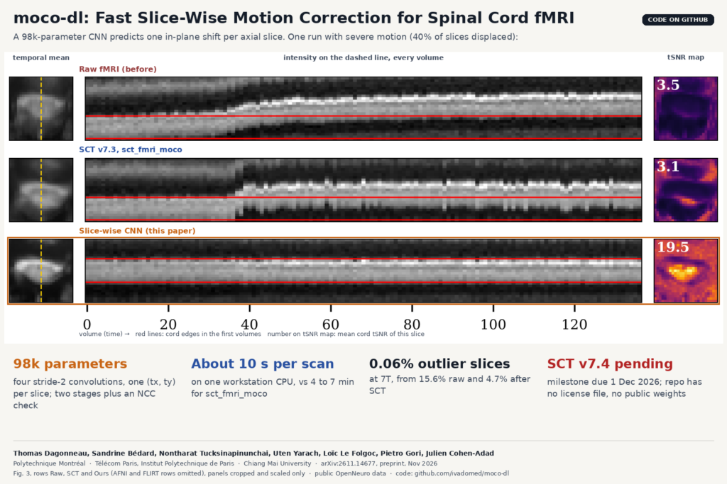
Fig. 3 of arXiv:2611.14677 by Dagonneau, Bedard et al. (Polytechnique Montreal, Telecom Paris, Chiang Mai University): temporal mean, intensity profile over time and tSNR map of one spinal cord fMRI slice with severe motion, for the raw data, SCT sct_fmri_moco and the proposed slice-wise CNN.

A tiny CNN aligns every axial slice of a spinal cord fMRI run on a CPU in seconds and beats SCT’s current tool at 3T and 7T.

LoopMR: Accelerated MRI Reconstruction With One Looped Block
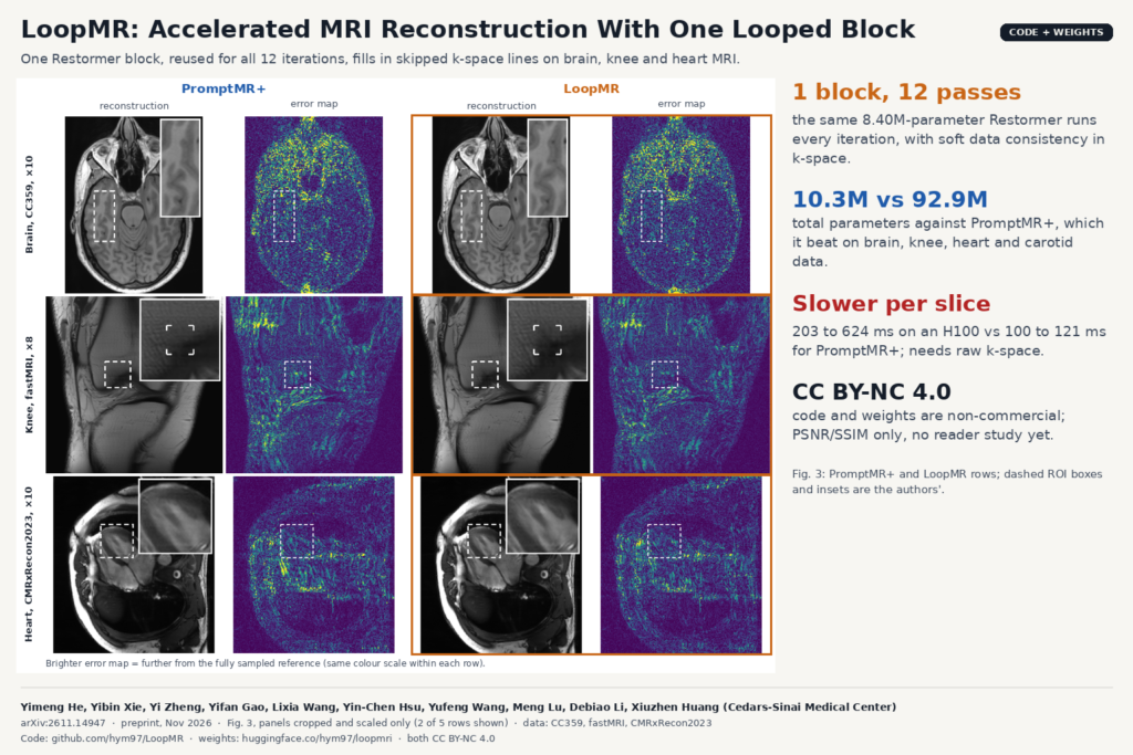
Fig. 3 of arXiv:2611.14947 by He, Xie et al. (Cedars-Sinai Medical Center): PromptMR+ and LoopMR reconstructions of undersampled brain (CC359 x10), knee (fastMRI x8) and heart (CMRxRecon2023 x10) MRI with their absolute error maps and the authors' zoomed regions of interest.

One Restormer block, run 12 times, rebuilds undersampled brain, knee, heart and carotid MRI with a ninth of PromptMR+’s parameters.

CADS: One Open Model Suite for 167 Whole-Body CT Structures
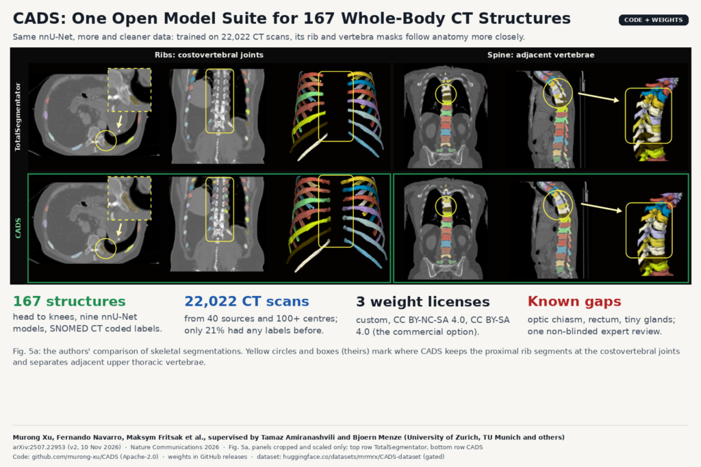
Fig. 5a of arXiv:2507.22953 by Xu et al. (University of Zurich, TU Munich and others): CT slices and 3D renders comparing TotalSegmentator (top) and CADS (bottom) rib and vertebra masks, with the authors' yellow circles at the costovertebral joints and upper thoracic vertebrae.

Trained on 22,022 curated CT scans from 100+ centres, CADS segments head to knees, runs in 3D Slicer and outputs SNOMED CT labels.

MicroBT: Finding and Counting Sub-3 mm Brain Tumors on MRI
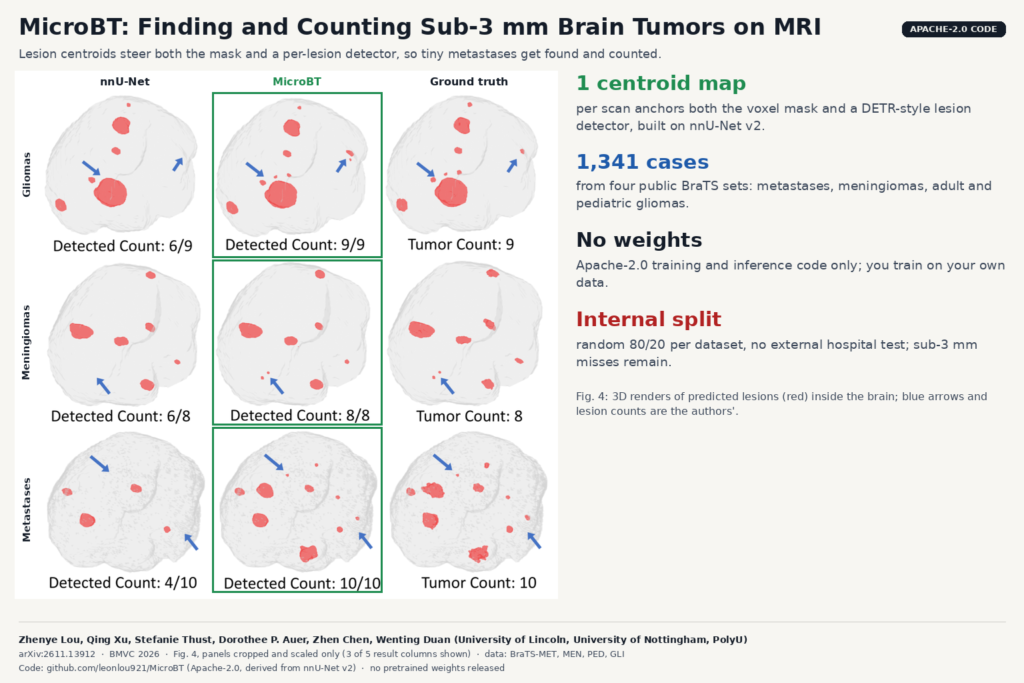
Fig. 4 of arXiv:2611.13912 by Lou, Xu et al. (University of Lincoln, University of Nottingham, PolyU): 3D brain renders with red tumor lesions predicted by nnU-Net and MicroBT next to ground truth, with the authors' lesion counts and blue arrows marking small lesions.

A centroid-guided nnU-Net extension segments brain tumors on MRI and counts each lesion, aimed at the tiny metastases that models miss.

P2Echo: One Text-Prompted Model Segments Four Echo Views
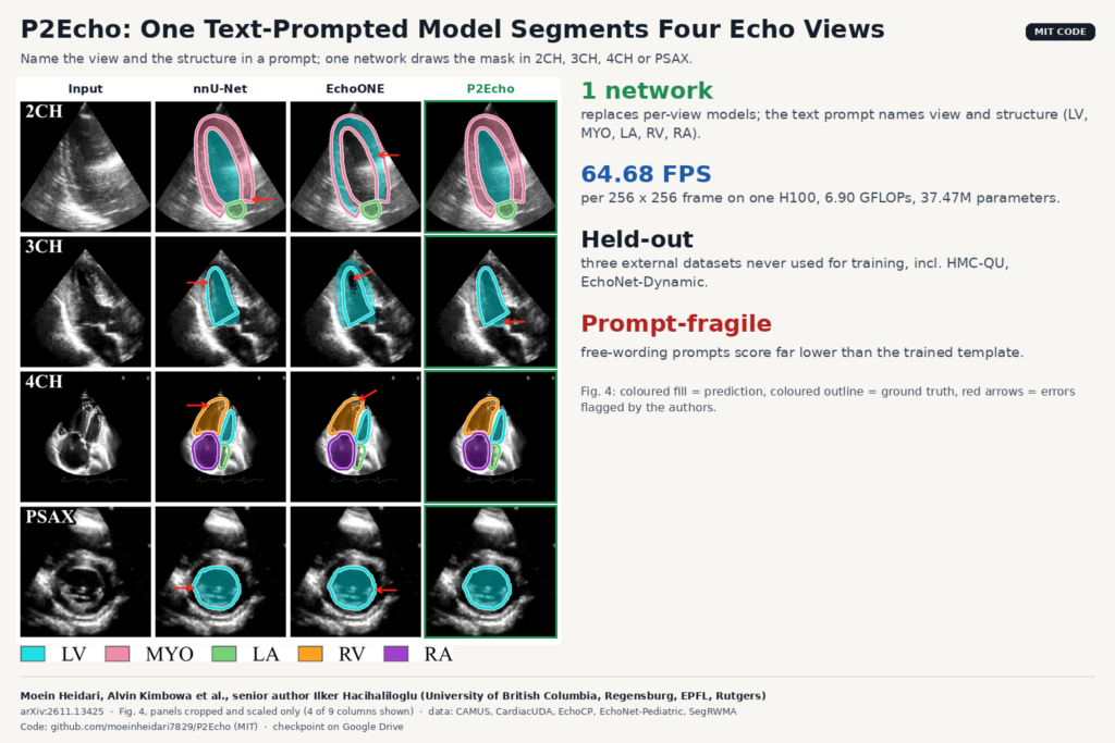
Fig. 4 of arXiv:2611.13425 by Heidari, Kimbowa et al. (University of British Columbia and co-authors): echocardiography frames in four views with LV, myocardium, LA, RV and RA masks from nnU-Net, EchoONE and P2Echo, ground-truth outlines and red arrows marking errors.

UBC’s P2Echo takes a view-and-structure prompt and draws LV, MYO, LA, RV or RA masks in 2CH, 3CH, 4CH and PSAX with one network.

UltraVoxelGS: 3D Ultrasound From Tracked Sweeps in Seconds
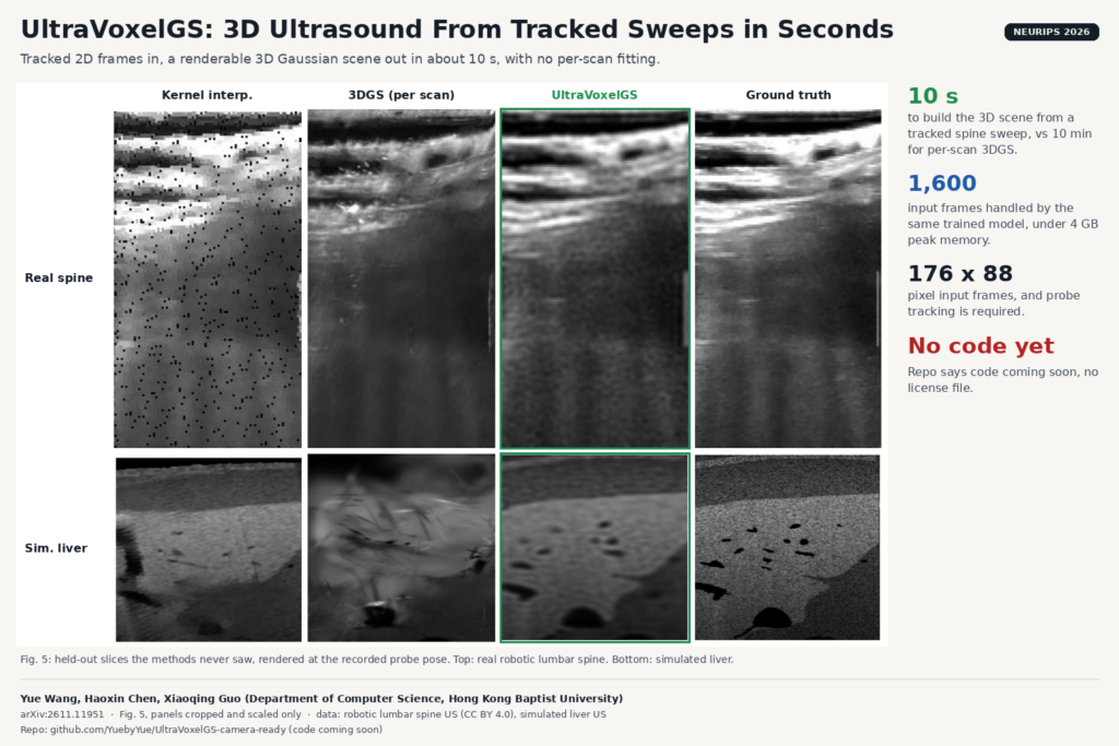
Fig. 5 of arXiv:2611.11951 by Wang, Chen and Guo, Hong Kong Baptist University: held-out ultrasound slices of a real robotic lumbar spine scan and a simulated liver scan, rendered by kernel interpolation, per-scan 3DGS and UltraVoxelGS next to the recorded ground truth.

Hong Kong Baptist University turns tracked 2D ultrasound frames into a 3D Gaussian scene in about 10 seconds, no per-scan fitting.

BrainGFM: Synthetic Brain Scans From Few Labels
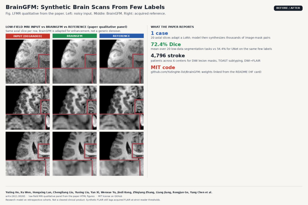
Comparison built from the low-field MRI qualitative panel of arXiv:2611.00265 by Yuting He, Xu Wen, Hongxing Lan et al.: three axial brain MRI rows with degraded input (left), BrainGFM enhancement (middle), and acquired reference (right).

One annotated case adapts a LoRA. BrainGFM then synthesizes image-mask pairs, class images, and sequence transforms for MRI and CT.

LungMend: Fix Incomplete Lung Masks on Severe TB CT
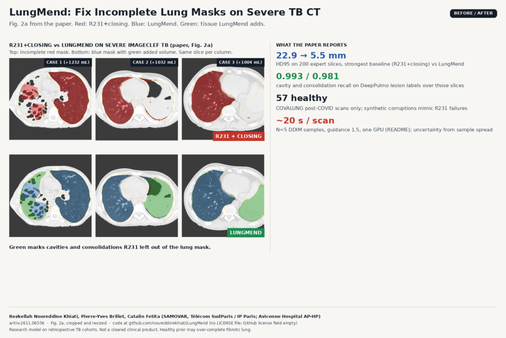
Comparison built from Fig. 2a of arXiv:2611.06556 by Rezkellah Noureddine Khiati, Pierre-Yves Brillet and Catalin Fetita: three severe ImageCLEF TB axial CT slices with R231+closing lung masks in red (top row, incomplete) and LungMend masks in blue with green added tissue (bottom row).

R231 drops cavities and consolidations on severe TB CT. LungMend fills them from a healthy shape prior and bone cues.

Virtual Contrast Breast MRI That Spares Gadolinium for Some Patients
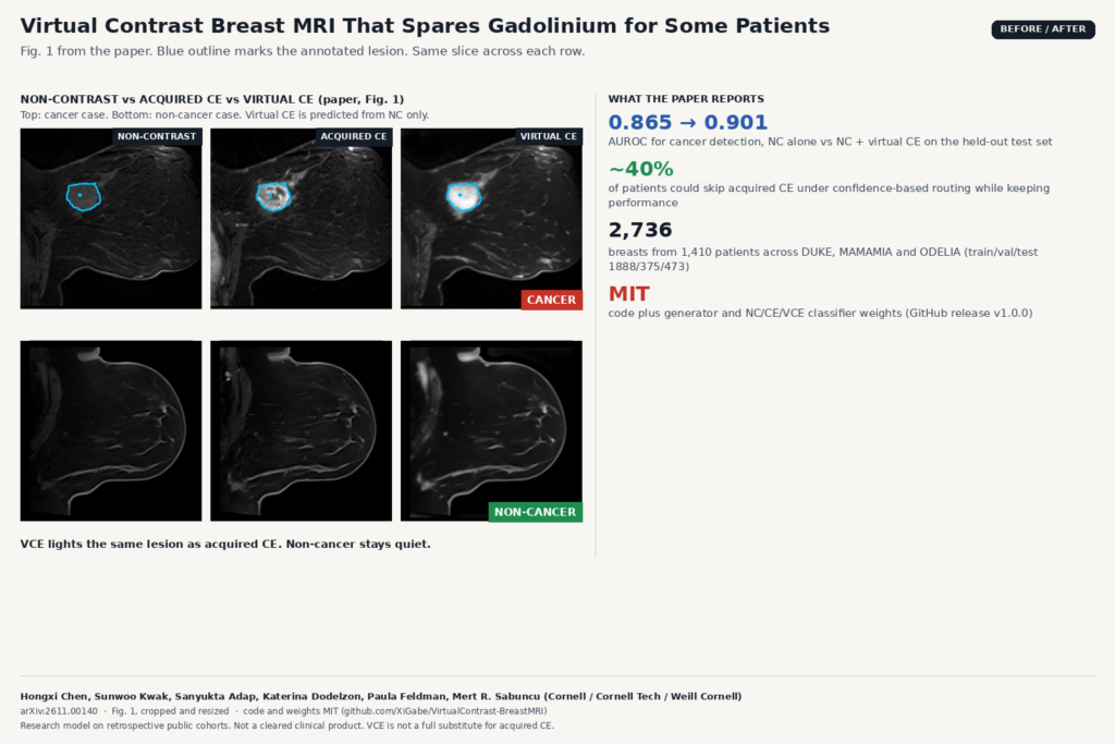
Comparison built from Fig. 1 of arXiv:2611.00140 by Hongxi Chen, Sunwoo Kwak, Sanyukta Adap, Katerina Dodelzon, Paula Feldman and Mert R. Sabuncu: breast MRI slices showing non-contrast, acquired contrast-enhanced and virtual contrast-enhanced images for a cancer case and a non-cancer case, with blue outlines on the annotated lesion.

A 3D U-Net predicts virtual CE from non-contrast breast MRI. NC+VCE reaches AUROC 0.901; about 40% of patients could skip acquired CE.

HoloCT: A CT Model That Backs Every Finding With a 3D Mask
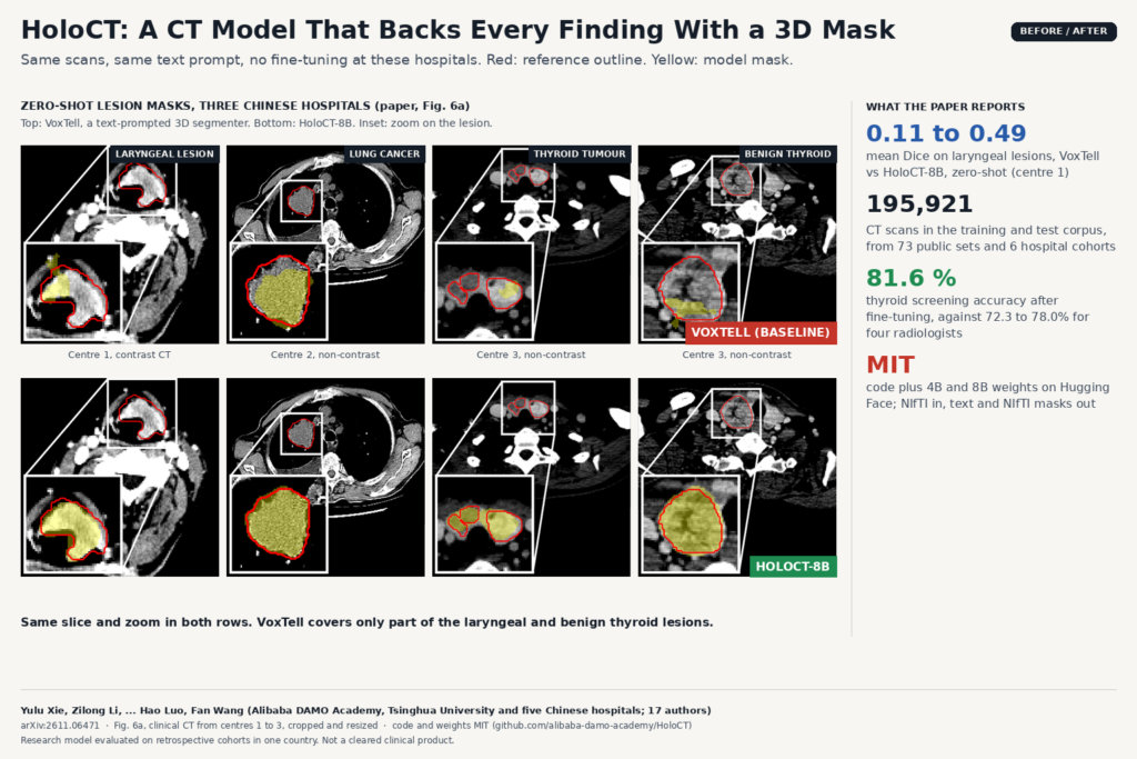
Comparison built from Fig. 6a of arXiv:2611.06471 by Yulu Xie, Zilong Li, Hao Luo, Fan Wang and co-authors: clinical CT slices of laryngeal, lung and thyroid lesions from three hospitals, with reference outlines in red and zero-shot masks in yellow from VoxTell (top row) and HoloCT-8B (bottom row).

Alibaba DAMO’s open CT model screens, answers and drafts reports, and draws a 3D mask for each finding so a reader can check it.

← Previous
Next →