Skip to content
  • Home
  • Services
    • All Services
    • Custom DICOM Viewer
    • Case Studies
  • Products
    • All Products
    • PYCAD DICOM Viewer
    • PYCAD Dental Viewer
    • PYCAD SSR Engine
  • Case Studies
  • Blog
  • About us
  • Home
  • Services
    • All Services
    • Custom DICOM Viewer
    • Case Studies
  • Products
    • All Products
    • PYCAD DICOM Viewer
    • PYCAD Dental Viewer
    • PYCAD SSR Engine
  • Case Studies
  • Blog
  • About us
Get in Touch

Category: DICOM & Viewers

Taurus: An LLM Reviewer for Aortic CT Annotation
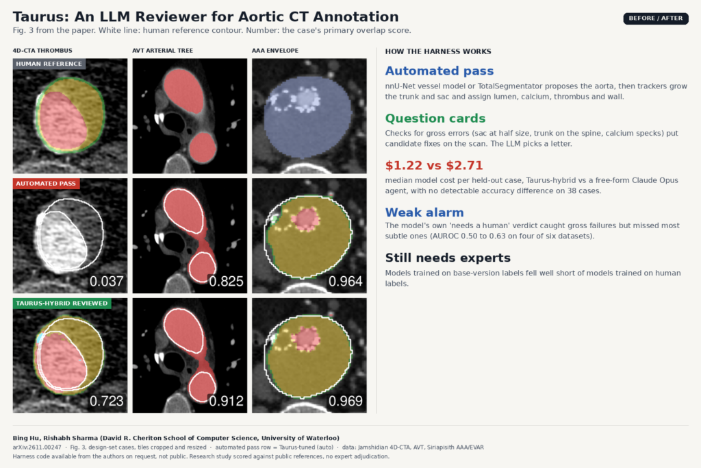
Comparison built from Fig. 3 of arXiv:2611.00247 by Bing Hu and Rishabh Sharma, University of Waterloo: axial CT angiography tiles from three public aortic datasets showing the human reference label, the Taurus automated pass, and the Taurus-hybrid result after LLM review.

An automated pass labels the aorta. A language model then picks among candidate fixes drawn on the CT, at under half the agent cost.

BrainGFM: Synthetic Brain Scans From Few Labels
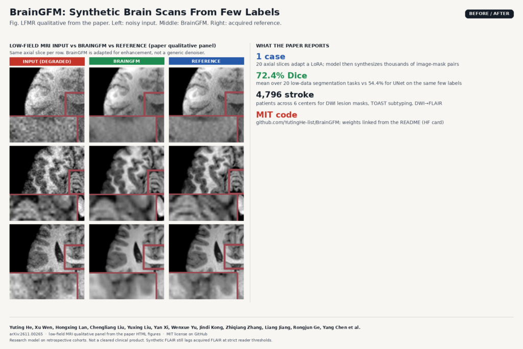
Comparison built from the low-field MRI qualitative panel of arXiv:2611.00265 by Yuting He, Xu Wen, Hongxing Lan et al.: three axial brain MRI rows with degraded input (left), BrainGFM enhancement (middle), and acquired reference (right).

One annotated case adapts a LoRA. BrainGFM then synthesizes image-mask pairs, class images, and sequence transforms for MRI and CT.

LungMend: Fix Incomplete Lung Masks on Severe TB CT
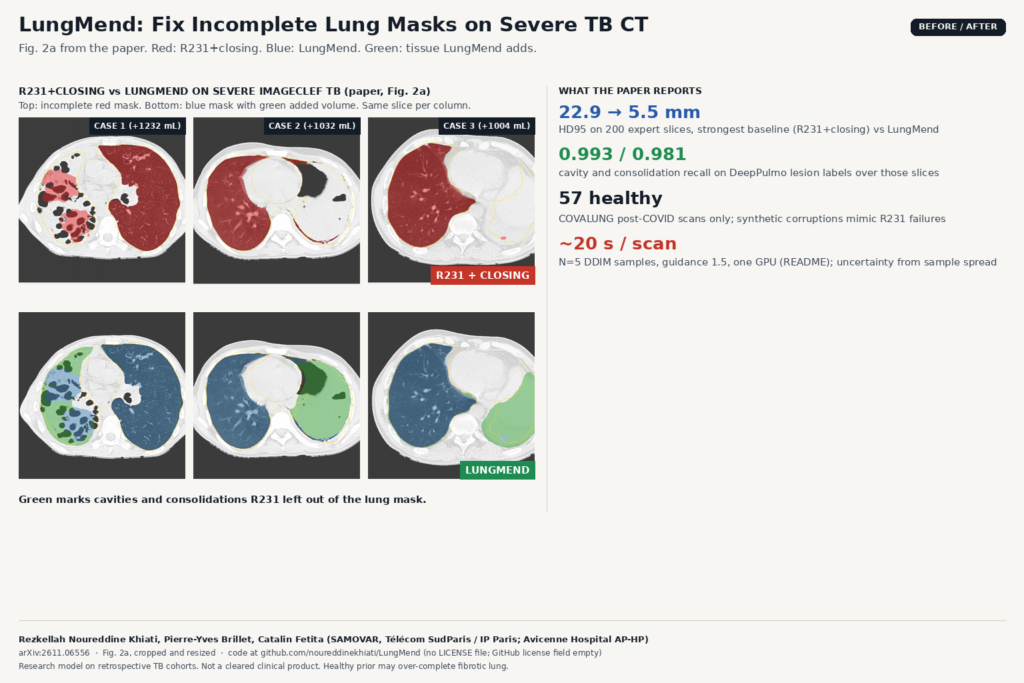
Comparison built from Fig. 2a of arXiv:2611.06556 by Rezkellah Noureddine Khiati, Pierre-Yves Brillet and Catalin Fetita: three severe ImageCLEF TB axial CT slices with R231+closing lung masks in red (top row, incomplete) and LungMend masks in blue with green added tissue (bottom row).

R231 drops cavities and consolidations on severe TB CT. LungMend fills them from a healthy shape prior and bone cues.

Virtual Contrast Breast MRI That Spares Gadolinium for Some Patients
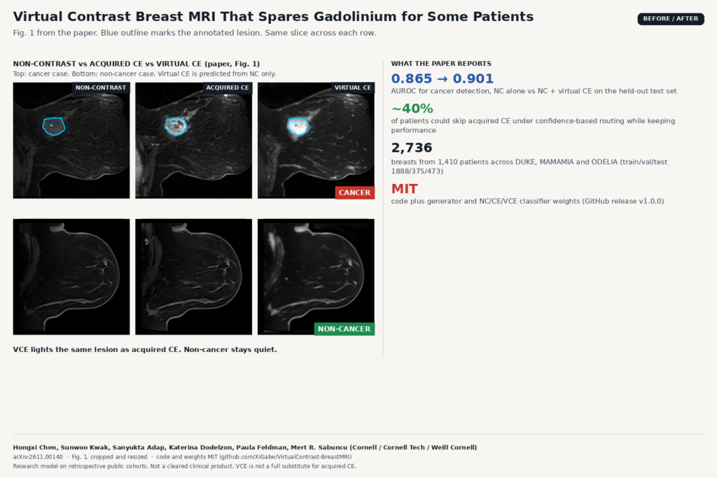
Comparison built from Fig. 1 of arXiv:2611.00140 by Hongxi Chen, Sunwoo Kwak, Sanyukta Adap, Katerina Dodelzon, Paula Feldman and Mert R. Sabuncu: breast MRI slices showing non-contrast, acquired contrast-enhanced and virtual contrast-enhanced images for a cancer case and a non-cancer case, with blue outlines on the annotated lesion.

A 3D U-Net predicts virtual CE from non-contrast breast MRI. NC+VCE reaches AUROC 0.901; about 40% of patients could skip acquired CE.

HoloCT: A CT Model That Backs Every Finding With a 3D Mask
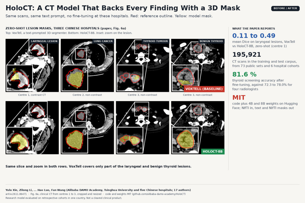
Comparison built from Fig. 6a of arXiv:2611.06471 by Yulu Xie, Zilong Li, Hao Luo, Fan Wang and co-authors: clinical CT slices of laryngeal, lung and thyroid lesions from three hospitals, with reference outlines in red and zero-shot masks in yellow from VoxTell (top row) and HoloCT-8B (bottom row).

Alibaba DAMO’s open CT model screens, answers and drafts reports, and draws a 3D mask for each finding so a reader can check it.

Saliency Maps Do Not Flag Chest X-Ray AI Errors at New Hospitals
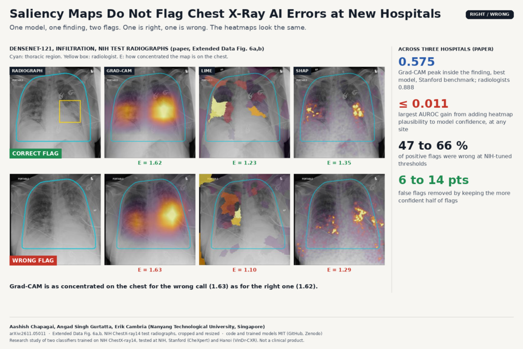
Comparison built from Extended Data Fig. 6a,b of arXiv:2611.05011 by Aashish Chapagai, Angad Singh Gurtatta and Erik Cambria: two NIH ChestX-ray14 test radiographs where DenseNet-121 flagged infiltration, one correct and one wrong, each shown with Grad-CAM, LIME and SHAP heatmaps of nearly equal concentration on the chest.

A three-site study finds Grad-CAM, LIME and SHAP look the same on right and wrong calls. Model confidence is the better alarm.

Dx-Rx: An MRI Agent That Measures Artifacts Before Fixing Them
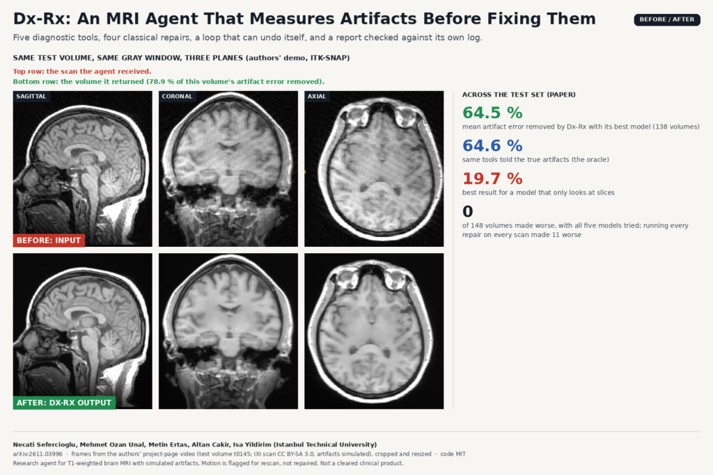
Before and after frames from the project-page video of arXiv:2611.03996 by Necati Sefercioglu, Mehmet Ozan Unal, Metin Ertas, Altan Cakir and Isa Yildirim: sagittal, coronal and axial slices of IXI test volume t0145 with simulated bias field, spike and noise, and the volume returned by the Dx-Rx agent.

Dx-Rx gives a language model five MRI artifact detectors and four classical repairs, lets it undo its own steps, and checks its report against the log.

EventMC: Training-Free Motion Correction for 3D Brain MRI
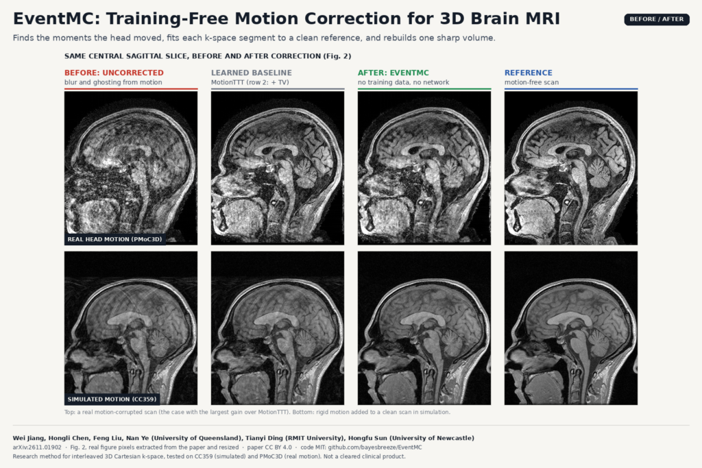
Before and after figure built from Figure 2 of arXiv:2611.01902 by Wei Jiang, Hongli Chen, Tianyi Ding, Feng Liu, Nan Ye and Hongfu Sun: central sagittal brain MRI slices from a real motion-corrupted scan and a simulated case, shown uncorrected, after the MotionTTT baseline, after EventMC, and the motion-free reference.

EventMC finds when the head moved, fits each k-space segment to a clean reference and rebuilds one sharp brain volume.

LAPIS: Chest X-Ray Anonymization Beyond the DICOM Header
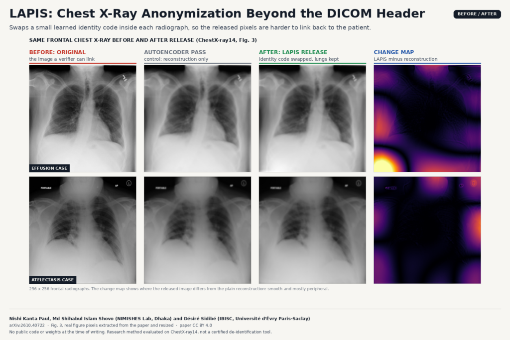
Before and after figure built from Figure 3 of arXiv:2610.40722 by Nishi Kanta Paul, Md Shihabul Islam Shovo and Désiré Sidibé: two frontal chest radiographs (effusion and atelectasis cases) shown as original, autoencoder reconstruction, LAPIS anonymized release and a change map that is smooth and mostly peripheral.

LAPIS swaps a small learned identity code inside each chest X-ray so released images are harder to link back to the patient.

EUReCA: One Network for Sparse-View Linac CBCT
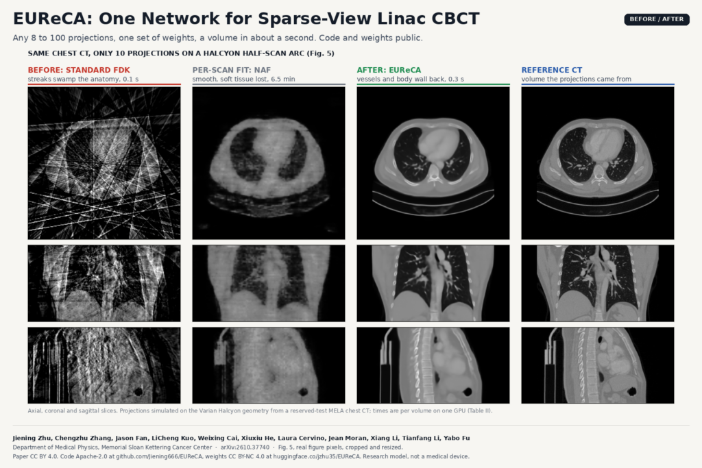
Before and after figure built from Figure 5 of arXiv:2610.37740 by Jiening Zhu, Yabo Fu and colleagues (Memorial Sloan Kettering Cancer Center): axial, coronal and sagittal chest slices reconstructed from 10 projections with standard FDK (streaks), NAF per-scan fitting (smooth, soft tissue lost), EUReCA (vessels and body wall recovered) and the reference CT.

Any 8 to 100 projections on a Halcyon arc, one set of weights, and a CBCT volume in about a second instead of minutes.

← Previous
Next →