
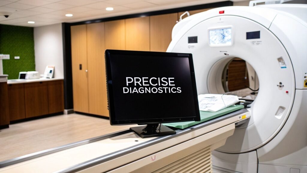
A CT scan reader, in the way this URL is used, is the CT scanner — the gantry that makes the volume. It is not the radiologist. It is not a DICOM viewer. It is not a PYCAD product.
If you meant window / HU / slice / artifacts → Hounsfield units, window width vs window level, apply window settings in Python, slice thickness, CT artifacts. If you meant open the files and paint a mask → Slicer + ITK-SNAP. If you meant a list of DICOM viewers → DICOM viewer software. If you meant how any camera turns a body into pixels → acquisition of images.
PYCAD builds custom web DICOM viewers and imaging models. It does not sell a gantry, a “CT-reader” app, or a scanner SKU.
What the machine actually does
An X-ray tube and a detector array sit opposite each other and rotate around the patient. Tissue absorbs the beam in proportion to density. The detector records what got through. A reconstruction algorithm turns those projections into slices. Stack the slices and you have a volume you can window, measure, and send to PACS.
Godfrey Hounsfield’s EMI scanner and the first patient study (Atkinson Morley, 1971, with James Ambrose) are why the density scale is named in Hounsfield units. The scale itself is the HU page. This page stops at the gantry.
| Piece | Job | What breaks it |
|---|---|---|
| X-ray tube | Makes the beam. kV and mA set energy and dose | Tube heat, a protocol copied from another vendor |
| Detectors | Count what got through. Energy-integrating on most installed bases; photon-counting on newer systems | Dead channels, scatter, a bow-tie mismatch |
| Reconstruction | Projections → slices (filtered back-projection or iterative / model-based) | Kernel too sharp (noise) or too soft (blur). Metal. Motion |
| Table / gantry | Helical (spiral) pitch vs axial step. Multi-slice detectors take many rows per rotation | Pitch that skips anatomy; a breath hold the patient cannot do |
| Contrast (optional) | Iodine raises the HU of blood and enhancing tissue so vessels and some lesions separate | Wrong phase, extravasation, a contrast allergy / kidney constraint |
Multi-slice is why a chest is seconds, not a minute of single-slice stepping. 16- / 64- / 128-row systems are the installed language. More rows is faster coverage and thinner slices if the protocol asks for them — it is not automatically a better study. Slice thickness as a number is its own page. Spatial resolution is resolution of a CT scan.
After the gantry
The file that leaves the console is DICOM. Someone still has to window it (lung vs bone vs brain), ignore the artifact, and sign. Those jobs are not this URL. Windowing is WW vs WL. Streaks, motion, and beam-hardening are CT artifacts. Opening the series on a desktop is Slicer / ITK-SNAP, not a “CT reader” SKU.
Photon-counting CT (individual X-ray photons, energy bins) is a real next detector, not a slogan. It does not make PYCAD a scanner vendor. Dose modulation and iterative recon are how most rooms already spend less dose for a given noise; they are console features, not an app we sell.
What this page is not
- Not how to read a CT. Search pattern, windows, and HU live on the 686 pages linked above.
- Not a viewer list. Horos / OsiriX / OHIF / Slicer sit on DICOM viewer software and the Slicer walkthrough.
- Not a gantry buyer’s guide. Slice-count / cost tiers are a vendor quote, not a blog table.
- Not a PYCAD CT-reader. No app, no console add-on, no “AI-powered reader” SKU.
If the work is a viewer or a model on the volume the gantry just made, that is the imaging piece. Case studies.