
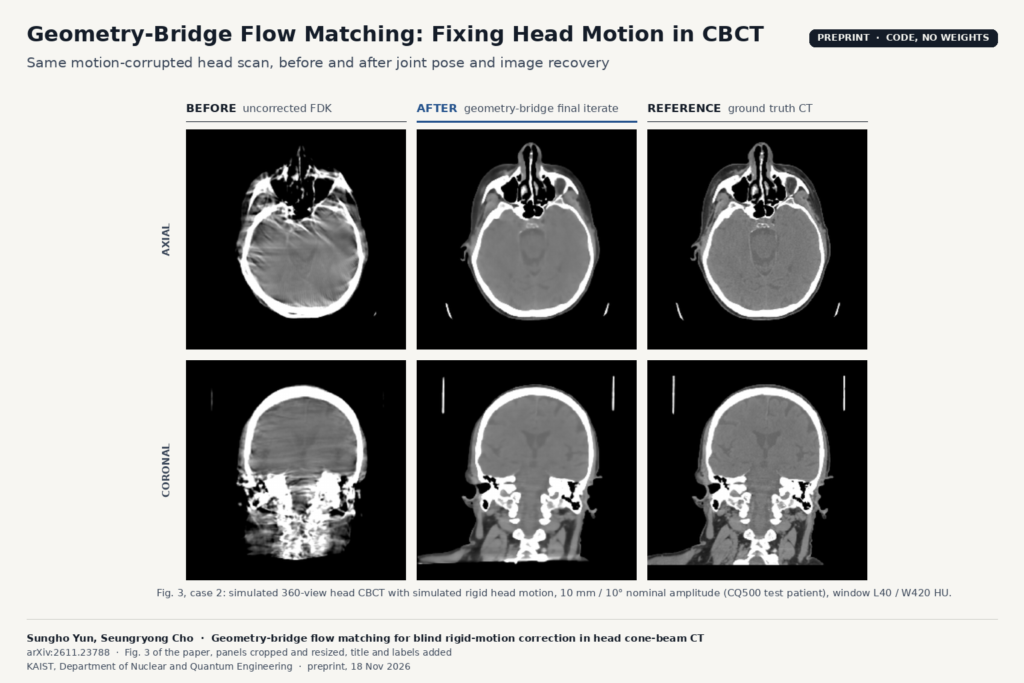
A patient who shifts their head during a cone-beam CT spin ruins more than one slice. Every projection after the movement was taken from a slightly different angle than the reconstruction assumes, so the whole volume picks up streaks, shading and doubled bone edges. In dental clinics, head-and-neck radiotherapy rooms and interventional suites, the fallback is often a rescan and a second dose. Sungho Yun and Seungryong Cho at KAIST (arXiv:2611.23788, preprint, 18 November 2026) recover the head pose for every projection from the raw data itself, then rebuild the image at those poses, without a tracker or markers.
The new part is how the image prior is trained. The network learns how a reconstruction changes as the motion in the scan gets smaller.
The problem with borrowing a denoiser
The strongest learned approach before this one, JRM-ADM from De Paepe and colleagues, pairs a wavelet diffusion prior with motion fitting. That diffusion prior was trained on a Gaussian denoising path that has nothing to do with motion. As the pose estimates improve, the measured data pull the image one way and the prior keeps pulling it toward its own smoothed guess, so the output ends up a compromise. In the authors’ figures that shows up as clean bone edges with washed-out soft tissue. The other family, learned autofocus (Thies et al.), tunes the poses to maximise a predicted image-quality score, without checking them against the projections. When the motion is large, the image it scores is too damaged for the score to guide it well.
What the geometry bridge is
Take one clean head CT and one simulated motion trajectory. Simulate the scan with the full motion, then with 90% of it, 80%, and so on down to zero, and reconstruct each version with standard FDK on the nominal circular orbit. The anatomy never changes; only the motion artifacts fade. That sequence of volumes is the bridge. Because FDK is linear in the projections, the authors can compute the exact direction the image moves at each point along it, using the derivative of the forward projector with respect to geometry. A 3D U-Net is trained with flow matching to predict that direction from the current image.
The simulated motion is used only to build training pairs. At inference the method needs neither the true motion nor a clean image.
What happens at reconstruction time
The loop starts from the uncorrected FDK volume and zero motion, and runs 50 rounds. In each round:
- The frozen network pushes the image one step along the learned direction.
- Six pose curves (three translations, three rotations), each an Akima spline through 30 nodes, are refitted by gradient descent so that the image reprojects onto the measured projections.
- Five conjugate-gradient iterations pull the image back toward the measured data at the new poses, followed by a light total-variation step.
You get two outputs: the refined volume, and a plain FDK reconstruction at the estimated poses. The second output contains no learned pixels; the network only helped find the geometry.
How well it works
Everything is simulated from CQ500 head CTs: 150 patients for training, 50 for validation and 30 for testing, with 360-view scans on a 256³ grid at 1 mm voxels and a nominal test motion amplitude of 10 mm and 10° per component. Two numbers carry the result. The mean detector-plane reprojection error falls from 6.12 mm uncorrected to 0.27 mm, against 0.84 mm for JRM-ADM and 2.26 mm for learned autofocus. Plain FDK at the recovered poses reaches 31.9 dB PSNR and looks close to FDK at the true poses in the two cases shown; the refined volume reaches 37.1 dB. The refined volume beat JRM-ADM in PSNR for all 30 test patients.
The ablation keeps the network, the endpoints and the inference budget fixed and swaps the training path for a straight blend between the corrupted and clean images. PSNR then drops by 1.6 dB and reprojection error rises by about half. Swapping the pose fitter for B-splines with RMSprop barely moves the numbers.
Runtime is 9.1 minutes per patient on an RTX A6000, against 4.6 hours for JRM-ADM and 1.9 minutes for learned autofocus.
Where it would fit
This is reconstruction-side work. It needs the raw projections and the scanner geometry, so it belongs in the scanner vendor’s or the reconstruction service’s pipeline, before anything reaches PACS. A DICOM viewer only ever sees the result. For a team building CBCT software for dental groups or radiotherapy, the useful part is the FDK-at-estimated-poses output: a motion-corrected volume made by a standard algorithm, which is easier to defend to a clinician than a network-generated image. Nine minutes is too slow for chairside use today, but fine for an offline “rescue this scan” job queued after acquisition.
Limits
- Simulated scans only. The source volumes are real head CTs, but every projection is simulated, with no noise, scatter or beam hardening. The authors say plainly that performance on acquired CBCT is not established.
- One geometry, one motion per patient. A single 360-view circular orbit (from Thies et al.) and one trajectory per test patient. Dental units with partial arcs or different detectors are untested.
- Rigid head only. The model treats the skull as one rigid body, so independent jaw or neck motion is outside it, which matters for dental scans.
- Motion range. Training covered up to 15 mm and 20°, testing used 10 mm and 10°. Behaviour beyond the training range is unknown.
- Soft tissue not read by clinicians. The image-quality claims are PSNR and SSIM against ground truth; there is no reader study.
- GPU-heavy. Training ran 500,000 iterations, and the experiments ran on a 48 GB RTX A6000.
Code and licence
The code is public at github.com/dbstjstod1/Flow_matching_motion_3D, with training, reconstruction, the baselines and the cohort scripts. As of 20 November 2026 the repository has no licence file, which by default means no permission to reuse the code, and the README states that pretrained weights are not provided: you train the prior yourself on CQ500. CQ500 is released under CC BY-NC-SA 4.0, so a prior trained on it is not cleared for a commercial product without other data or permission.
For related reading on our side, see AD-GS for sparse-view dental CBCT, EventMC for motion correction in brain MRI and how to share a CBCT scan as a link.
Source
Paper: Sungho Yun and Seungryong Cho, “Geometry-bridge flow matching for blind rigid-motion correction in head cone-beam CT”, arXiv:2611.23788 (v1 18 November 2026, v2 19 November 2026), preprint. Affiliation: Department of Nuclear and Quantum Engineering, Korea Advanced Institute of Science and Technology (KAIST). Featured image: case 2 of Fig. 3 (axial and coronal views: uncorrected FDK, the proposed final iterate and ground truth), cropped from the paper and resized, with labels added.