
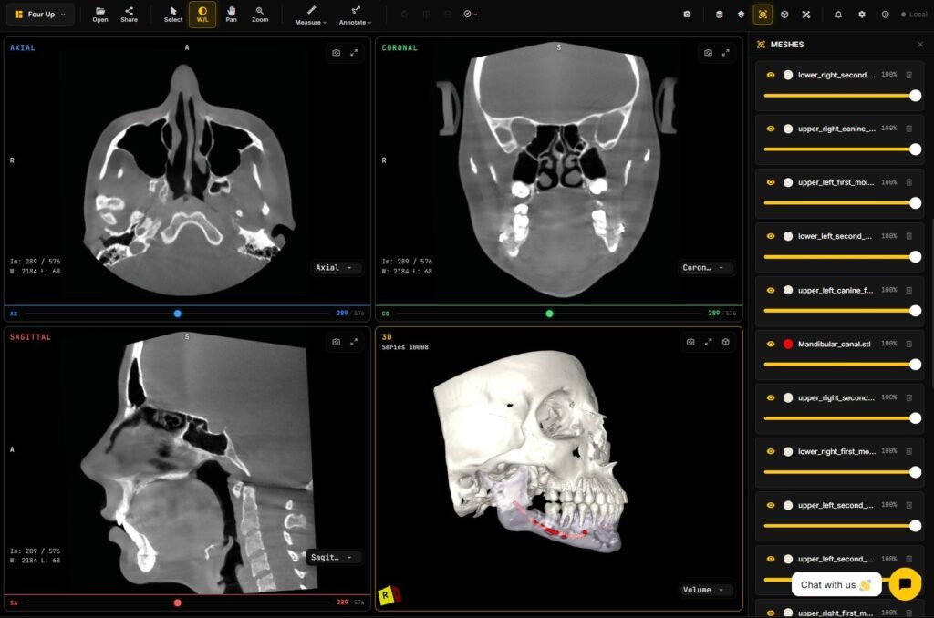
A patient walks in with a CBCT from an imaging center. It might be a disc in an envelope, a USB stick, a download link from the center’s patient portal, or a ZIP sent through a file transfer service. Inside is a stack of DICOM files, usually several hundred of them for one scan.
Opening that scan should take a minute. In many practices it takes much longer. We have a dental DICOM viewer that opens CBCT in the browser, and the handoff between imaging center and dentist comes up in most of our conversations with dental clinics and imaging partners. This guide covers what arrives, where it goes wrong, and the two ways to open it.
If you want background on the scan itself first, read what is a CBCT.
What arrives from the imaging center
Most imaging centers send a CBCT in one of these forms:
- A CD or DVD with a DICOM folder and, often, a small Windows viewer program bundled on the disc.
- A USB stick with the same contents.
- A patient portal download, where the center emails a link and you download a ZIP.
- A ZIP sent through email or a file transfer service.
- A share link that opens the scan in a browser.
The disc, USB, and ZIP versions usually contain a DICOMDIR file, folders of image files with names like IM000001 or no file extension at all, and sometimes a viewer executable. One scan can run to several hundred megabytes.
Why dentists get stuck
These are the problems we hear about most often:
- The viewer on the disc runs only on Windows, and the practice uses Macs.
- The practice laptop has no disc drive.
- The ZIP contains another ZIP, or extracts to a temporary folder that disappears.
- The image files have no extension, so double-clicking does nothing.
- Only one file gets copied off the disc, so the viewer shows a single slice.
- A general DICOM viewer opens the scan but has no arch or panoramic view, so the dentist scrolls axial slices to find the tooth.
- The practice’s planning software is licensed for one computer, which someone else is using.
- Some scanners export in a compressed or vendor-specific layout that a general viewer will not read.
One orthodontic software partner told us their dentists stalled at exactly this step. Doctors did not want to unpack compressed CBCT exports themselves. The fix they asked for was for the imaging center to upload the scan once and assign it to the doctor.
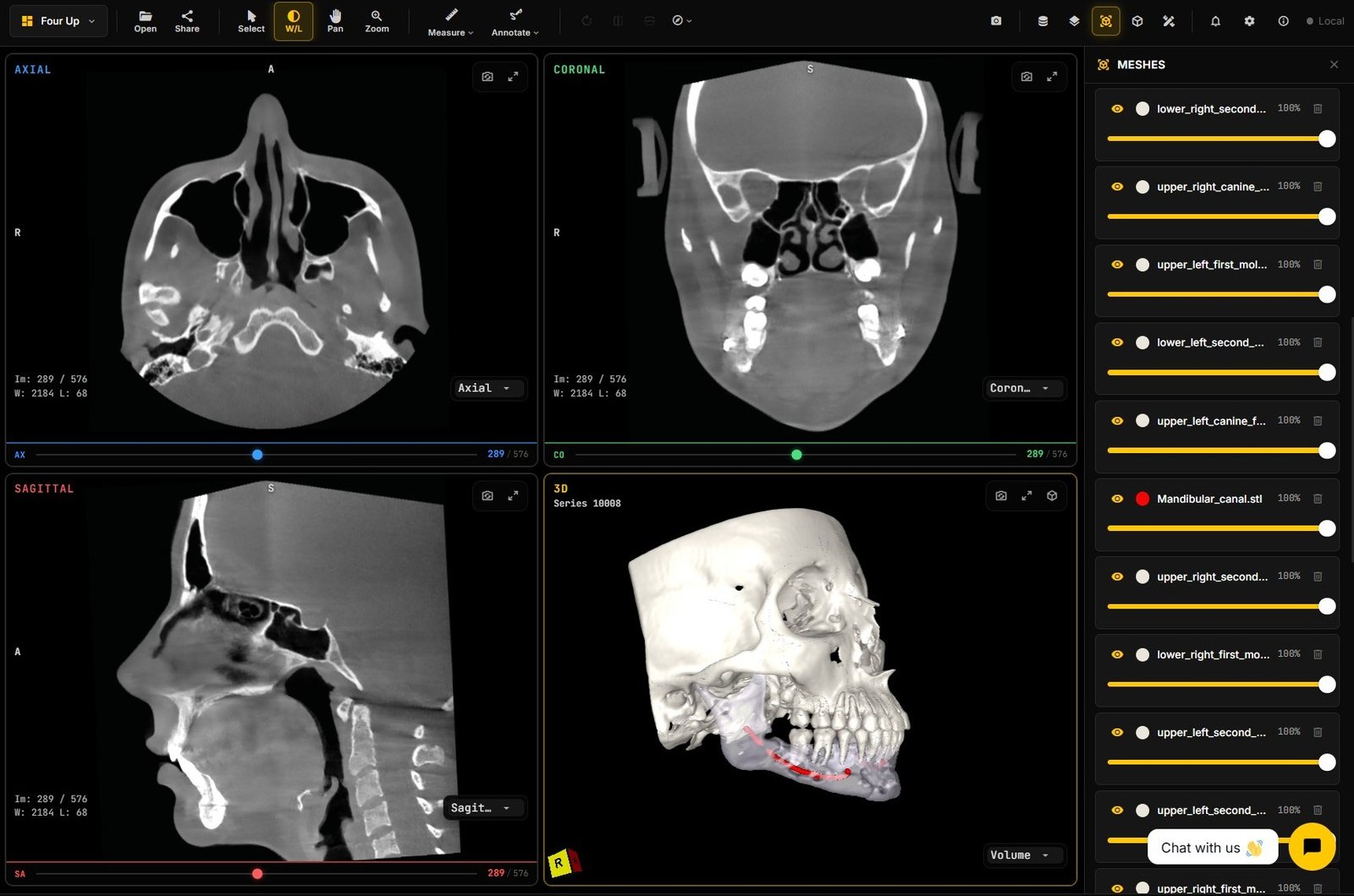
Path 1: open it in a viewer you already have
If the practice already owns a DICOM or dental planning viewer, this path works with a few habits:
- Copy the whole disc or extracted folder to the local drive, keeping the folder structure. Do not copy single files.
- Open the scan through the viewer’s import or “open folder” option, or point it at the DICOMDIR file if one exists.
- Check that you loaded the full volume. Some exports include a scout image or a dose report as separate series. The CBCT volume is the series with hundreds of slices.
- If the viewer refuses the files, ask the imaging center to re-export as standard uncompressed DICOM.
Our how to open a DCM file guide covers general DICOM files in more detail. For CBCT, the extra need is a dental layout: a curved panoramic along the arch and cross-sections at the tooth site.
Path 2: open a share link in the browser
With a share link, the imaging center uploads the scan once and sends a URL. The dentist clicks it and the CBCT opens in the browser. There is no disc, no unzipping, and no second install.
This path also suits the imaging center. It stops burning discs and couriering USB sticks to referring practices. When a referring dentist calls, the center can open the same link and look at the same scan.
What a good share-link handoff looks like
Not every share link gives the dentist what they need. Check for these:
- It opens on Windows, Mac, and a tablet with nothing to install.
- The full CBCT volume loads, at a usable resolution first and sharper as more data arrives.
- Dental layouts are available: axial, coronal, and sagittal views, plus a curved panoramic along the arch with cross-sections.
- Measurements read in millimetres from the DICOM geometry.
- The patient name and study date are visible, so you know you have the right scan.
- The dentist can pass the case on to an endodontist or surgeon with another link.
General clinic-share portals handle the first two points well. Many stop at a generic slice viewer without the dental layouts, so the dentist still needs a second tool to plan.
Open a CBCT in our dental viewer
We have a dental DICOM viewer that opens CBCT scans in the browser. It shows MPR views and a curved panoramic synchronized with cross-sections. You can place and adjust implants, take distance, angle, and density measurements, and load segmentation masks as colour overlays. Large scans are served at server-derived resolution levels, so it stays responsive on a phone, a tablet, or a slow connection. With a free account you can save a viewing session and share a link with a colleague or a patient.
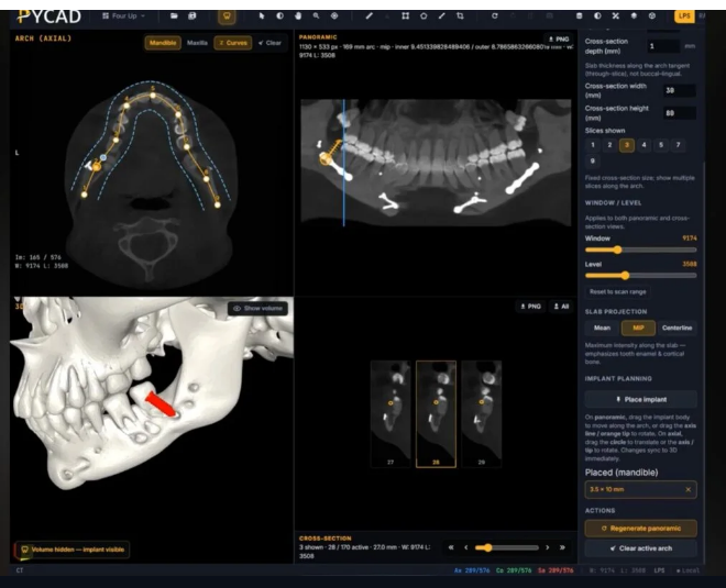
Try it with a sample scan in the dental DICOM viewer demo.
If you run an imaging center or a clinic group and want referring dentists to receive scans as share links under your own brand, Contact us with the number of referring practices you serve and how you send scans today.