
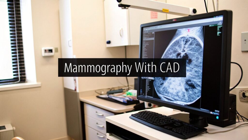
Mammography with CAD is computer-aided detection as a second reader on a screening mammogram: after the exam, software marks candidates and the radiologist reviews those marks with the originals. It does not sign the report. It does not replace the reader.
If you meant AI for medical diagnosis in general → AI for medical diagnosis. If you meant AI in radiology as a field → artificial intelligence in radiology. If you meant PACS / DICOM handshake → PACS integration. If you meant FDA 510(k) → FDA medical device approval process. If you meant CDS / alerts → what is clinical decision support.
If the work is overlaying a CAD mark on a mammogram inside a web viewer, PYCAD is the imaging stack (viewer / model), not a mammography-CAD vendor.
What it flags
The job is a safety net on a screening read. The system scans the digital mammogram and highlights regions that deserve a second look. Three findings dominate the mark set:
- Microcalcifications. Tiny calcium deposits. Many are benign. Tight clusters or certain patterns are an early red flag.
- Masses. Dense regions, especially irregular shapes or spiculated margins — the tentacle-like edges that raise suspicion.
- Architectural distortion. A shift in the normal tissue structure. Easy for a tired eye to miss; the algorithm is trained to look for it.
| Aspect | What it is |
|---|---|
| Primary goal | Second reader: highlight candidates on the mammogram for the radiologist to accept or drop. |
| What it looks for | Microcalcifications, masses, architectural distortion. |
| What it is not | A signed diagnosis. The radiologist still reads the study. |
| How the mark arrives | A DICOM object overlaid in the viewer, not a separate portal. |
CADe vs CADx
Not every “CAD” box does the same job. The split is the mammography one, not a costume paragraph on a generic diagnosis page.
| Class | Name | What it returns | What the radiologist still does |
|---|---|---|---|
| CADe | Computer-aided detection | Marks — here, look here. No judgment on malignancy. | Decide which marks are real. Sign the report. Goal is sensitivity: fewer missed cancers. |
| CADx | Computer-aided diagnosis | A likelihood or risk score on a finding already in view. | Use the score as one input to classify and decide next steps (including biopsy). Still a person. |
Classic second-reader CAD is CADe. CADx is the later layer. A product that does both is still assistive: a mark or a score is a candidate, not the chart note. The generic detection-vs-diagnosis explainer is 669. This page is the breast-screening job.
Sensitivity, specificity, and the ROC
A CAD system is only as useful as the trade it makes. Sensitivity is how often it flags a cancer that is actually there. Specificity is how often it leaves healthy tissue alone. A metal detector that beeps on every coin is sensitive; one that stays quiet over rocks is specific. Early CAD bought sensitivity with a pile of false positives — more callbacks, more biopsies that came back benign.
The comparison tool is the ROC curve: sensitivity plotted against the false-positive rate (1 − specificity) across operating points. A guess is the diagonal. A useful system hugs the top-left. You pick a threshold for the clinic you actually run; you do not copy a paper’s AUC onto the workstand.
Two published screening results, not market decks:
- Chang et al., Nature Communications 2025 (AI-STREAM) — prospective, multicenter, South Korea’s national program, 24,543 women, single-read. Breast radiologists with AI-CAD: cancer detection rate 5.70 per 1,000 vs 5.01 without — a 13.8% lift, no significant change in recall. Paper: doi:10.1038/s41467-025-57469-3.
- Wakelin et al., Nature Health 2025 (ASSURE) — US digital breast tomosynthesis plus an AI CADe/x mark and a safeguard second review on high-suspicion cases. AI workflow vs prior 3D-only standard of care: cancer detection rate +21.6% (5.6 vs 4.6 per 1,000); +22.7% in dense breasts. Cohort included more than 150,000 Black women. Paper: doi:10.1038/s44360-025-00001-0.
Those are two studies, two workflows, two countries. They are not a reason to buy a named vendor, and they are not “AI diagnoses cancer.”
How the mark lands on the mammogram
A useful CAD finding is a DICOM object: location, size, suspicion — packaged so any compliant viewer can draw it on the original study. A basic viewer shows the picture. A working one reads the mammogram and the CAD object and overlays the marks. The radiologist toggles them, zooms, and keeps or drops each flag in one workspace.
The rest of the pipeline is ordinary radiology plumbing, not a second mammography article:
- CAD software — the engine that writes the marks.
- PACS — the archive. The study and the CAD object have to live together.
- RIS — scheduling, the worklist, the report.
Scan → CAD → archive with the marks attached → open the case and both layers are there. The handshake (PACS / RIS / DICOMweb, worklist, viewer) is PACS integration. A separate portal is how tools die.
Clearance is a step
A mammography CAD box that touches patients is a medical device. In the US that is FDA clearance (typically 510(k) for assistive imaging AI); in Europe, CE. The submission path — what you file, what a predicate is, what “safe and effective” actually means — is FDA medical device approval process, not a second chapter here.
Training data has to be labeled and de-identified. HIPAA (US) and GDPR (EU) apply because the pixels are PHI, not because a vendor said “compliant.” After go-live the failure mode is alert fatigue: too many marks and the reader starts ignoring them. Write a rule for disagreement (radiologist vs CAD) before you turn the flags on. Worklist alerts as a product class are clinical decision support, not this URL.
What this page is not
Personalized risk scoring from a mammogram, or a model that claims to predict chemo response before the first dose, is a different research job. It is not second-reader CAD. It is not this article.
Generic AI diagnosis is 669. AI in radiology as a field is 671.
FAQ
Does CAD replace the radiologist?
No. A mark is a candidate. The signed report is still a person who has the history and the rest of the study. Most cleared mammography CAD is assistive, not autonomous.
What is the difference between old CAD and modern AI-CAD?
Early systems were hand-coded rules: look for this shape, this density. They were noisy. Modern systems learn from labeled mammograms and pick up patterns a rule list never named. That can mean better sensitivity and fewer false marks. It does not remove the reader.
How does it show up on the screen?
The CAD output is a separate DICOM object. The viewer overlays circles or arrows on the mammogram. Toggle, zoom, accept or dismiss. If that overlay is the piece you are building, that is a viewer / model job — not a “mammography CAD product.”
Where does PYCAD sit?
The imaging stack: a web DICOM viewer or a model when the mark has to live inside a clinic app. Not a screening-mammography CAD engine, not a CADe/CADx box, not a second-reader vendor.