
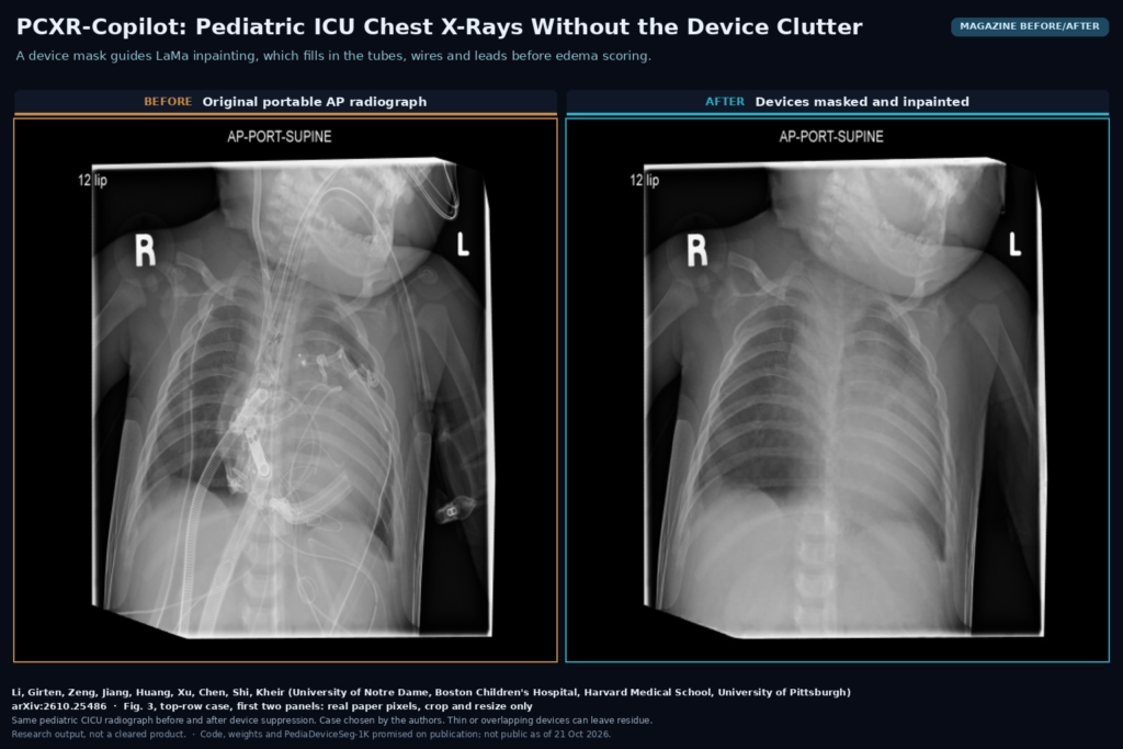
A chest X-ray from a pediatric cardiac ICU shows sternal wires, pacing wires, chest tubes, a nasogastric tube, an endotracheal tube, central lines, ECG leads and stickers, all laid over a small chest. The intensivist still has to judge lung water and body-wall swelling through all of it, often every day. Xueyang Li, Alec Girten, Peng Zeng, Mingze Jiang, Shaoyang Huang, Gelei Xu, Danny Z. Chen, Yiyu Shi and John N. Kheir from the University of Notre Dame, Boston Children’s Hospital, Harvard Medical School and the University of Pittsburgh posted arXiv:2610.25486 on 18 October 2026 (revised 20 October) with two things: PediaDeviceSeg-1K, 1,000 pediatric CICU radiographs with device annotations, and PCXR-Copilot, a pipeline that masks the devices, optionally paints them out, and turns what is left into edema cues. It already runs inside TWIST, the clinical data platform at Boston Children’s.
Why this matters for a chest X-ray platform
Most public chest X-ray datasets are adult, and most pediatric ones (PediCXR, VinDr-PCXR, the pediatric pneumonia set) are labeled for disease, not for the hardware in the image. Devices cause two problems. They hide anatomy, and a model can learn to use the devices as shortcuts, since sicker children carry more of them. In this cohort each radiograph has a median of 19 device instances and 6 device categories per radiograph, and 90.4% of images with at least two categories. Sternal wires appear in 732 of the 1,000 images, nasogastric tubes in 578, ECG leads in 536, temporary pacing wires in 427.
What goes in and what comes out
The input is one frontal portable chest radiograph, ideally as DICOM so the pipeline can read pixel spacing and exposure tags. The outputs are a lung mask, a device mask, an optional device-suppressed copy of the radiograph, a lung-field edema heatmap with a scalar pulmonary edema (PE) index, and a body-wall edema mask with left, right and average chest-wall width in millimetres. Every output is shown next to the original image.
How it works
There are two branches. The pulmonary edema branch starts with lung segmentation, a U-Net++ trained on 300 hand-annotated pediatric radiographs with a cross-entropy plus active-contour loss. Next comes device segmentation. The annotators drew thin devices as polylines and bulkier ones as polygons in CVAT, and the team rasterised the polylines with fixed widths (10 px for endotracheal tubes, 8 px for nasogastric tubes, 4 px for the rest). An nnU-Net trained as a single device-versus-background task at 512 x 512 then predicts one union mask of all hardware. They also tried per-category masks, but the label distribution has a long tail (several categories appear in one or two images), so the pipeline uses the binary mask.
That mask feeds a LaMa inpainting model, fine-tuned from the public big-lama checkpoint, which takes the radiograph plus the device mask and fills the masked pixels with plausible tissue. A rib segmenter (U-Net++ pretrained on VinDr-RibCXR, fine-tuned on 50 local images) removes ribs from the calculation. The PE index is the mean grayscale of the lung field, ribs excluded, on the device-suppressed image. It measures how white the lungs look. Because whiteness also depends on technique, the index is corrected with a linear regression on three DICOM tags, Exposure (0018,1152), KVP (0018,0060) and entrance dose in mGy, fitted on training data. The heatmap comes from Grad-CAM on a ReXNet-150 edema classifier trained on 1,000 adult NIH ChestX-ray14 images, clipped to the lung mask.
The body-wall branch is a separate U-Net++ (EfficientNet-B0 encoder) trained on 60 radiographs with hand-drawn lateral soft-tissue bands. A post-processing step keeps at most two connected regions, one per side, and the width per side is converted to millimetres from DICOM pixel spacing, with patient left and right mapped correctly from the display convention.
What the evaluation shows
For device segmentation, nnU-Net reached a binary Dice of 77.8% in five-fold cross-validation, ahead of GLCP, two U-Net++ variants and DeepLabv3+. Lung masks were easier, at 92.7%. There is no device-free ground truth for suppression, since nobody takes a second X-ray with the tubes removed, so the authors used proxies. Mean brightness inside device regions dropped from 151.1 to 132.0 on a 0 to 255 scale, and device-to-tissue contrast fell by 67%. Outside the mask the image changed by 0.83 gray levels, which the authors put down to resampling between the native image (up to about 2,450 px wide) and the 512 x 512 inpainting grid.
The PE index has thinner evidence behind it. In one patient it fell from 0.406 to 0.290 over 15 radiographs in 14.7 days, with heatmaps that fade in step. A pediatric cardiac intensivist, blinded to the index, compared the first and last radiograph of 97 admissions and called a direction in 66 pairs. The signed index difference matched his call with an AUC of 0.806. Suppression also cut the index’s link to device burden (Spearman correlation with device-lung overlap from 0.126 to 0.049). On 648 adult MIMIC-CXR radiographs with edema grades, the plain lung-whiteness index, without suppression or rib removal, still rose with severity, though weakly (Spearman 0.211, AUC 0.681 for grade 0 against grade 3).
What it would look like in a viewer
Fig. 2 of the paper shows the TWIST radiology viewer with two radiographs of the same child 28 hours apart, toggles labelled Body Wall Edema and Lung Parenchyma under each image, and a “NOT FOR DIAGNOSTIC USE” note next to the link that fetches the DICOM. With the lung overlay on, the drop in lung water between the two films is visible at a glance, and a PICC line and a pilot balloon are subtracted from the overlay. The viewer sits beside ventilator settings, fluid balance and medications in the same platform. Inference runs in a container with fixed model versions, and outputs come back as overlays and numbers.
Where it breaks
The authors list the failure cases themselves. Suppression leaves residue or artifacts when the device mask misses thin wires, when several devices overlap inside the lung fields, and near strong mediastinal edges where the inpainter has to guess how texture continues. The device masks are rasterised polylines with assumed widths, so they approximate the real device edges. The authors present the PE index as a relative cue for trends in one patient and do not claim it measures lung water, and its Grad-CAM heatmap comes from a classifier trained on adults. The reader study used one intensivist, who is also a co-author, and 66 pairs. Many images are portable supine films with variable positioning. There is no prospective study yet of whether clinicians decide faster or better with it. The data come from one hospital.
Code, data and license
As of 21 October 2026 there is no public repository. The paper says the PCXR-Copilot code, the trained segmentation and suppression weights, PediaDeviceSeg-1K and the smaller lung (300), rib (50) and body-wall (60) mask sets will be released in a public repository upon publication, with data-use terms to be described there. During review they are available to editors and reviewers on request. nnU-Net and LaMa, the two main building blocks, are both Apache 2.0 on GitHub. MIMIC-CXR and its edema grades need PhysioNet credentialing. The arXiv paper itself is under arXiv’s non-exclusive distribution license.
How we would use it
We build DICOM viewers and clinical imaging platforms, and the TWIST integration is close to how we would wire this. We would never overwrite the original radiograph. The device-suppressed image would go out as a separate derived series (new Series Instance UID, Image Type DERIVED\SECONDARY, a series description that says devices were removed by inpainting) so nobody reads tube tips off it. Device and lung masks would travel as DICOM SEG, and the PE index and chest-wall widths as a structured report, so the trend over an admission can be plotted. The regression on exposure tags would need refitting per site and per detector, as the authors say. For a pediatric site, the dataset may end up worth more than the pipeline, since device masks are what you need to check whether any chest X-ray model is reading the lungs or the hardware. Background reading: what a DICOM viewer does and our post on DeBoneDiT bone suppression for chest X-rays.
Sources
- Li, X., Girten, A., Zeng, P., Jiang, M., Huang, S., Xu, G., Chen, D. Z., Shi, Y., Kheir, J. N. PCXR-Copilot: Device-aware pediatric chest radiographic analysis with segmentation, suppression, and edema-related cues. arXiv:2610.25486, 2026. https://arxiv.org/abs/2610.25486. PDF: https://arxiv.org/pdf/2610.25486.
- Jiang, M., Li, X., Kheir, J., Girten, A., Shi, Y. LightRefine-PCXR: device suppression in pediatric chest X-rays (same group): proceedings.mlr.press.
- nnU-Net (Apache 2.0): https://github.com/MIC-DKFZ/nnUNet.
- LaMa inpainting (Apache 2.0): https://github.com/advimman/lama.
- MIMIC-CXR pulmonary edema severity grades: physionet.org.
- Featured image: Fig. 3 (top row, first two panels) of arXiv:2610.25486, real paper pixels, cropped and resized only.