
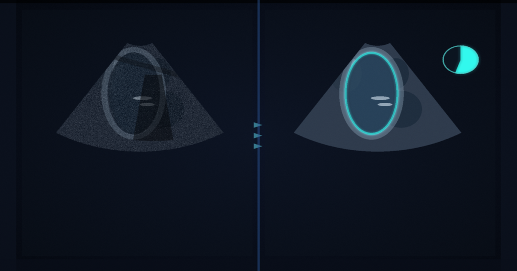
You hang a parasternal long-axis cine and the apical four-chamber window is foreshortened, dropped out, or never acquired at the bedside. This pipeline classifies that clip as PLAX, maps the cine to a study-level left-ventricular ejection fraction, and, when an A4C clip exists in the same study, averages the two predictions. Zhiyuan Gao and Yaser S. Abu-Mostafa (Caltech Electrical Engineering) and Dominic Yurk (Asari AI) posted arXiv:2609.02969 around 4 September 2026. The paper is Learning from Scarce Labels: Multi-View Echocardiography for Ejection Fraction Prediction, the MELBA camera-ready of a MIDL 2025 oral (DOI 10.59275/j.melba.2026-8194, published 27 August 2026). Labels, pretrained A4C and PLAX weights, a Hugging Face Space, and a Colab notebook are at github.com/Jeffrey4899/PLAX_EF_Labels_202509 (HTTP 200 on 4 September 2026). Raw MIMIC-IV-Echo videos stay behind the PhysioNet data-use agreement.
Public EF labels live on A4C. EchoNet-Dynamic has about 10,000 A4C cines with expert EF. EchoNet-LVH has about 12,000 PLAX cines labeled for hypertrophy, not EF. MIMIC-IV-Echo has roughly 500,000 unlabeled videos. PLAX is the view you often get on a focused bedside scan, and until this release there was no public PLAX-to-EF benchmark you could rerun.
Mine the views, borrow A4C EF, test on notes
They train a ResNet-34 on TMED-2 stills, then an X3D-s video classifier on EchoNet-Dynamic (A4C), EchoNet-LVH (PLAX), and MIMIC clips mined as OTHER. Two reviewers on 300 MIMIC videos showed the stills model was weak on PLAX (72 to 78 of 100). The video model is what they keep.
An R(2+1)D A4C teacher trained on EchoNet-Dynamic hits 4.37% MAE on that test split. They run it on MIMIC A4C clips, average those predictions per study, and write that number onto every PLAX clip in the same study. That yields 25,532 PLAX videos across 4,822 studies (80/20 study-level split). Patients in the test set never enter train or val.
Ground truth is independent of the teacher. They time-match MIMIC-IV-Echo to MIMIC-IV-Note, extract EF from discharge text with GPT-4, and keep a 1-day window plus view-confidence, duration over 1 s, and no color Doppler. That leaves 295 studies (1,017 A4C clips, 1,320 PLAX clips). Three extractors (GPT-4, GPT-5.5, Claude Opus 4.8) agreed exactly on 99.5% of notes that all three parsed. They keep the GPT-4 numbers so the released split does not move. On that cohort the A4C teacher sits at 6.95% MAE versus the notes, close to EchoNet-Dynamic’s 6.0% out-of-sample figure.
They also used A4C error to tighten the view classifier: clips with MAE under 3% labeled A4C, over 20% labeled OTHER. After that fine-tune, A4C MAE on the 60-day pool dropped from 6.83% to 5.14%. For mining they take PLAX from the stricter pre-fine-tune classifier and A4C from the post-fine-tune one.
PLAX alone, then a mean with A4C
Two R(2+1)D PLAX models (batch 16 and 32) ensemble to 6.86% study-level MAE and Pearson 0.670 on the 295-study note cohort. Bland-Altman bias is -0.62%, with limits of agreement 16.87% and -18.10%. Dispersion grows at high EF. That MAE sits next to typical A4C video methods at 6% to 7%, ExoAI’s disclosed PLAX figure of 7.29%, and an LVID-landmark PLAX pipeline at 8.45%.
On 284 studies that have both views, unweighted late fusion of A4C and the PLAX ensemble reaches 6.37% MAE and Pearson 0.709. A4C-only was 7.01%, PLAX-only 6.77%. Those two single-view errors are not statistically different (Wilcoxon p=0.66). A learned convex weight recovered w≈0.44 and did not beat the mean. There is proportional bias: overestimate low EF, underestimate high EF, steepest on PLAX (slope -0.88). Inter-view disagreement is a weak reliability signal (Pearson r=0.07).
How this lands in a viewer
If you already hang TTE cines in a DICOM viewer, treat this as an EF rail. It returns a study-level scalar you hang next to the series. Classify the clip as A4C, PLAX, or OTHER. Drop OTHER and color Doppler. Run the matching R(2+1)D, average clips inside the study, and if both views exist, average those two study numbers. Show the scalar next to the series. Fail closed when the view classifier is unsure, when the clip is under a second, when you need a diagnostic EF rather than a research estimate (the authors mark every artifact research and education only, not a medical device), when you lack a PhysioNet DUA and were hoping to rebuild the MIMIC cohort, or when you need calibrated EF at the tails (the Bland-Altman spread and the shrinkage toward the mean are the failure modes). Do not treat 6.86% MAE as a bedside number until you re-validate on your own lab’s notes and scanners.
Cine short-axis aorta contours hold through systole. CT or MRI whole-heart overlays skip a modality retrain. This paper is PLAX cine in, a study-level ejection fraction out when the apical window is the thing you do not have.
Rebuild from arXiv:2609.02969. As of 4 September 2026 the abstract, PDF, GitHub repo, Hugging Face models, and Space all respond (HTTP 200). MIMIC videos are not in the repo. The MELBA paper is the source of record.
Sources
- Gao, Z., Yurk, D., Abu-Mostafa, Y. S. Learning from Scarce Labels: Multi-View Echocardiography for Ejection Fraction Prediction. MELBA 2026:025, DOI 10.59275/j.melba.2026-8194, published 27 August 2026 (MIDL 2025 special issue). arXiv:2609.02969, posted ~4 September 2026. https://arxiv.org/abs/2609.02969 (HTTP 200 on 4 September 2026). PDF: https://arxiv.org/pdf/2609.02969 (HTTP 200 on 4 September 2026). Code and labels: https://github.com/Jeffrey4899/PLAX_EF_Labels_202509 (HTTP 200 on 4 September 2026). Models: https://huggingface.co/Jeff4899/202509_PLAX_EF, https://huggingface.co/Jeff4899/202509_A4C_EF. Demo: https://huggingface.co/spaces/Jeff4899/202509_PLAX_EF_Demo (HTTP 200 on 4 September 2026).