
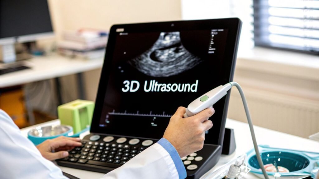
3D ultrasound is a volume made of sound: many B-mode planes (or a steered pyramid) stacked so you can re-slice and render anatomy that a single plane cannot show. 4D is that volume over time. It is not “3D medical imaging” the field. It is not reconstruction software. It is not a PYCAD ultrasound product.
If you meant CT / MRI / 3D US / PET as cameras that make a volume → 3D medical imaging (247 already names 3D ultrasound as one row). If you meant MPR / MIP / volume-rendering software → 3D imaging software. If you meant Couinaud segments on a transducer → ultrasound liver segments. If you meant how any scanner makes pixels → acquisition of images.
247 is the field. This URL is the probe. PYCAD builds custom web DICOM viewers and imaging models. It does not sell a 3D ultrasound scanner or a “4D baby” studio.
How the volume is acquired
A 2D probe images one plane. A 3D acquisition is a way to know where that plane sat, many times, and to build a grid. Three common ways:
| Method | How the slices are collected | What you trade |
|---|---|---|
| Freehand + tracking | The operator sweeps. A position sensor (or, worse, an assumed constant speed) tells the software how to stack | Cheap probe. Gaps and shear if the sweep is uneven. Harder to reproduce |
| Mechanical sweep | A motor inside the housing rocks the array through a known arc | Repeatable volume. Bulkier head. A pause while it sweeps — not a live pyramid |
| Matrix array | A 2-D grid of elements steers the beam electronically. A pyramidal volume without moving parts | Real-time 3D / 4D (echo, fetal face). Expensive. Smaller footprint; edges of the pyramid fall off |
After the grid exists, the machine can re-slice (any plane, not only the one the operator held), measure a volume, or volume-render a surface (the “baby face”). Rendering is a transfer function on echo intensity, not a photograph. Speckle, shadow, and gel are still there.
2D, 3D, 4D — same physics, different grid
- 2D. One B-mode plane, live. Still the workhorse: biometry, a vessel, a needle in-plane.
- 3D. A volume you can hang after the sweep (or from a matrix freeze). Useful when the question is shape: a cleft, a valve, a fibroid, a volume you will re-cut.
- 4D. 3D repeated fast enough to be a loop. Fetal movement, a beating heart, a valve that only fails in systole. Frame rate and volume size fight each other; a big pyramid is a slower movie.
None of this ionises. ALARA still applies: thermal and mechanical index, not a “3D is safer than X-ray” slogan. CT / PET dose lives on those cameras — 247 already says “3D is not a safety rating.”
Where a volume actually changes the read
- Obstetrics (diagnostic). Face and palate, spine, some limb and cardiac questions that a lucky 2D plane misses. Boutique “keepsake” 3D is a photo session, not a report. This page is the diagnostic one.
- Echo. 3D / 4D TEE or transthoracic for valve geometry and ventricular volumes. Function, not a baby souvenir.
- GYN, prostate, some MSK and interventional. A fibroid volume, a gland, a needle you want to see off-plane. Liver segments as Couinaud anatomy are 7675, not this explainer.
Bone and air still block sound. A 3D acquisition does not see through rib or gas that 2D could not. The operator is still the machine: a bad window is a bad volume. Handheld probes with helpers (Butterfly-class) are a hardware+AI kind on medical AI companies / devices, not a second 3D-US article.
What this page is not
- Not 247 restated with an ultrasound costume. That URL is four modalities. This one is how a 3D / 4D probe is acquired and used.
- Not 7604. Reconstruction software (MPR / VR on a CT stack) is a different job. US volumes can land in a viewer; the field explainer is still this slug.
- Not 7675 (Couinaud on US) and not 7360 (4D CT).
- Not a market CAGR. Precedence / Coherent / “USD 3.7 billion” and “74% of deaths” were unsourced-as-used. Dropped.
- Not a patient FAQ (“will my baby look like the picture,” “best week for a 3D keepsake”). Dropped.
- Not a PYCAD 3D-ultrasound SKU.
If the missing piece is hanging a DICOM-US volume in a clinic app, that is the imaging piece. Case studies.