
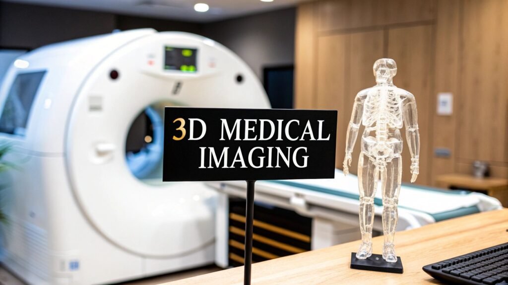
3D medical imaging is the field: modalities that capture a volume (CT, MRI, 3D ultrasound, PET) so you can re-slice and spin anatomy instead of guessing from one projection. It is not a software list. It is not a PACS. It is not “what is AI in radiology.”
If you meant MPR / MIP / volume-rendering software → 3D imaging software. If you meant Blender / Unreal / KeyShot → 3D visualization software. If you meant how the scanner makes the pixels → acquisition of images. If you meant the hospital stack (PACS / RIS / viewer) → medical imaging software. If you meant painting a volume in a transfer function → volume rendering of CT. If you meant sinogram → slices → mesh → 3D reconstruction from CT. If you meant the future of imaging as a field (radiomics / ethics) → the future of medical imaging.
PYCAD builds the imaging piece (a web DICOM viewer or a model) when the volume has to live in a clinic app. It is not a 3D-imaging vendor and not a gantry.
What “3D” adds
A radiograph is a shadow. Organs stack on one plane; a vessel can hide behind a bone. A volume is hundreds of slices you can cut in any plane and spin. The spatial question — where the fragment sits, which vessel the tumor touches — is the reason the extra acquisition exists.
3D is not automatically better. A good two-view radiograph still answers a lot of fractures. You pay for a volume with time, dose (CT / PET), or money (MRI). Use it when the plane you need is not the plane the machine acquired.
Four ways to get a volume
| Modality | What it measures | What 3D is for |
|---|---|---|
| CT | X-ray attenuation (Hounsfield units) | Bone, lung, vessels with contrast, trauma. Fast. Dose is the cost. Window / HU sit on the CT pages, not here |
| MRI | Tissue response in a magnetic field (mostly water) | Soft tissue: brain, cord, joints, myocardium. No ionizing dose. Slow, expensive, metal is a problem |
| 3D ultrasound | Echoes of high-frequency sound | A live volume: fetus, heart, a needle. Cheap and portable. Bone and air block it; the operator is the machine |
| PET | A tracer’s uptake (function, not anatomy) | Where metabolism is hot. Low spatial detail alone — almost always fused to CT or MRI |
These are complementary cameras, not a leaderboard. CT for the fracture. MRI for the ligament. Ultrasound when you need the needle now. PET when the question is “is this metabolically active,” not “how long is it.” How each scanner actually acquires is acquisition of images. A glossary of “what is a modality” (including therapy) is a different leftover.
From volume to a decision
A volume sitting in an archive is not 3D imaging. Someone has to reconstruct, hang, and sometimes label it:
- Re-slice (MPR). Axial / sagittal / coronal / an oblique that matches the vessel. The cheap 3D.
- Project (MIP) or paint (volume render). Brightest voxels, or a transfer function. Visualization, easy to over-trust. That software job is 3D imaging software / volume rendering of CT.
- Segment. A mask so a volume, a print, or a dose field has a boundary. Methods: medical image segmentation.
Clinic uses that are not a second article: a corridor through cortex, a valve case on a patient-specific heart, a tumor against a vessel before radiation, fragments in a joint. The software that does MPR / VR is 7604. The field is this URL.
What AI actually changes here
A model can reconstruct faster, denoise a low-dose CT, or draft a mask. It does not acquire photons and it does not sign. “Will AI replace the 3D read?” is the same answer as the rest of imaging: assistive, not autonomous. That argument lives on artificial intelligence in radiology, not a second field explainer.
A market CAGR is not a reason to buy a scanner. Dropped.
FAQ
Is a 3D scan safer than an X-ray?
Depends which 3D. MRI and ultrasound: no ionizing radiation. CT and PET: yes, radiation — justified when the volume changes the decision. “3D” is not a safety rating.
Is this the same as 3D imaging software?
No. This page is the modalities and why a volume exists. The reconstruction product (MPR / MIP / VR) is 3D imaging software.
Does PYCAD sell 3D medical imaging?
No. The imaging piece inside someone else’s stack: a viewer or a model. Case studies.