
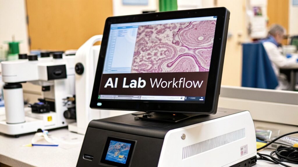
Digital pathology software is the stack that turns a glass slide into a file a pathologist can open: a scanner, a whole-slide image (WSI), a tiled viewer, an image-management system, and a hook into the LIS. This page is that stack. It is not artificial intelligence on a slide. It is not a PYCAD product.
If you meant triage / quant / second-reader on a WSI → artificial intelligence in pathology. If you meant generic diagnosis / CAD → AI for medical diagnosis. If you meant the hospital imaging stack (PACS / viewer) → medical imaging software. If you meant FDA class / 510(k) → FDA medical device approval process.
PYCAD builds custom web DICOM viewers and imaging models. A pathology WSI is a different file and a different viewer. We do not ship a digital-pathology LIS, a slide scanner, or a pathology suite.
Glass to file
A microscope ties the diagnosis to a piece of glass in one room. A second opinion means a courier. Digital pathology is the conversion: a whole-slide scanner walks the tissue, writes a gigapixel pyramid (often .svs / .tiff; sometimes wrapped as DICOM), and a viewer tiles it so you are not downloading the planet.
Until the lab scans, there is nothing for software to open. That is why this page starts at the scanner, not at a model. The model job — after the file exists — is 289.
| Piece | Job | What breaks it |
|---|---|---|
| Scanner | Glass → WSI. Throughput and a file the viewer can open | A stain / compression the next room cannot read. Drift across scanner vendors |
| WSI file | A pyramid, not a JPEG. Often gigabytes per case | A CT DICOM viewer. Wrong tool |
| Viewer | Pan / zoom / tile. Side-by-side stains. Annotations that travel with the case | Loading the whole slide into RAM. No sync across H&E and IHC |
| IMS | The library: who can open which slide, linked to the patient | A folder of files with no case |
| LIS hook | The flag and the report land on the same case the lab already runs | A sidecar portal. Second login, re-key, demo |
What the software actually adds (vs a microscope)
| Job | Glass | Digital |
|---|---|---|
| Access | One person, one room, the slide in a tray | The same pyramid, more than one viewer, if the IMS allows it |
| Compare | Swap slides. Rely on memory | Two windows, same coordinates, H&E next to IHC |
| Measure | An eyepiece estimate | A length or area on the pyramid, if the calibration is honest |
| Consult | Ship the glass, or a consensus meeting | A shared viewport. Still a person who signs |
| Archive | A cabinet. Fade, break, lose | A file. Storage cost replaces shelf cost. The file still needs a backup |
Tiling / pyramiding is why a gigabyte slide does not freeze the workstation: the viewer asks for the tile at the zoom you are on, the way a map asks for one city block. That is engineering, not a slogan.
AI is a later layer
A model that flags a region, counts mitoses, or drafts a percentage is useful only after the stack above exists. That layer is artificial intelligence in pathology, not this URL. Do not buy a scanner because a slide deck said “AI.” Buy a scanner because the glass has to become a file.
Clinical use of a diagnostic overlay still needs a cleared / CE-IVD claim for that intended use. The pathway is 7568. An unvalidated research heat map is a research heat map.
What this page is not
- Not pathology AI. Triage / quant / second reader → 6219.
- Not a PYCAD pathology suite, LIS, or scanner SKU.
- Not a market CAGR. Dropped the $9.1B / $1.30B / Asia-Pacific theater and the Precedence / MarketsandMarkets outbounds.
- Not a YouTube embed, a chatiant knowledge-base outbound, or a 42coffeecups AI-services page. Dropped.
- Not /portfolio. Case studies live at case-studies.
If the work is a viewer or a model on medical images — annotation through deployment — that is the imaging piece. Pathology WSI is a different file. Case studies.