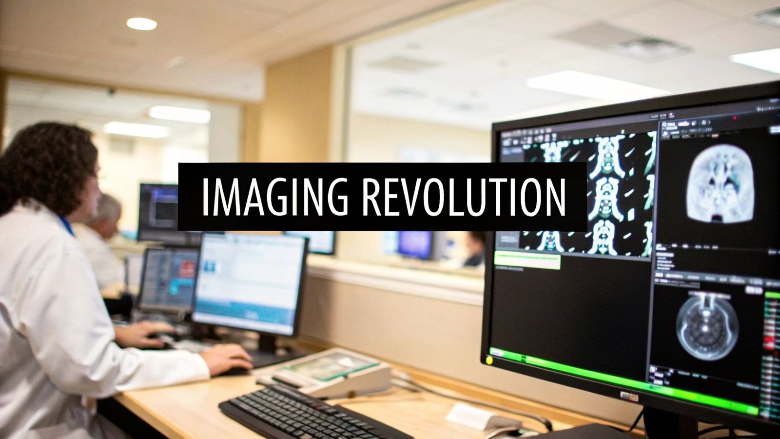
Imagine a technology that can look inside the human body and not just see a picture, but truly understand what it's seeing. That's the incredible promise of imaging processing software. These brilliant tools are like digital interpreters, translating complex medical scans into clear, actionable insights that help doctors forge a new path in patient care.
Unlocking the Stories Hidden in Pixels
At its core, every medical image tells a story—the story of a bone healing, a disease taking hold, or the complex, beautiful pathways of the brain. But the raw data from an MRI, CT scan, or X-ray is like a story written in a language we can't immediately understand. It’s a dense collection of pixels and data points, packed with information but completely lacking context.
This is where imaging processing software comes in. It’s the essential translator between the scanning machine and the medical expert.
The software takes this raw, unprocessed data and meticulously refines it, transforming flat, grayscale images into vibrant, three-dimensional models. Think of it like a high-tech darkroom, where an expert developer brings a photograph to life. The software cleans up digital "noise," sharpens crucial details, and highlights areas of concern that would otherwise be invisible. This process gives healthcare professionals the power to spot diseases earlier, plan surgeries with pinpoint accuracy, and design truly personalized treatment plans.
From Data Acquisition to Diagnosis
The journey from a simple scan to a life-saving insight follows a clear and powerful workflow. It begins when a scanner acquires the raw data. From there, the software takes over, processing every pixel before delivering insightful predictions that clinicians can act on.
This flow is the engine of modern diagnostics.
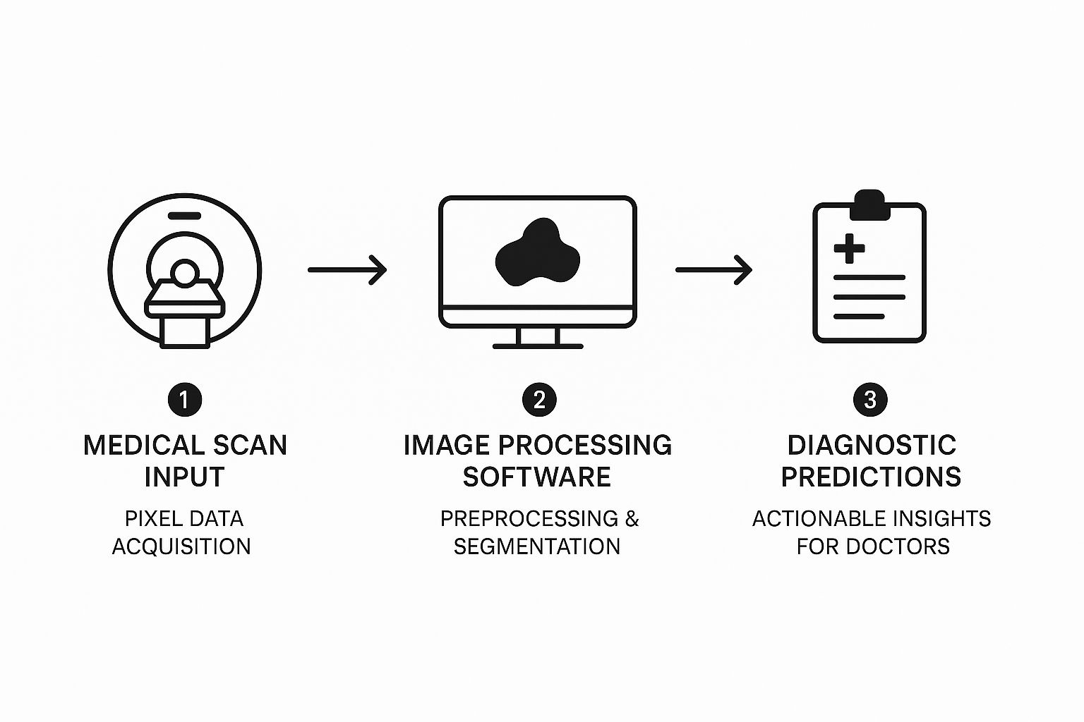
As you can see, the software sits at the very heart of this process, turning a flood of unstructured data into structured, understandable medical intelligence.
The Foundation of Modern Medical Platforms
This entire diagnostic ecosystem is built upon a foundation of specialized tools designed to handle and display complex medical files, like the industry-standard DICOM format. The ability to view, manipulate, and analyze these images isn't just a feature; it's the whole point.
The true power of this technology isn't just in looking at pictures. It's in creating an interactive space where clinicians can explore patient data dynamically, leading to more confident and informed decisions.
At PYCAD, we build the very bedrock for these systems. Our expertise lies in creating custom web DICOM viewers and integrating them into sophisticated medical imaging web platforms. We engineer the seamless and secure environments where these breakthroughs happen every day. By changing how medical professionals see and interact with visual data, we help turn silent pixels into a powerful voice that guides them in their mission to save lives.
You can see how our custom solutions are making a difference by exploring our portfolio.
How Medical Imaging Software Actually Works
Let's pull back the curtain on the technology that powers today's most crucial diagnostics. At its heart, imaging processing software is a sophisticated engine designed to take raw, often messy data from a medical scan and turn it into a crystal-clear visual story for clinicians. This isn’t a one-and-done action; it's a careful, multi-stage workflow where each step builds on the last to reveal the insights hidden within the data.
Imagine the initial scan from an MRI or CT machine as an undeveloped photograph. All the vital information is there, but it's often clouded by digital noise, inconsistent lighting, or tiny distortions from the hardware itself. The very first job is to prepare this raw data for a proper analysis.
The Preprocessing Stage
This first phase, called preprocessing, is all about cleaning up and enhancing the image. The goal here is simple: improve the quality so that any analysis done later is far more accurate and trustworthy. Think of it like a sound engineer meticulously removing static from a recording; this software filters out digital imperfections.
A few key tasks happen here:
- Noise Reduction: This process smooths out the random pixel variations that show up as graininess. Modern algorithms are smart enough to do this without blurring the important anatomical details.
- Contrast Enhancement: Here, the software adjusts the brightness and darkness levels across the scan, making it much easier for a doctor—or an AI—to tell different types of tissue apart.
- Normalization: This standardizes the intensity values, either within a single image or across a whole set of them. This is critical for making apples-to-apples comparisons, especially when tracking how a condition is changing over time.
Getting this foundational step right is absolutely non-negotiable for diagnostic accuracy. To really dig into this, you can learn more by exploring our guide on image preprocessing, which unpacks the specific techniques that make clear analysis possible.
The Segmentation and Analysis Stage
Once the image is clean and sharp, the next big step is segmentation. This is the digital version of carefully tracing an object in a photo to isolate it from the background. In medicine, that "object" might be an organ, a specific blood vessel, or a tumor.
Segmentation is where raw data begins its transformation into measurable intelligence. By isolating a region of interest, the software enables precise quantitative analysis, moving from "what does this look like?" to "how large is it, and how is it changing?"
After an area is segmented, the software can dive into a detailed analysis. This could mean calculating the volume of a lesion, measuring the rate of blood flow through an artery, or assessing the density of a specific tissue. These numbers provide objective data that supports a clinician’s expert assessment, leading to more confident, data-driven decisions.
DICOM: The Universal Language of Medical Imaging
For any of this magic to happen seamlessly between different hospitals, clinics, and equipment brands, everyone needs to speak the same language. That language is DICOM (Digital Imaging and Communications in Medicine). It’s the global standard for storing, viewing, and sharing medical images.
DICOM acts as a universal translator. It ensures that an MRI scan taken on one machine can be perfectly read and analyzed by software on a completely different system, potentially thousands of miles away. This standard is the very backbone of modern radiology and telemedicine.
At PYCAD, we live and breathe this standard. We specialize in building custom web DICOM viewers and integrating them directly into medical imaging web platforms. Our work provides the foundational tools that make this entire process—from the initial scan to the final diagnosis—possible, secure, and accessible from anywhere.
Building high-performance, reliable imaging software demands a disciplined approach. Using proven practices like Test-Driven Development (TDD) for robust software is key to ensuring the accuracy and stability required in these critical medical applications. You can see how we put these principles into action by checking out the innovative solutions in our portfolio.
The Transformative Role of AI in Medical Imaging
Artificial intelligence has officially stepped out of science fiction and into the clinic, becoming one of a doctor's most powerful new allies. AI and machine learning are supercharging imaging processing software, turning it from a simple visualization tool into an intelligent partner in diagnostics. This evolution is fundamentally changing what's possible in medicine.
Think of an AI algorithm as a highly trained assistant who has studied millions of medical scans. This digital expert can work tirelessly, 24/7, scanning thousands of images to spot subtle anomalies that even a seasoned human eye might miss after a long shift. This isn't about replacing radiologists—it's about augmenting their expertise and giving them a second set of eyes that never gets tired.
From Detection to Prediction
The integration of AI takes imaging software far beyond basic analysis and into the realm of predictive diagnostics. Instead of just identifying what’s already visible, machine learning models can recognize incredibly complex patterns that point to future health outcomes. This gives clinicians a precious window to intervene earlier and more effectively.
This shift is a cornerstone of proactive medicine, moving the focus from just treating existing diseases to preventing them from ever taking hold. AI helps answer not only, "What does this scan show?" but also, "What does this scan tell us about the patient's future health?"
AI empowers imaging processing software to learn from every single scan it analyzes, creating a continuously improving cycle of diagnostic accuracy. It's an engine for medical knowledge that gets smarter with each new piece of data, directly benefiting patient care.
The impact of this technology is unmistakable. The global image processing software market is projected to hit USD 38.54 billion by 2026, growing at a 7.4% compound annual growth rate. This incredible growth is largely driven by the adoption of AI, which is unlocking new potential in these essential medical tools.
Automating the Mundane to Focus on the Critical
A significant chunk of a radiologist's day can be spent on repetitive, manual tasks like measuring tumors or segmenting organs. AI is brilliant at automating these meticulous jobs with superhuman speed and consistency. For example, an AI can precisely calculate a tumor's volume from a series of CT scans in seconds, a task that could take a human expert a considerable amount of time.
This automation frees up clinicians to concentrate on what they do best: interpreting complex findings, consulting with other specialists, and crafting treatment plans. It allows them to transition from data gatherer to data interpreter.
Here’s how AI-driven automation is making a difference:
- Quantitative Analysis: AI delivers objective, repeatable measurements, tracking disease progression or treatment response with a level of precision that was once unimaginable.
- Workflow Efficiency: It drastically cuts down the time needed to process and analyze scans, helping to clear backlogs in busy radiology departments.
- Standardized Reporting: Automated measurements ensure consistency in reports, which is absolutely vital for clinical trials and long-term patient monitoring.
Of course, with great power comes great responsibility. The need for transparent and understandable AI models is paramount. For anyone curious about the ethics and mechanics behind these systems, we have an insightful article on explainable AI in healthcare.
AI's power isn't just confined to analyzing visual data; it’s also making waves in other areas of healthcare, like documentation. For instance, the advancements in AI in medical voice recognition are changing the game for how doctors capture patient notes efficiently.
At PYCAD, we are right at the heart of this movement. We build custom web DICOM viewers and integrate them into medical imaging web platforms, often equipping them with intelligent AI features. Our mission is to create systems that don't just display images but provide deep, actionable insights that can change patient outcomes.
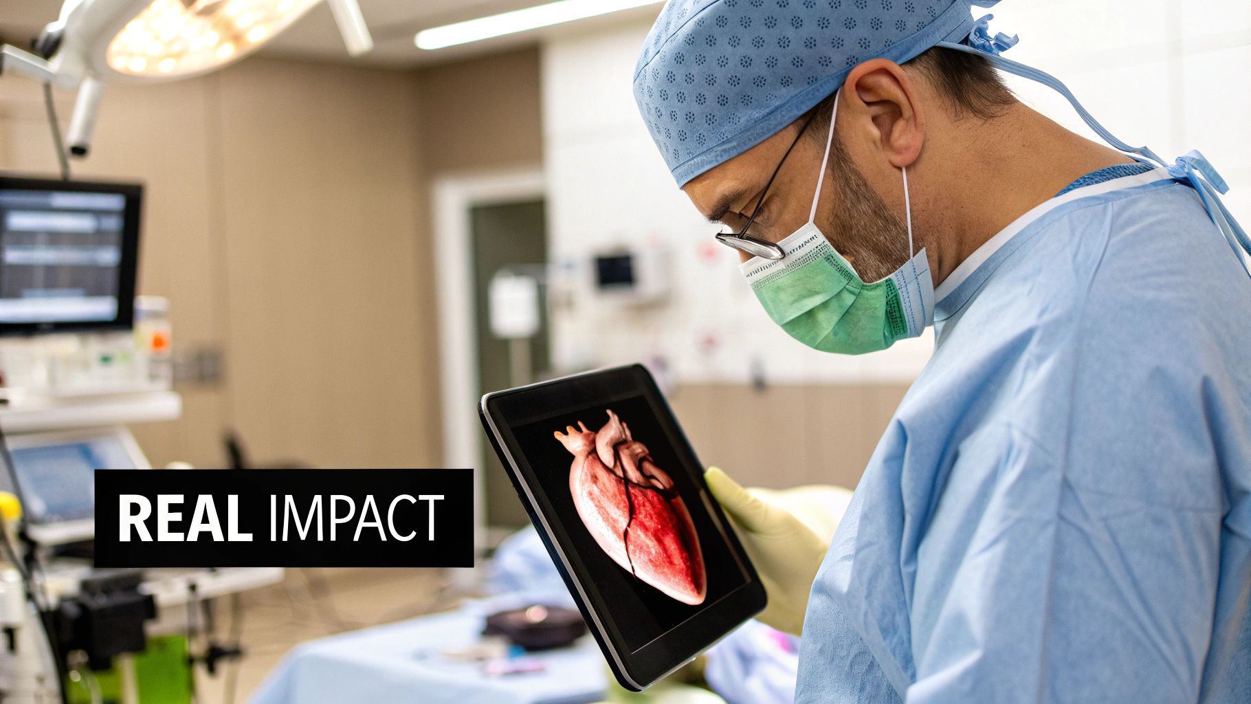
Where Software Meets Humanity: Real-World Breakthroughs
It's one thing to talk about algorithms and technical specs, but where this technology truly shines is in its impact on real lives. Imaging processing software isn't just about pixels on a screen; it’s about giving a doctor the clarity to save a life, the precision to plan a delicate surgery, and the insight to stop a disease before it progresses.
Let’s step into the shoes of an oncologist. They're managing a patient with a complex tumor, and every week, they face the same critical question: Is the chemotherapy working? Is the tumor shrinking, or is it holding its ground? Advanced imaging software delivers the answer not as a guess, but as undeniable visual proof.
A New Era for Oncology and Cardiology
In an oncology ward, this software can take a series of CT scans and stitch them together into a dynamic, 3D timeline of the patient's cancer. It isolates the tumor, calculates its volume with incredible accuracy, and maps its changes over weeks or months. This hard data gives doctors the confidence to stick with a successful treatment or pivot quickly to a new one if the first isn't working.
Cardiology is another area seeing incredible gains. For someone with a potential heart condition, cardiologists can now watch blood flow through arteries in real-time using dynamic MRI scans. The software creates a living model of the beating heart, highlighting dangerous blockages or weaknesses long before they might cause a catastrophic event.
This technology transforms a flat, static medical image into a living, breathing model of a patient's own body. It’s the difference between looking at a paper map and exploring a rich, interactive globe.
The impact of these tools is fueling major growth. The medical image analysis software market is on track to hit a revenue of USD 4.26 billion by 2025. We're also seeing a clear trend in adoption across different imaging types, with MRI's market share projected to grow from 12% to 14% in that same timeframe. To get a better sense of these trends, you can explore detailed statistics on medical imaging software.
Giving Surgeons a Crystal Ball
Perhaps the most powerful application is what happens before a patient even enters the operating room. Surgeons can now conduct a full dress rehearsal of a complex procedure, all thanks to software. By converting a patient's CT or MRI scans into a high-fidelity 3D model, imaging processing software creates a perfect digital twin of the patient's anatomy.
Surgeons can spin this model on screen, look at it from every conceivable angle, and even walk through the surgery step-by-step. This kind of planning is a game-changer for:
- Complex Reconstructions: Figuring out the perfect placement for implants in an orthopedic procedure.
- Tumor Removal: Mapping the exact borders of a tumor to ensure every bit is removed while sparing healthy tissue.
- Vascular Surgery: Navigating a delicate web of blood vessels to repair an aneurysm or clear a clot.
This virtual roadmap dramatically reduces surprises during surgery, which means shorter operating times, fewer risks for the patient, and far better outcomes. It’s a perfect illustration of how software is directly elevating surgical skill and patient safety.
At PYCAD, this is exactly what we live and breathe. We build custom web DICOM viewers and integrate them into medical imaging platforms, giving specialists the precise tools they need to visualize, plan, and act with total confidence. To see how we’ve helped our partners, feel free to explore our work in the PYCAD portfolio.
Choosing the Right Imaging Software Solution
Picking the right imaging processing software is one of the most important decisions a healthcare organization will ever make. This isn't just about buying a new tool. It’s about choosing the very heart of your diagnostic, research, and clinical operations. Get it right, and you accelerate innovation. Get it wrong, and it becomes a source of daily frustration.
For hospital administrators, visionary researchers, and medical innovators, this choice demands real foresight. The perfect solution should feel like a seamless extension of your team, fitting naturally into your existing electronic health records (EHR) and Picture Archiving and Communication Systems (PACS). Of course, it also needs to be rock-solid, with bulletproof compliance for regulations like HIPAA and all necessary FDA clearances.

Navigating the Build vs. Buy Decision
Sooner or later, every organization faces a fundamental question: do we buy an off-the-shelf product or invest in building a custom solution? A generic software package might tick a few boxes, but what happens when your workflow is anything but generic? Or when your research is pushing the very limits of what’s possible? This is where the "build vs. buy" debate really matters.
An off-the-shelf solution can feel like the easy path. It’s quick and seems straightforward at first. But more often than not, it forces your team to bend their processes to fit the software’s rigid structure, instead of the other way around. For truly innovative work or highly specialized clinical pathways, a one-size-fits-all approach rarely fits anyone perfectly.
This is where a bespoke solution truly comes into its own. Building custom software means creating a tool sculpted precisely to your needs.
A custom solution turns your technology from a simple utility into a powerful strategic asset. It’s an investment in a platform that grows with you, adapts to new challenges, and frees your team to do their best work.
This approach is especially critical as the global image processing systems market explodes in value, from USD 19.07 billion today to a projected USD 49.48 billion by 2032. The rising tide of chronic diseases is creating a massive demand for systems that can handle huge imaging datasets with absolute precision—a challenge that custom engineering is uniquely positioned to solve. You can learn more about the growth drivers in the image processing market to get a better sense of these trends.
To help clarify this decision, we've outlined the key factors to weigh when comparing a ready-made product against a custom-built platform.
Key Criteria for Selecting Imaging Processing Software
| Evaluation Criterion | Off-the-Shelf Software Considerations | Custom Solution (PYCAD) Considerations |
|---|---|---|
| Functionality & Workflow Fit | Does it meet 80% of your needs? Be prepared for workarounds. | Built from the ground up to match your 100% specific workflow. |
| Scalability | May have limitations on user count, data volume, or feature expansion. | Designed to scale with your organization's growth and data needs. |
| Integration Capabilities | Limited to standard integrations (EHR/PACS). Custom connections are often difficult or impossible. | Can be integrated with any proprietary or legacy system you rely on. |
| Competitive Advantage | You're using the same tools as everyone else. | Creates a unique, proprietary asset that sets your research or practice apart. |
| Long-Term Cost | Lower initial cost, but ongoing subscription fees and costs for unmet needs can add up. | Higher upfront investment, but greater long-term ROI and no recurring license fees. |
| Support & Maintenance | Reliant on the vendor's general support and roadmap priorities. | Dedicated support and a development roadmap that you control. |
Ultimately, the choice depends on your long-term vision. If you’re looking to simply manage standard tasks, an off-the-shelf tool might suffice. But if you’re aiming to innovate and lead, a custom solution is the only way to build a true competitive edge.
Finding the Right Partner for Your Vision
Deciding to build a custom solution is a commitment to excellence, and it demands a partner who brings both deep technical skill and a genuine understanding of the medical world. This is exactly where we at PYCAD shine. We specialize in building custom web DICOM viewers and integrating them into complete medical imaging platforms. Our entire process starts by listening—truly listening—to what you need to achieve, then architecting a solution that takes your work to the next level. You can see how we approach this in our overview of custom medical imaging software development.
Whether you need to automate a groundbreaking analysis technique or design an incredibly intuitive interface for a complex diagnostic tool, our team has the experience to make it happen. By partnering with us, you get technology that isn't just functional, but inspirational—an asset that actively fuels your mission to advance patient care.
To see how we’ve helped organizations just like yours, we invite you to explore our portfolio of successful projects.
The Future of Medicine Is Visual
We're standing on the brink of a new era in healthcare, one where imaging processing software isn't just a tool, but the very language of discovery. What we're witnessing today isn't the finish line; it's the starting block for a future where medicine is more predictive, deeply personal, and, above all, visual.
Think about the next generation of medical breakthroughs. Picture a surgeon in the operating room, their vision enhanced by an augmented reality (AR) display. This transparent overlay, constructed from the patient's own pre-op scans, would project the precise location of vital nerves and blood vessels directly onto the patient. It’s a roadmap for the human body, delivered in real-time.
Weaving Data into a Health Tapestry
Looking beyond the OR, the real magic happens when we start combining imaging data with other biological information. The true potential is unlocked when we fuse a patient's CT scan with their specific genomic data, creating a form of medicine that's genuinely personal.
This means treatment will no longer be based on just what a tumor looks like, but on its unique genetic signature. It allows clinicians to predict how a patient will respond to therapy before a single dose is given, shifting the entire paradigm from reactive treatment to proactive, individualized care.
The future of imaging isn't just about seeing inside the body with greater clarity. It's about understanding the intricate story our biology tells at every level, from our genes to our anatomy.
Making Advanced Diagnostics Available to All
Another profound change is the rise of cloud-based platforms that put world-class diagnostics within reach of everyone, everywhere. A small rural clinic could upload a complex scan to a secure cloud service and, in minutes, receive an analysis from powerful AI algorithms that equals the expertise of a top-tier urban hospital. This is how we begin to close the healthcare gap worldwide.
We are actively building a future where AI constantly scans routine images, flagging disease risks years before symptoms ever emerge. This isn't science fiction; it's the reality being engineered today by dedicated teams pushing the limits of what we thought was possible.
At PYCAD, we live and breathe this work. Our passion is crafting the custom web DICOM viewers and medical imaging platforms that will drive this future forward. We work alongside the innovators who are turning these incredible ideas into reality.
To see what this looks like in practice, we invite you to explore our portfolio and see what’s truly possible.
Your Questions About Imaging Software, Answered
Diving into the world of medical imaging can feel a bit overwhelming, and it often brings up some great questions. Getting a handle on the key differences and the processes behind the magic will really open your eyes to what’s possible with today's diagnostic tools. Let's tackle some of the most common things people ask.
Image Processing vs. Image Analysis
So, what’s the real difference between image processing and image analysis? It's a great question. Think of it like a skilled photo editor working on a masterpiece.
Image processing is the first part of the job. It’s all about enhancing the raw image—adjusting brightness, sharpening edges, and removing noise—to make every detail pop. In medical imaging, this means cleaning up a scan so a radiologist can see everything with absolute clarity.
Image analysis is what comes next. This is where we go beyond just looking at the picture and start pulling out concrete, measurable information. It's like the photo editor measuring the exact distance between two points in the image. In a clinical setting, analysis software might calculate the precise volume of a tumor or measure the rate of blood flow, turning visual information into hard data for decision-making.
How Does the Software Handle Different Scan Types?
It's amazing how this software can work with so many different kinds of scans, right? The secret is that modern imaging platforms aren't a one-size-fits-all solution. They're built for incredible versatility.
These systems use highly specialized algorithms, each one fine-tuned for a specific imaging modality. An algorithm designed for a CT scan works differently from one meant for an MRI or an X-ray. This ensures that no matter the source, the final image is perfectly optimized to answer the specific clinical question at hand, giving doctors the clearest possible view for a confident diagnosis.
Integrating a Custom DICOM Viewer
What does it take to get a custom DICOM viewer integrated into a platform? The journey really starts with a deep understanding of your clinical workflow and your current tech setup. It's not just about plugging in a new tool; it's about building a seamless bridge from what you have now to a far more powerful and intuitive viewing experience.
Integration isn't just about adding a feature. It's about fundamentally elevating the user experience, making complex data accessible, interactive, and truly insightful.
The key steps involve careful API integration planning, working together on the user interface design, and, of course, a ton of testing to make sure everything runs flawlessly. This is exactly what we live and breathe at PYCAD. We build custom web DICOM viewers from the ground up and integrate them into medical imaging web platforms, helping turn your vision into a functional reality.
If you'd like to see how we bring these powerful custom solutions to life, I invite you to explore our portfolio.


