
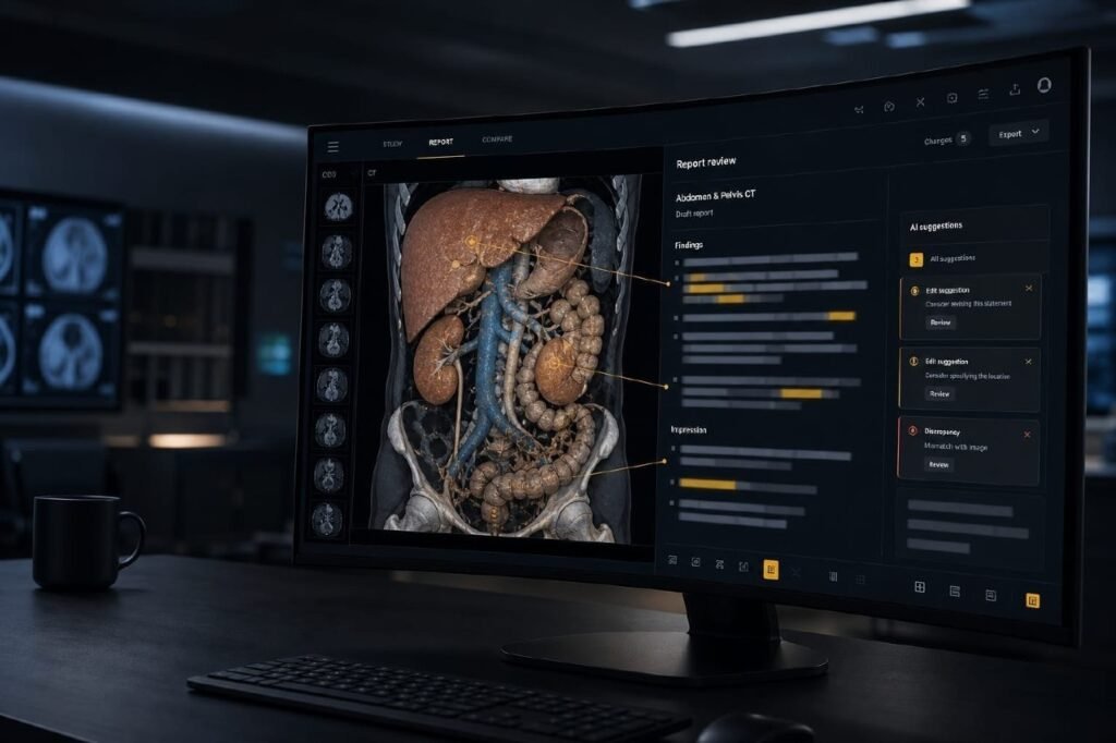
Radiology AI has become very good at generating text, answering image questions, and scoring well on benchmark datasets. A more practical question for real-world deployment is whether a system can review and improve a draft radiology report against the actual 3D study.
That is why RADAR stands out. Published in 2026 as RADAR: A Multimodal Benchmark for 3D Image-Based Radiology Report Review (arXiv 2603.06681), it focuses on discrepancy analysis between a preliminary report and the underlying study. Instead of asking whether a model can generate a report, RADAR evaluates whether it can detect, assess, and support edits using the imaging evidence itself. The generic explainer for what radiology AI does in a clinic is artificial intelligence in radiology; this page is the RADAR benchmark.
For teams building medical imaging software, reporting tools, or AI-enabled workflow products, that shift is the useful one. It also connects to the radiology AI workflow automation problems product teams hit in real deployments.
Why report review matters
In actual radiology workflows, reporting is rarely a one-shot act of text generation. A draft is created, revised, checked, corrected, or refined before final sign-off. Subtle wording changes can reflect meaningful clinical differences. An omitted finding, an overcalled abnormality, an incorrect laterality, or a mismatch between the report and the images can all affect downstream care. Report review is not only a language task. It is image-grounded reasoning. RADAR benchmarks that gap.
What RADAR evaluates
RADAR pairs 3D abdominal CT studies with a preliminary radiology report and candidate edits tied to the same exam. The model is not writing from scratch. It has to review a draft against the study, judge whether a proposed change is supported by the images, and reason about image-text alignment at a finer level than standard report generation.
| Report generation | RADAR (report review) | |
|---|---|---|
| Input | 3D study | 3D study + preliminary report + candidate edits |
| Job | Write findings from pixels | Judge whether an edit is image-grounded |
| Output | A draft report | Discrepancy / edit support, not a new narrative |
| Closest clinic analog | AI author | Second-reader QA before sign-off |
That makes the benchmark relevant for radiology report QA, AI-assisted discrepancy detection, second-reader review, report refinement for structured or narrative reporting, and multimodal clinical documentation products.
Why this is different from familiar imaging AI benchmarks
Many medical imaging benchmarks still focus on diagnosis prediction, VQA, captioning, or broad vision-language scores. Those are useful. They do not fully capture the reasoning needed when a model reviews a draft and decides whether it should be corrected. The model must connect a written claim to slices, findings, and anatomy; distinguish a plausible edit from a clinically justified one; and do it in a setting closer to how radiology work is actually checked and finalized. That is a more deployment-oriented way to evaluate radiology AI. It also complements radiology AI agents inside real DICOM workflows, where performance depends on operating in the workflow, not only on an isolated score.
From generation to verification
Report generation gets the attention because drafting findings from images is ambitious and visible. In practice, verification may be just as important. A system that flags discrepancies, verifies edits, and improves report consistency can fit a clinic more naturally than one that tries to replace the whole reporting process. For many environments, an AI reviewer is easier to trust, validate, and integrate than an AI author operating alone.
Why this matters for product teams
RADAR highlights a category of capabilities that matter in imaging platforms: image-grounded report checking, discrepancy flagging before sign-off, clinical text review tied to the visual study, human-in-the-loop QA for drafts, and tools that connect viewer context to reporting context. That is relevant for hospitals and radiology groups, and for companies building DICOM viewers, reporting systems, and AI-enabled imaging workflows. If the next wave is less “produce fluent text” and more “make clinical documentation reliable,” benchmarks like RADAR tell you what to build toward.
Why now
Multimodal models are getting better at connecting images and language. Stronger general capability creates a new requirement: better evaluation. Without a focused benchmark it is hard to tell whether a model is ready for a high-value clinical review task or just good at producing convincing text. RADAR centers the review process itself. That helps researchers, and it helps product teams decide where to invest.
Source
- RADAR: A Multimodal Benchmark for 3D Image-Based Radiology Report Review — arXiv 2603.06681
PYCAD builds the imaging side of this — custom pipelines and web DICOM viewers when the study has to live in a clinic app. Case studies.