
Most deep-learning MRI reconstruction models are trained on one big public dataset, usually fastMRI from New York University on Siemens scanners, and lose quality, with aliasing coming back, when the vendor, field strength or protocol changes. SIM (Scalable Inverse Model), from the Shenzhen Institutes of Advanced Technology, Peking University Shenzhen Hospital and Tencent, trains mostly on natural photos turned into simulated multi-coil k-space and scales the model to about one billion parameters. The paper is arXiv:2611.15758 (eess.IV, 11 November 2026).
What SIM does
SIM reconstructs accelerated 2D MRI slices from undersampled multi-coil k-space in a single forward pass. The training pairs come from a physics simulator. The simulator takes images from SA-1B, the open photo set released with Segment Anything, turns them into complex-valued “subjects”, multiplies them by random coil sensitivity maps, adds k-space noise and applies the undersampling mask. The target is the fully sampled, noisy multi-coil image combined by root-sum-of-squares, the same way a clinical reference is formed.
The authors argue that what a reconstruction network has to learn is the undersampling physics, how aliasing folds signal from one place onto another, and that this does not depend on anatomy. Photos give far more variety of edges, textures and contrasts than any MRI archive. In their scaling experiments, a model trained on fastMRI Brain stopped improving and overfit as it grew, while the same architecture trained on simulated data kept improving up to the largest size they tested.
How it works
- Random subjects. Two channels of a photo (taken from RGB, HSV or YCbCr) become the real and imaginary parts of the signal. A phase-alignment step spreads the phase over the full circle, and magnitudes are rescaled with a log-normal factor, so the simulated signals do not all look alike.
- Random coils. Every training sample gets its own coil array: Gaussian blobs for sensitivity magnitude, linear ramps for phase, normalised with root-sum-of-squares. The aim is to cover so many layouts that the coils of an unseen scanner fall inside what the model has already seen.
- Matched noise and masks. SNR is drawn over a wide range to cover low-field and high-field noise. The undersampling masks used in training must include the masks used at test time; the authors report a marked drop when a mask configuration is left out of training.
- mcViT. The network is a plain Vision Transformer adapted from the Diffusion Transformer (DiT) with 2D rotary position embeddings. It takes the aliased zero-filled image of each coil, predicts the aliasing residual for each coil separately, and combines the coils with root-sum-of-squares at the end. There is no explicit sensitivity-map estimation and no data-consistency block, unlike unrolled networks such as E2EVarNet, which build in both.
- Scale. The main model has 1.01 billion parameters, patch size 5 (4,096 tokens per channel), trained on about 1.01 million simulated samples mixed with 70,748 real fastMRI Brain slices, with a mean absolute error loss. Training ran on two eight-GPU nodes of consumer RTX 4090 and RTX 5090 cards. A variant trained only on simulated data is called SIM-Zero.
What they tested it on
The benchmark has 14 datasets with raw multi-coil k-space: 8 from public sources and 6 in-house, from seven centres and five vendors, from 0.3 T (M4Raw) to 7 T (AHEAD), across more than ten anatomies and more than ten protocols. fastMRI Brain is the only in-distribution test set; everything else is zero-shot. Undersampling is retrospective, with 1D equispaced masks and an 8% fully sampled centre, at effective accelerations of 3.2x, 4.2x and 5.1x.
Against nine baselines covering end-to-end networks, unrolled networks, a diffusion model, untrained (scan-specific) networks and L1-ESPIRiT compressed sensing, SIM was best on all 8 out-of-distribution benchmarks, by 1.06 to 3.95 dB PSNR over the second-best method. The authors also measured the drop when moving off fastMRI: up to 10.01 dB PSNR for E2EVarNet, at most 5.64 dB for SIM. SIM-Zero, which never saw a real scan in training, was only slightly behind SIM and still ahead of every baseline out of distribution.
The paper also checks quantitative outputs. On T2* mapping from multi-echo data (AHEAD at 7 T and an in-house MULTIPLEX sequence at 3 T), each echo was reconstructed separately and then fitted; SIM’s maps had at least 1.83 ms lower mean absolute error than E2EVarNet’s at every acceleration. On a dynamic creatine CEST study of calf muscle at 5 T, the recovered phosphocreatine time constant was 282 s against 279 s from the fully sampled data.
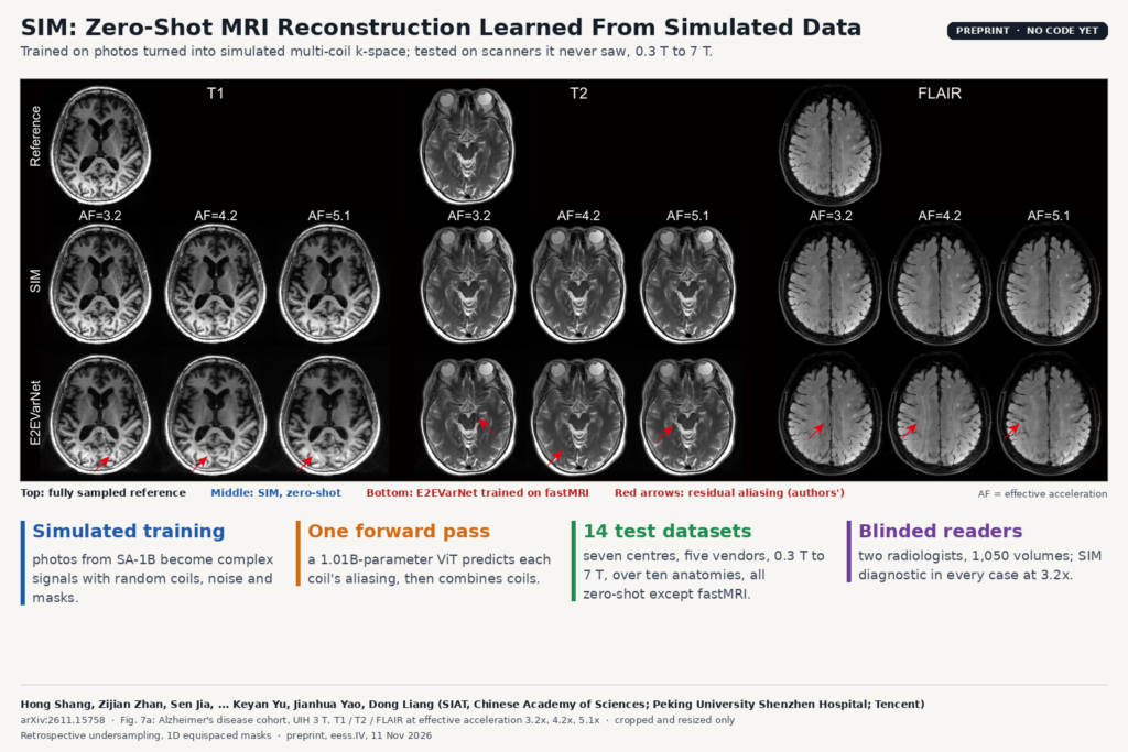
The clinical test is a blinded reader study on an Alzheimer’s disease cohort from Peking University Shenzhen Hospital (50 subjects, 25 patients, UIH 3 T, T1 MPRAGE, T2 and FLAIR, so 150 scans). Two radiologists with more than ten years of experience rated 1,050 volumes: the reference plus SIM and E2EVarNet at three accelerations. At 3.2x, SIM was rated diagnostic in every case, like the reference. At 5.1x, E2EVarNet was non-diagnostic in 100% (T1), 66% (T2) and 99% (FLAIR) of ratings, SIM in 4%, 0% and 1%. Inter-reader agreement was 0.91 weighted kappa for image quality and 0.73 for usability.
The featured image is Fig. 7a of the paper: reference, SIM and E2EVarNet on that cohort at the three accelerations, with the authors’ red arrows on the residual aliasing in the E2EVarNet images.
What it would change in a reconstruction pipeline
For a team that ships reconstruction or works with raw data from several sites, the practical promise is one model that does not need retraining per scanner, coil or protocol. The input is the same thing a scanner already has, undersampled multi-coil k-space, and the output is a magnitude image that can be written to DICOM like any other series. One forward pass is much faster than diffusion models or scan-specific networks that iterate hundreds of times per slice, and that speed decides whether the reconstruction can run on the scanner side or in a service next to PACS.
The quantitative results are what we would look at first for a clinical platform. A reconstruction that looks fine but shifts T2* values or CEST curves would quietly break any mapping or perfusion tool built on top of it. The T2* check covers two sequences and the CEST check one exercise study, so they are a first test of quantitative fidelity.
For groups that cannot move patient data between sites, the training set is photos plus a simulator, so the model and its data can be shared without patient data, and sites can add their own real slices on top, as the authors did with fastMRI.
What it does not tell you yet
- All undersampling is retrospective. Nothing was acquired prospectively on a scanner with an accelerated protocol, and the authors list prospective multicentre studies as the next step.
- It is 2D multi-slice and magnitude-only. 3D acquisitions were reformatted into 2D slices, and phase-sensitive imaging is not supported.
- Only 1D equispaced Cartesian masks were tested, and the masks must be in the training distribution. Radial, spiral, variable-density or vendor-specific patterns are untested.
- Several in-house references were themselves undersampled at acquisition (STAR 2D at 2.2x, STAR 3D at 2.6x, DCE at 2.5x, Multi-Contrast at 5.3x) and were turned into pseudo-fully-sampled data with L1-ESPIRiT. On those sets SIM is scored against a compressed-sensing reconstruction, not true full sampling.
- The reader study compares SIM with E2EVarNet trained only on fastMRI, a model that had never seen UIH data. It does not compare against the vendor’s own accelerated reconstruction, which is what a radiologist would see today.
- Everything is cropped or padded to a 320 by 320 matrix, so higher-resolution protocols were handled at reduced size.
- A one-billion-parameter transformer with 4,096 tokens per channel is heavy. The paper reports runtime on an RTX 5090 but no memory figures for deployment hardware.
How to try it
The paper links no code or weights, and searches on GitHub and Hugging Face on 16 November 2026 found no SIM repository from the authors, so there is nothing to run yet. The discussion mentions sharing open-source simulation code instead of patient data, which suggests the simulator may be released. The methods section is detailed enough to rebuild the simulation engine (photo channels, phase alignment, random Gaussian coils, noise, masks), and the training schedule is spelled out. The public test sets (fastMRI, CC-359, M4Raw, AHEAD, Stanford 2D and 3D) are available from their own sources. On the blog, see also LoopMR, accelerated MRI reconstruction with one looped block and LGGS, faster follow-up brain MRI using the patient’s last scan.
Source and license
Paper: Hong Shang, Zijian Zhan, Sen Jia, Xiang Fan, Jintao Meng, Yang Zhou, Xiang Zhou, Tianyu He, Xinran Chen, Qiaoling Zeng, Keyan Yu, Jianhua Yao and Dong Liang, “Physics-based simulation unlocks scaling laws for generalizable MRI reconstruction”, arXiv:2611.15758 (eess.IV, 11 November 2026), preprint, 45 pages. Affiliations: Shenzhen Institutes of Advanced Technology, Chinese Academy of Sciences; School of Biomedical Engineering, Shenzhen University; Department of Medical Imaging, Peking University Shenzhen Hospital; AI for Life Sciences Lab, Tencent; State Key Laboratory of Biomedical Imaging Science and System, Chinese Academy of Sciences (corresponding authors Hong Shang, Jianhua Yao and Dong Liang). Code and weights: not released, licence not stated. Data: public raw k-space benchmarks plus six in-house cohorts that are not public. Featured image: Fig. 7a of the paper, cropped only.