
Brain MRI lesion detectors usually work on the contrast and resolution they were trained on, and they learn from real lesion masks. A hospital archive holds T1-weighted, T2-weighted and FLAIR scans from many scanners, at 1 mm or 6 mm slices, with every kind of pathology. PathoDraw, a preprint from Harvard Medical School and Massachusetts General Hospital with MIT, the Medical University of Vienna and Microsoft (arXiv:2611.21975, cs.CV, 16 November 2026), trains one 3D network that flags stroke, glioma and white matter hyperintensities on any of those inputs, zero-shot, without ever seeing a real lesion.
What it does
PathoDraw is a generator of synthetic brain MRI with pathology, and a network trained only on its output. The authors start from healthy brain segmentations, draw a random lesion into the label map, then render the label map as an image with random contrast, resolution, bias field and noise, in the style of SynthSeg. Rendering the same anatomy with and without the lesion gives a pair of images that differ only where the lesion is, and that difference is the training target.
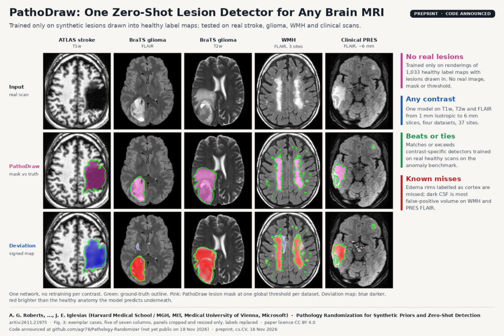
At test time the network takes a real scan and returns two things: the healthy anatomy it thinks lies under the lesion (56 labels), and a signed deviation map showing where and how the scan departs from that healthy anatomy, darker or brighter. A lesion mask is a global threshold on the size of that deviation. The same generator also trains networks for super-resolution and registration that keep the lesion intact.
How it works
- Healthy anatomy. 1,033 label maps of young adults from the Human Connectome Project and the Chinese HCP, segmented with FreeSurfer under manual quality control and extended with extracerebral labels. No image of these subjects enters training, only their labels.
- Pathology randomization. Each draw picks one of three geometric modes: tract-aligned lesions in periventricular white matter (small-vessel disease), vascular-aligned lesions in deep gray nuclei or a cortical territory (infarcts), and local lesions in any brain label (tumors and focal lesions). Each other mode adds a second lesion with probability 0.15. The lesion shape comes from a thresholded random field, it is split into a core and a shell, and mass effect pushes the surrounding anatomy aside.
- Visible by construction. Every synthetic lesion must differ from its surroundings by between k and 2k times the intensity spread in a band around it, with k drawn between 1 and 2.5, so the network is never asked to find a lesion that cannot be seen. Lesion brightness is mixed from the tissue it replaced and either CSF or gray matter, so it comes out dark on T1-like draws and bright on FLAIR-like draws. Half the draws use dark CSF, as on FLAIR.
- The network. A 3D UNet after SynthSeg with attention gates, an anatomy head and a deviation head, trained from scratch for 500,000 steps with two seeds. A control network with the same recipe but no pathology in its training data is the ablation for every result.
What they found
The detection test follows a published unsupervised anomaly detection benchmark, on ATLAS stroke (210 scans from nine unseen scanner cohorts), BraTS 2023 adult glioma on T1w, T2w and FLAIR (1,249 cases from 24 sites), the WMH challenge on FLAIR and T1w (60 scans, three sites) and 43 clinical FLAIR scans of posterior reversible encephalopathy syndrome (PRES) from the hospital archive, acquired at roughly 0.5 by 0.5 by 6 mm. The benchmark’s main score is a ceiling Dice, with one oracle threshold per dataset, so read the numbers as upper bounds. On BraTS T1w PathoDraw reaches 0.537 against 0.437 for FAE and 0.274 for DISYRE, both trained on real healthy scans of that one contrast; on BraTS T2w it is level with DISYRE (0.657 against 0.646); on ATLAS it sits at 0.369 against DISYRE’s 0.386. The control network trained without pathology scores lower on most rows, for example 0.228 on BraTS T1w and 0.072 on WMH T1w, which ties the gain to the synthetic lesions.
The featured image is Fig. 3 of the paper. The pink masks and the red and blue deviation maps come from one model with no retraining, across a 1 mm T1w stroke, glioma on FLAIR and T2w, white matter hyperintensities and a thick-slice clinical PRES scan. Absolute Dice stays low on white matter lesions (0.259 on WMH FLAIR, 0.225 on PRES), where lesions are small and scattered.
The authors also check that the synthetic shapes cover real ones. Using 3D shape descriptors, 0.86 to 0.93 of real lesion components across the four datasets fall inside the generator’s shape distribution, against 0.49 to 0.74 for a control set of random ellipsoid blobs. Registration to the MNI152 atlas keeps more of the lesion than the published SynthMorph model, with lesion Dice through the warp of 0.757 against 0.644 on ATLAS.
Why a viewer or clinic team should care
If you build a DICOM viewer or a research platform on top of a hospital archive, most of the work goes into mixed protocols, thick slices and contrasts nobody labelled. A single detector that accepts any structural brain MRI and returns a signed map of tissue that does not look normal fits that archive. You could use it to surface studies with focal pathology before running a disease-specific model, to flag cases for a radiologist to annotate, or to exclude lesioned brains from a healthy reference cohort. The healthy-anatomy head also gives you a label map under the lesion, which is what a volumetry or registration step downstream needs.
In a viewer, the signed deviation map works as an overlay on the native series, blue for darker than expected and red for brighter, with the threshold exposed as a slider.
Limits and failure modes
- Detectable pathology has to lie on the generator’s manifold of shape, placement and intensity. Disease that does not look like a local, vascular or tract-aligned lesion is outside what it was trained to flag.
- Most false-positive volume on WMH FLAIR and PRES comes from dark CSF. The authors expect this is because synthetic CSF has one mean intensity per label while a real ventricle is darker at its center than at its walls. Mildly deviant white matter adds more false positives, mostly on ATLAS.
- Misses are mainly intermediate-intensity tumor rim and edema that the anatomy head labels as cortex, mostly on BraTS T2w and FLAIR.
- The headline score is a ceiling Dice with an oracle threshold per dataset. At thresholds set on healthy HCP-Aging scans the numbers are lower, and the paper reports them separately at each comparison model’s false-positive rate.
- It covers structural brain MRI only, T1w, T2w and FLAIR. No diffusion, susceptibility, contrast-enhanced series, CT or other organs were tested, and no reader study was run.
- It is a preprint and has not been peer reviewed.
Code and licence
The paper states that the code is available at github.com/agr78/Pathology-Randomizer. When we checked on 18 November 2026 that repository was not yet public, and no licence or weights were listed. The paper itself is released under CC BY 4.0. The test sets are public (ATLAS, BraTS, the WMH challenge, ADNI3, HCP-Aging) except the PRES cohort, which comes from the authors’ hospital archive under IRB approval.
On the blog, see also BrainGFM, synthetic brain scans from few labels, ReSeg, organ masks trained without real scans and joint WMH and stroke lesion overlays from incomplete brain MRI.
Source
Paper: Alexandra G. Roberts, Tonmoy Hossain, Mark Olchanyi, Stefan Brandstätter, Jonathan Williams Ramirez, Ana Lawry Aguila, Matthew Rosen, Annabel Sorby-Adams, W. Taylor Kimberly and Juan Eugenio Iglesias, “Pathology Randomization for Synthetic Priors and Zero-Shot Detection”, arXiv:2611.21975 (cs.CV, 16 November 2026), preprint, 25 pages with appendices. Affiliations: Harvard Medical School and Massachusetts General Hospital, Massachusetts Institute of Technology, Medical University of Vienna, Microsoft. Featured image: Fig. 3 of the paper, five of seven columns, panels cropped and resized only.