
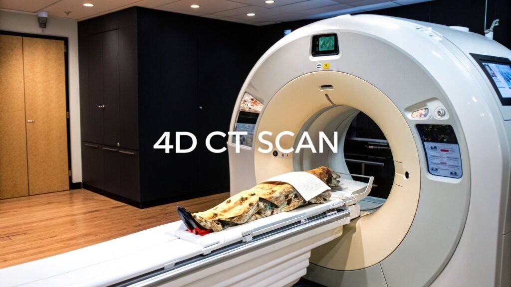
A 4D CT scan is a 3D CT with time as the fourth axis: a loop of volumes binned by a physiological phase, usually the breath. A 3D scan is a still of a moving car. A 4D scan is the movie. This page is that movie — phase bins, RPM, respiratory gating. It is not “what CT is.”
If you meant temporal resolution as a pillar (65 ms freeze) → resolution of a CT scan. If you meant 3D reconstruction from slices → 3D reconstruction from CT. If you meant volume rendering → CT volume rendering.
Time as the fourth dimension
The scanner still takes 3D volumes. A second signal — a camera on a reflective box, or a diaphragm track with no external marker — stamps each volume with where the patient is in the cycle. Software sorts those volumes into phase bins (10% inhale, 50%, full exhale, …). Play the bins in order and the organ moves.
Tumor motion on that loop is tracked on the order of 1–2 mm in published 4D-CT work. That figure is why oncology uses the scan: a target that walks with the breath is no longer a guess plus a fat margin.
In the room
You lie on the table and breathe normally. That is the point — a breath-hold is a 3D trick. A common external tracker is Varian RPM (Real-time Position Management): a camera watches a small reflective box on the chest or abdomen. Rise and fall = phase. Some rooms skip the box and track the diaphragm on the images (“deviceless”).
Prep is ordinary CT prep when the protocol asks for it (fasting, contrast, meds). The 4D-specific part is the tracker and the instruction: do not hold your breath unless they say otherwise.
What the loop is for
| Field | Job | What 4D adds |
|---|---|---|
| Radiation oncology | Lung / liver / upper-abdomen tumor | Respiratory gating: beam on only while the tumor is in the window; smaller margins, less lung/liver in the field |
| Cardiology | Wall motion, valves, output | A beating volume, not a still chamber |
| Interventional radiology | Biopsy / ablation in a moving organ | A map of where the target will be on the next breath |
| Pulmonology | Diaphragm, chest wall | Excursion you can measure, not infer |
Gating is the oncology headline. A 3D plan has to assume the tumor could be anywhere in its breath path, so the PTV grows. A 4D plan sees the path and can gate — or can build an ITV from the real excursion instead of a textbook centimetre. Either way the still scan is the wrong input.
Cardiology uses the same idea on the ECG instead of (or with) the breath. Wall-motion and valve leaflets are function questions. A static chamber answers anatomy only.
Cost of the fourth axis
More phases = more data = more dose than a single 3D pass, and a longer appointment. Complexity sits on the therapist and the physicist (binning, a bad RPM trace, a patient who cannot breathe regularly). Those are real cons. They are why not every chest CT is 4D.
Safety vs 4D ultrasound is a different comparison: CT is ionizing; ultrasound is not. You do not order 4D CT because it is “safer video.” You order it because you need CT contrast and HU in motion.
This is not a history of CT from 1971, and it is not a 3D-reconstruction tutorial. Reconstruction of a still volume is 3D reconstruction from CT. Painting that volume as a rendered surface is volume rendering. The 65 ms cardiac freeze without phase bins is the temporal pillar on resolution of a CT scan.
PYCAD builds the imaging side of products that have to treat a phase bin as a first-class volume. Case studies.
Keep Reading
Related Articles
Explore the full Quantitative Imaging HubAll services, tools, and guides on this topic — in one place.
Visit Hub →