
Two MRI Fields of View In, Fewer False Brain-Met Flags Out
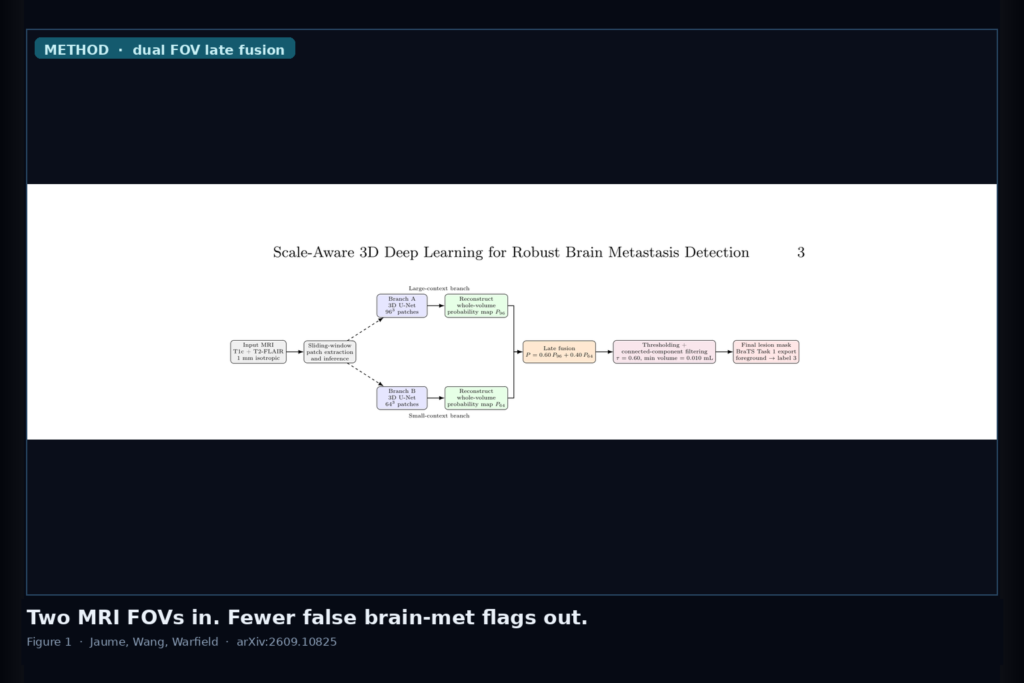
T1c + T2-FLAIR in; independently trained 96³ and 64³ 3D U-Nets; weighted late fusion cuts false brain-met flags on BraTS-METS. Jaume, Wang, Warfield, arXiv:2609.10825.
Partial Spine Ultrasound In, Complete Vertebra Geometry Out
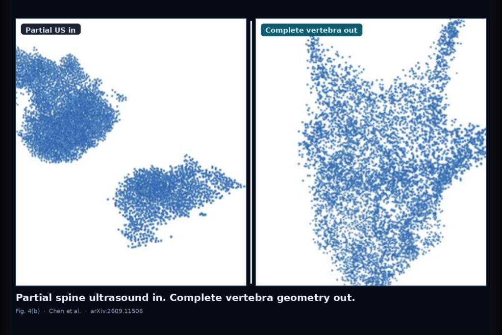
Hang a sparse tracked ultrasound bone cloud. UBone3D physics-rectified flow matching returns a complete vertebra you can hang for scoliosis follow-up without another CT. Chen et al., arXiv:2609.11506.
Loose Point or Box In, Colorectal Liver-Met Overlay Out on Contrast CT

Hang a portal-venous liver CT, drop a loose point or box (or none). SiGA/DiSECT spectral adapters on frozen SAM return colorectal liver-met overlays. Mojtahedi et al., arXiv:2609.11703.
Live Fluoro In, Preop CT Pose Out Without Patient Retrain
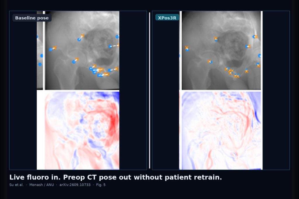
XPos3R takes a live X-ray and a preoperative CT and regresses an intraoperative pose with one anatomy-specific pretrained model, then optional seconds of test-time optimization. Su et al., arXiv:2609.10733.
Click One Lung Tumour Slice, Fix the Ones You Never Touched

LeCor meta-learns case adapters so each clinician click on a planning CT updates the lung tumour overlay on untouched slices, not only the session memory. arXiv:2609.09477.
Sparse FLAIR In, Fusion That Does Not Spoil the Overlay

CoReFuse-Med filters resampling junk on sparse T2/FLAIR in shallow layers and rebalances deep fusion so multi-modal brain overlays stay usable when naive fusion would otherwise lose to T1-only. arXiv:2609.10261.
When Medical Device Animation Wins vs Interactive Implant 3D
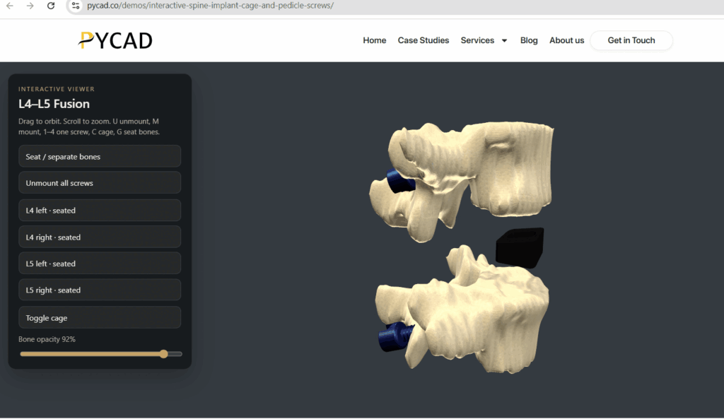
Implant manufacturers often choose between two strong formats for 3D on the web and at congress: a film-style rendered medical animation, and a manipulable interactive 3D embed of the commercial SKU (Stock Keeping Unit). We at PYCAD build the interactive embed side for marketing, med-ed, and sales enablement. Animation studios cover the directed film side. […]
CBCT Arch Cross-Sections and Panoramic Rebuild in a Web Viewer
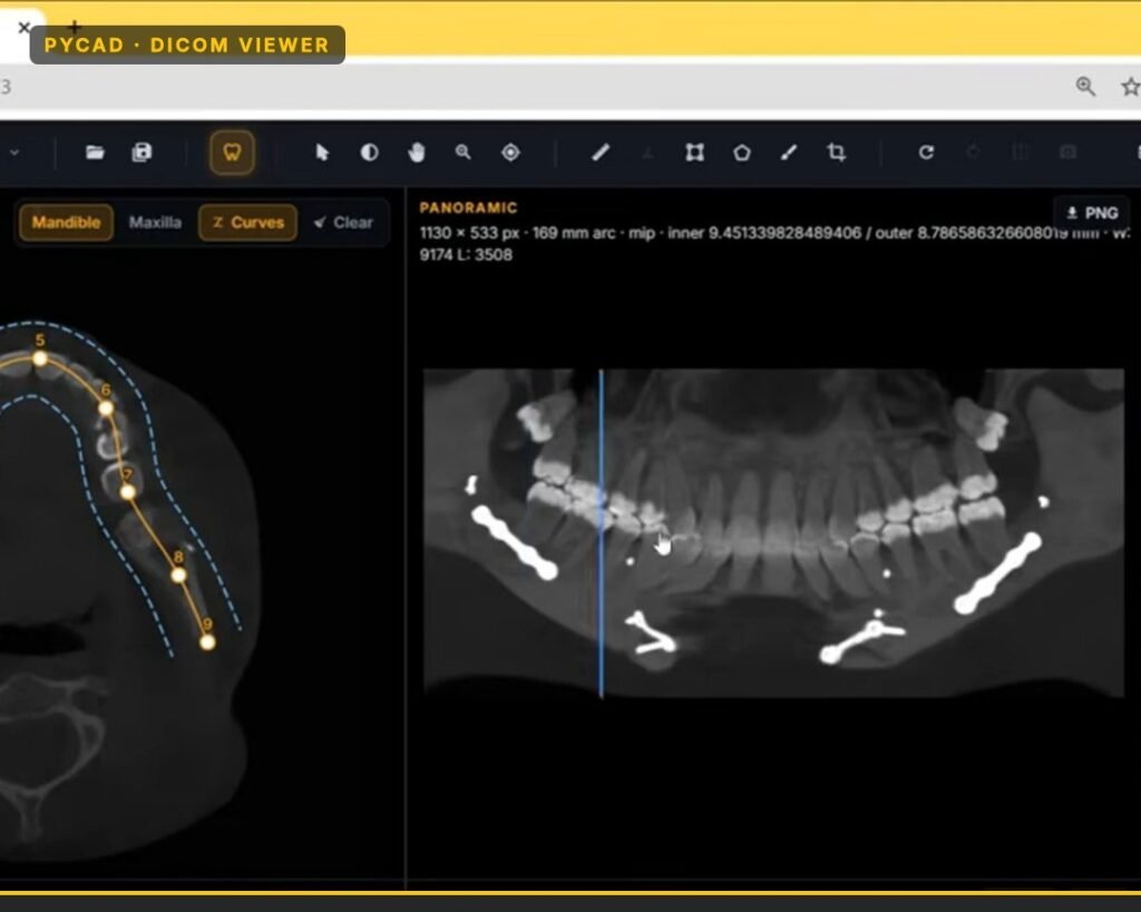
Plain multiplanar reformats open a CBCT on axial, coronal, and sagittal planes. That is useful. It is not how most dental users want to read an arch. We have a dental DICOM viewer that treats the arch as the control surface: you set a curve along the mandible or maxilla, get a panoramic-style rebuild from […]
Free OHIF Is the Silent Objection
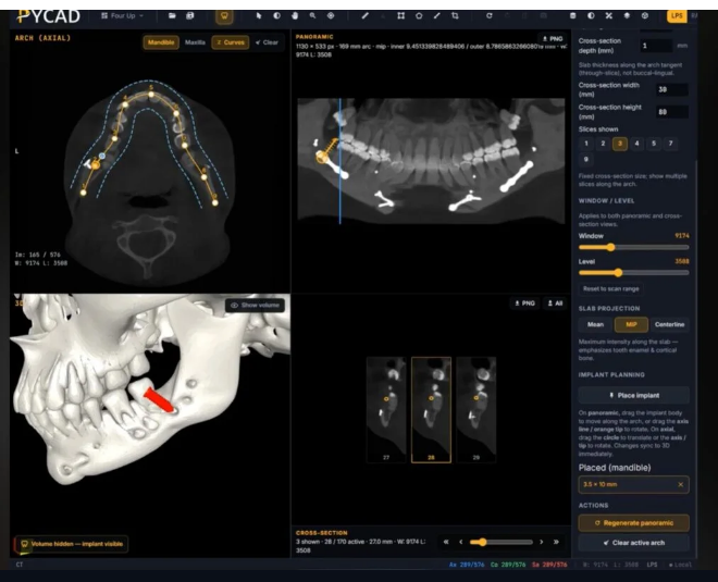
When a medical device OEM asks us at PYCAD about a custom DICOM viewer, someone in the room usually brings up OHIF. Sometimes they name another strong open radiology viewer in the same breath. The question underneath is the same: why pay for specialty work if a free viewer already opens CT, MRI, and CBCT […]
Mandibular Canal From CBCT: Segmentation → STL → Web Overlay
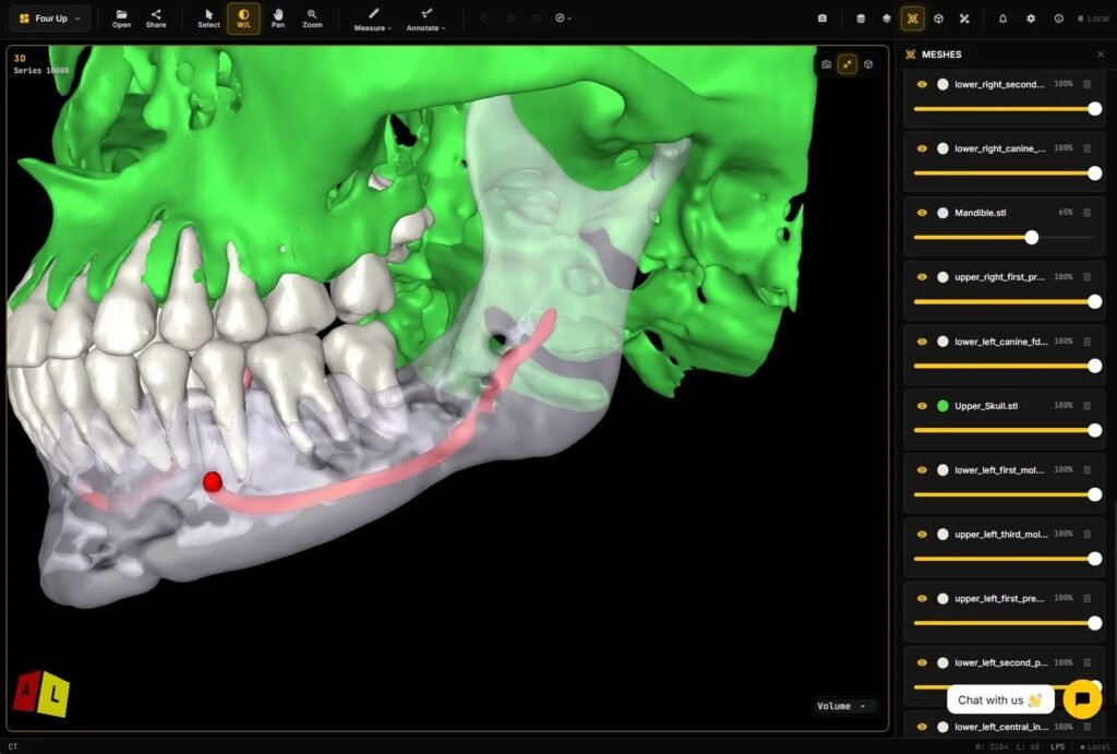
I get asked about mandibular canal work more than almost any other dental structure. Teams want the canal labeled on CBCT, turned into a mesh, and shown with the scan in a browser. That path is clear: CBCT → labels → surface → overlay. This post walks that path only. How the stages fit together, […]