Skip to content
  • Home
  • Services
    • All Services
    • Custom DICOM Viewer
    • Case Studies
  • Products
    • All Products
    • PYCAD DICOM Viewer
    • PYCAD Dental Viewer
    • PYCAD SSR Engine
  • Case Studies
  • Blog
  • About us
  • Home
  • Services
    • All Services
    • Custom DICOM Viewer
    • Case Studies
  • Products
    • All Products
    • PYCAD DICOM Viewer
    • PYCAD Dental Viewer
    • PYCAD SSR Engine
  • Case Studies
  • Blog
  • About us
Get in Touch

Category: Medical Imaging

NV-Generate-Body: Synthetic Whole-Body CT Masks You Can Dial In
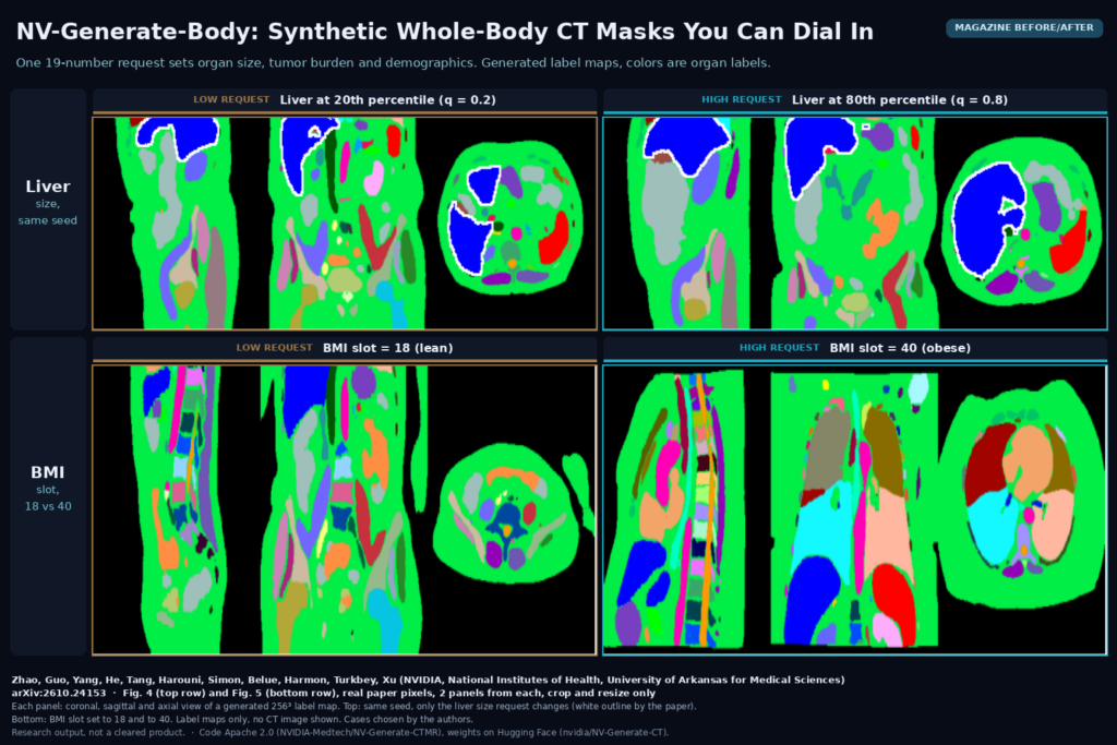
Magazine before/after from Figs. 4 and 5 of NV-Generate-Body: coronal, sagittal and axial views of generated 256-cube CT label maps, top row liver requested at the 20th and 80th percentile with the same seed, bottom row BMI slot set to 18 and 40. Zhao, Guo, ..., Turkbey, Xu, NVIDIA, NIH, UAMS, arXiv:2610.24153

NVIDIA’s NV-Generate-Body draws whole-body CT label maps with the organ sizes, tumors, sex and BMI you ask for. Code Apache 2.0, weights public.

GSMR3D: One Model Turns Thick-Slice Brain MRI Into 1 mm Volumes
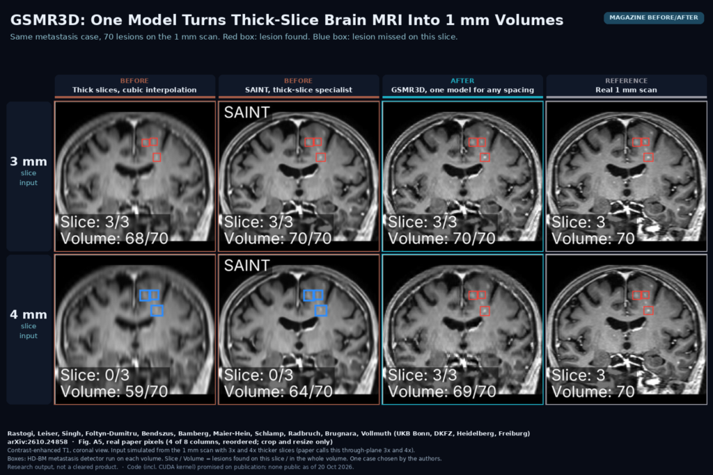
Magazine before/after from Fig. A5: coronal contrast-enhanced T1 of one brain-metastasis case rebuilt from 3 mm and 4 mm slices by cubic interpolation, SAINT and GSMR3D next to the real 1 mm scan, with detected (red) and missed (blue) lesion boxes. Rastogi, Leiser, ..., Brugnara, Vollmuth, arXiv:2610.24858

GSMR3D rebuilds thick-slice brain MRI on a 1 mm grid at any spacing, one model per contrast, and kept small metastases detectable.

LGGS: Faster Follow-Up Brain MRI Using the Patient’s Last Scan
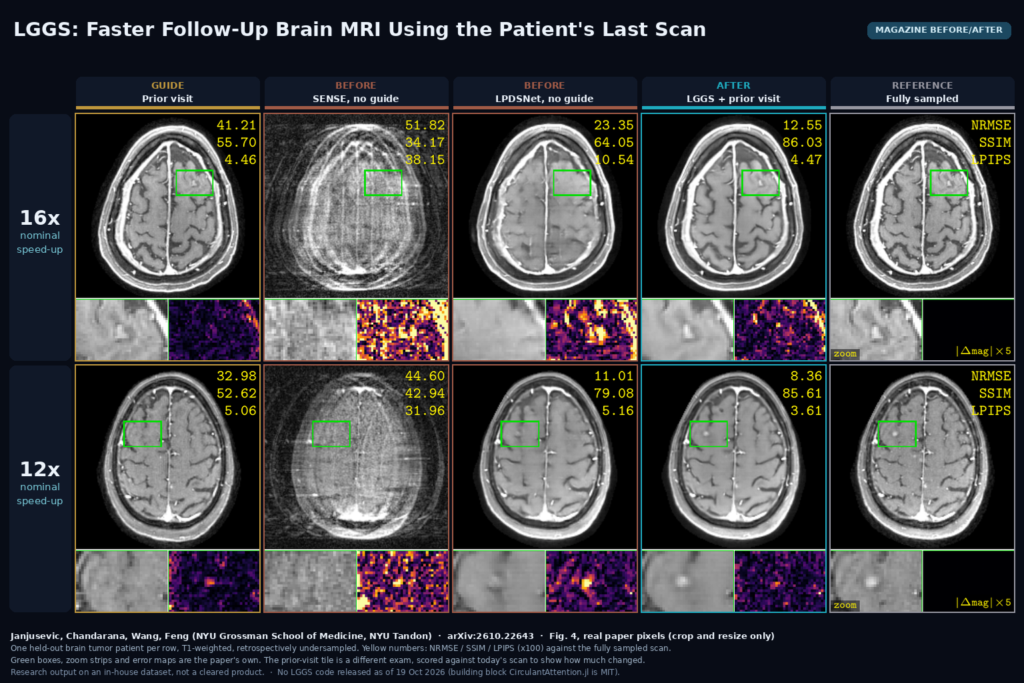
Magazine before/after from Fig. 4: two held-out brain tumor patients at 16x and 12x acceleration, each shown as the prior visit used as guide, SENSE, unguided LPDSNet, LGGS and the fully sampled scan, with zoom and error strips. Janjusevic, Chandarana, Wang, Feng, arXiv:2610.22643

LGGS rebuilds heavily undersampled follow-up brain MRI using the patient’s prior exam as a guide, without copying it into the new image. arXiv:2610.22643.

UniField++: Making Low-Field Brain MRI Look Like 3T and 7T
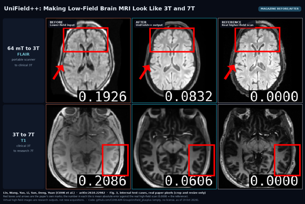
Magazine before/after from Fig. 3: two brain MRI test cases, a 64 mT FLAIR and a 3T T1, each shown as the lower-field input, the UniField++ virtual higher-field output and the real 3T or 7T scan. Lin, Wang, Yao, Li, Sun, Deng, Yuan, arXiv:2610.22982

UniField++ turns 64 mT brain MRI into virtual 3T and 3T into virtual 7T, with a vision-language planner that flags regions likely to go wrong. arXiv:2610.22982.

NoduleFlow: Automatic Lung Nodule Masks Across Follow-Up CT
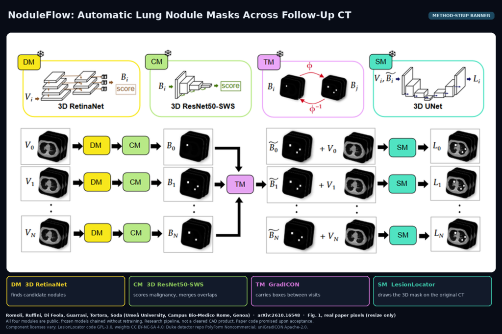
Method-strip banner from Fig. 1: chest CT volumes at several time points pass through a 3D RetinaNet detector, a ResNet50-SWS malignancy classifier, GradICON box transfer between visits and LesionLocator segmentation. Romoli et al., arXiv:2610.16548

NoduleFlow chains four public, frozen models to detect, score and segment lung nodules across a patient’s follow-up CTs with no clicks. arXiv:2610.16548.

MoFD: Synthetic Contrast-Enhanced Brain MRI From T1, T2 and FLAIR
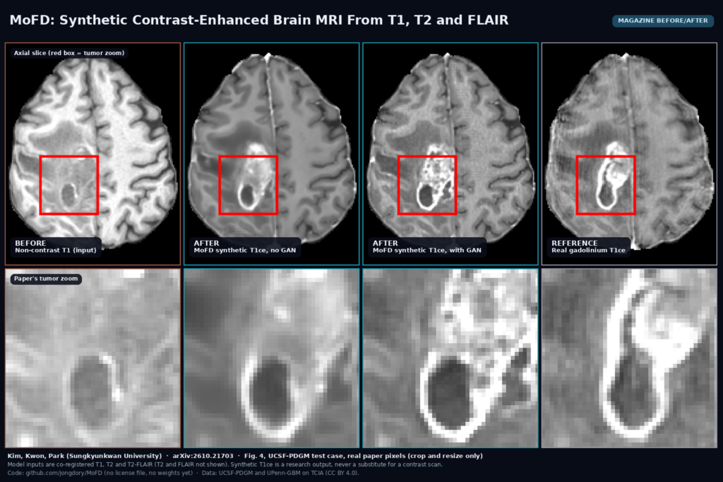
Magazine before/after from Fig. 4: a glioma case shown as the non-contrast T1 input, the MoFD synthetic T1ce without and with GAN loss, and the real gadolinium T1ce, with zoomed tumour views below. Kim, Kwon, Park, arXiv:2610.21703

MoFD predicts a contrast-enhanced T1 brain MRI from non-contrast T1, T2 and FLAIR with a 3D U-Net that mixes sequence features and their differences. arXiv:2610.21703.

DeBoneDiT: Soft-Tissue Chest X-Rays Without Dual-Energy Hardware
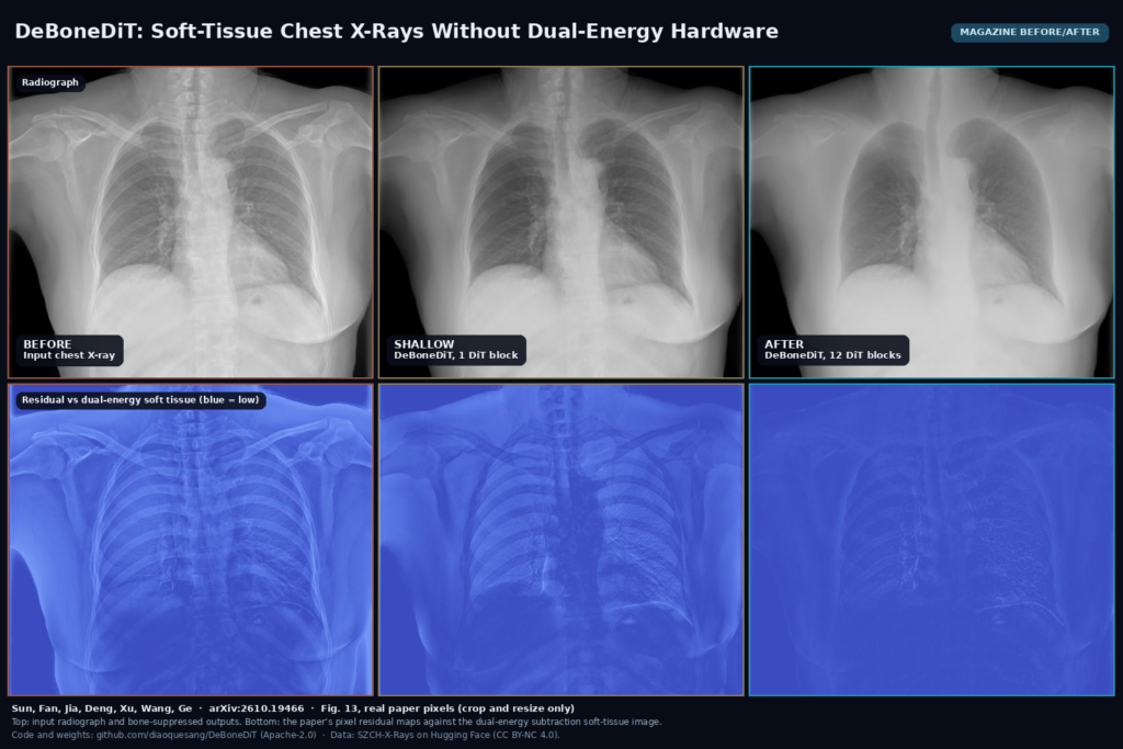
Magazine before/after from Fig. 13: an input chest X-ray beside DeBoneDiT bone-suppressed outputs with 1 and 12 DiT blocks, and below each the residual map against the dual-energy soft-tissue image. Sun, Fan et al., arXiv:2610.19466

DeBoneDiT turns one standard PA chest X-ray into a dual-energy style soft-tissue image in about 3.4 seconds. Apache-2.0 code and weights, arXiv:2610.19466.

AD-GS: Sharper Dental CBCT From Fewer Projections
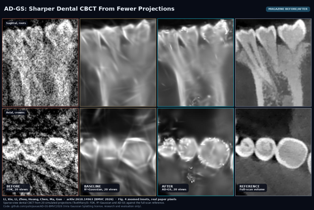
Magazine before/after from Fig. 4: zoomed sagittal roots and axial crowns of a dental CBCT reconstructed from 20 projections with FDK, R2-Gaussian and AD-GS, beside the full-scan reference. Li, Xie et al., arXiv:2610.14963

AD-GS fits each dental CBCT scan with 3D Gaussians, steering them toward enamel and bone so 20 to 30 projections keep root and crown edges. arXiv:2610.14963, BMVC 2026.

PSMAReg: Follow-Up PET/CT Registration That Keeps Tumor Burden
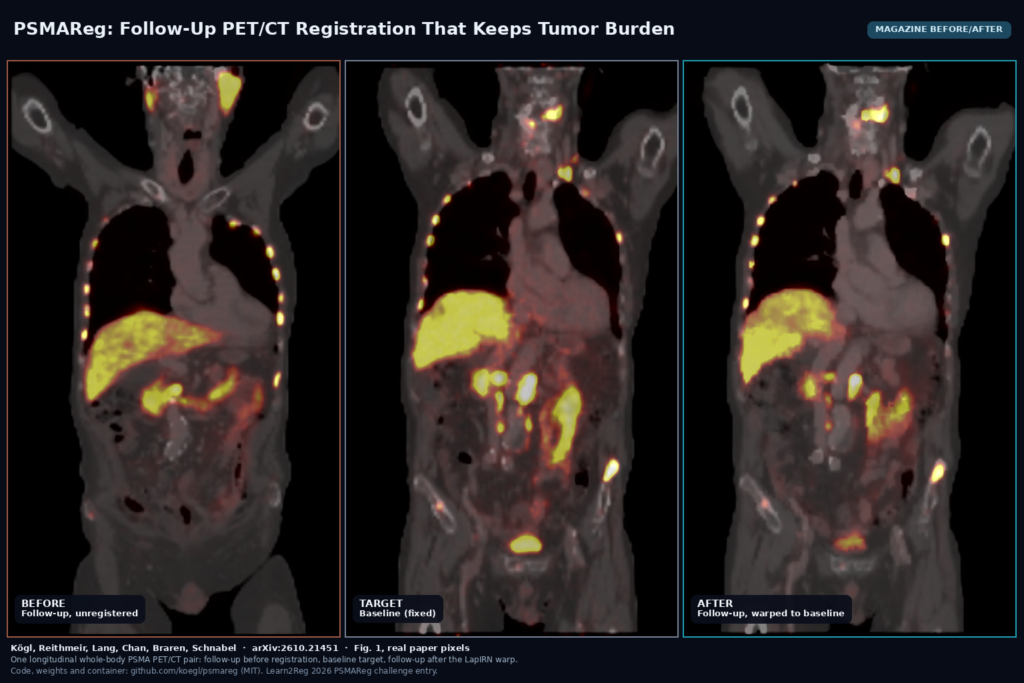
Magazine before/after from Fig. 1: coronal whole-body PSMA PET/CT of one pair, follow-up unregistered, baseline fixed, and follow-up warped by the LapIRN pipeline. Koegl et al., arXiv:2610.21451

A LapIRN pipeline from Helmholtz Munich and TUM aligns follow-up PSMA PET/CT to baseline while holding MTV and TLG steady. arXiv:2610.21451, MIT code and weights.

MMA-LTS: Trust Tumor Overlay Confidence When MRI Sequences Are Missing
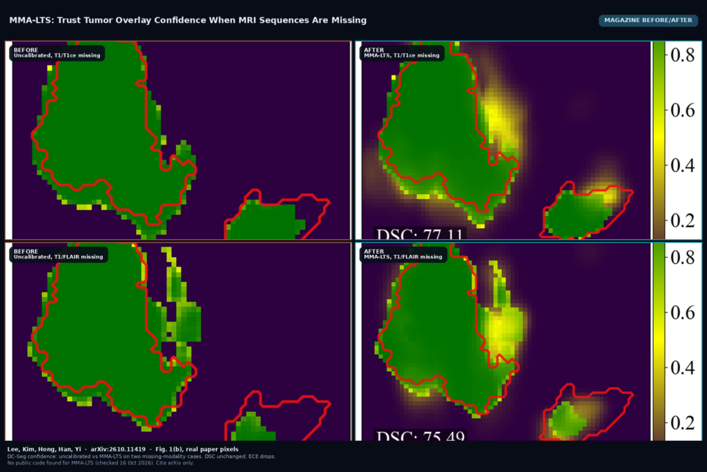
Magazine before/after from Fig. 1(b): DC-Seg uncalibrated confidence versus MMA-LTS for T1/T1ce missing (top) and T1/FLAIR missing (bottom). Lee et al., arXiv:2610.11419

MMA-LTS (arXiv:2610.11419) post-hoc calibrates voxel-wise tumor overlay confidence when MRI sequences are missing, cutting ECE while keeping Dice. KAIST. No public code yet.

← Previous
Next →