
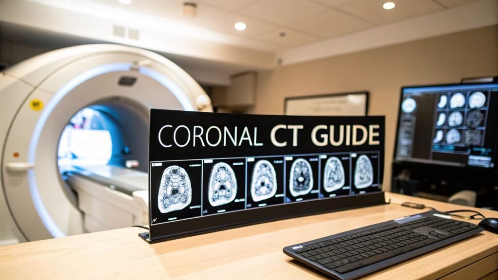
Coronal CT images are the face-on plane: anterior to posterior, like looking in a mirror at the inside. They are almost never a second scan. They are an MPR — a reslice of thin axials. This page is that one plane. It is not “what a cross-section is.”
If you meant what a cross-section is (CT vs MRI vs PET vs ultrasound) → cross-section analysis. If you meant thin slices so the MPR is not a stair-step → CT slice thickness. If you meant WW/WL → window width vs window level.
Axial and sagittal are the other two standard planes. This article does not invent sister posts for them. Cross-reference them at the workstation; do not look for a second PYCAD URL.
Face-on, not a new acquisition
Loaf of bread: top-to-bottom cuts are axial, side cuts are sagittal, front-crust-to-back are coronal. The scanner still acquires axials. Software stacks those slices and cuts the stack from front to back. No extra radiation. Scroll anterior → posterior the way you scroll feet → head on the axials.
That reslice is multiplanar reformation (MPR). Quality is almost entirely the thickness of the source axials. 0.625 mm cubes make a coronal that looks like the axial. 5 mm axials make a stair-step — organs with jagged edges that can hide a small finding. The mm table is on the slice-thickness page; the plane that suffers is this one.
Why this plane
- Side-to-side and cranio-caudal in one view. Liver next to spleen next to kidneys, top to bottom, without paging through fifty axials to rebuild the relationship.
- Symmetry. Sinuses, orbits, lungs — left vs right is the first look.
- Alignment. Scoliosis is a lateral curve. The coronal is the plane you measure it in (Cobb).
Where it earns the click
| Specialty | What you look at | Why coronal |
|---|---|---|
| ENT | Sinuses, ostiomeatal complex, polyps | Drainage paths stack vertically; axial floor-plans miss the stairwell |
| Trauma | Orbital floor, facial bones | Vertical displacement is the question |
| Oncology | Abdomen / pelvis tumor | Cranio-caudal extent — kidney into adrenal, liver into diaphragm |
| Orthopedics | Scoliosis, spinal balance | Cobb angle; head over pelvis |
| Urology | CT urogram | Kidneys, ureters, bladder on one map |
Sinuses. Fluid levels, an ostiomeatal blockage, an orbital-floor drop — the face-on cut is the one ENT and trauma actually use.
Abdomen. Staging a mass: how far it runs head-to-tail, and which organ is the neighbour. An axial tells you the slice; the coronal tells you the height.
Spine. Cobb is a coronal measurement. Fusion planning uses that curve as the blueprint.
How to read one without inventing a course
Scroll front to back. Follow one structure (a ureter, the IVC, the length of the liver) instead of hopping. Switch window when the question switches: bone window for a facial fracture, soft-tissue for organs, lung window for parenchyma. Those presets are WW vs WL, named here so you do not evaluate a rib in a soft-tissue window and call it intact.
Stair-step means the source axials were thick. Metal (clips, fillings) throws streaks on the coronal the same way it does on the axial — that catalog is artifacts in computed tomography.
A nodule on one coronal slice that is a vessel on the axial is why you never sign out a single plane. Confirm on axial and sagittal. That is a reading habit, not a reason to publish two more explainers from this body.
PYCAD builds viewers that have to offer MPR as a plane change, not a new study. Case studies.
Keep Reading
Related Articles
Explore the full DICOM HubAll services, tools, and guides on this topic — in one place.
Visit Hub →