
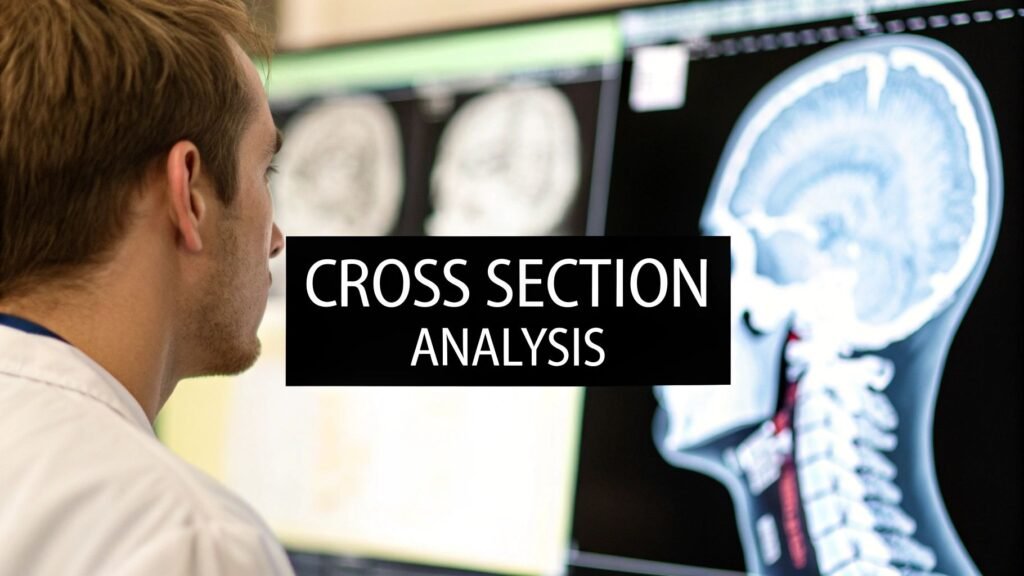
A medical cross-section is a thin digital slice through the body — the same idea as cutting a cake to see the layers, without an incision. CT, MRI, PET, and ultrasound each make those slices a different way. Stack the slices and you get a volume. This page is that survey. It is not one named plane, and it is not muscle architecture.
If you meant one named plane (coronal via MPR) → coronal CT images. If you meant skeletal-muscle architecture / PCSA → cross section of skeletal muscle. If you meant thin slices for reformats → CT slice thickness. If you meant window width / window level → window width vs window level. If you meant a 3D volume render → volume rendering of CT. If you meant imaging software to buy → medical imaging software.
What the slice is
A plain radiograph collapses bone and organ onto one film. A cross-section does not. Each image is one thin layer: organ, vessel, bone, or air, without the overlay. The scanner (or the probe) captures many of those layers. Reconstruction software stacks them in order and you can scroll, reformat, or build a 3D model.
The four named anatomical planes are axial (top / bottom), sagittal (left / right), coronal (front / back), and oblique (any other angle). This page is the slice itself — what it is, which modality made it, and how a stack becomes a volume. The coronal-via-MPR job lives on coronal CT images.
Why the slice matters
Overlap is the problem a projection cannot solve. A tumor behind a rib, a bleed next to bone, a fracture through a joint — those are layer questions. Cross-section analysis is how you answer them:
- Find it. Location, size, and shape of a lesion, a bleed, or a break.
- Plan it. A surgeon maps the approach. A radiation oncologist draws a target and spares the neighbor.
- Watch it. Same anatomy, later date: grew, shrank, or stayed.
How the modalities make the slice
Each modality is a different physical measurement. You do not use a CT the way you use an MRI. The table is the survey; the rest of this page does not become a protocol manual.
| Modality | Imaging principle | Best for | Key advantage |
|---|---|---|---|
| CT | X-rays from many angles, reconstructed into slices | Bone, trauma, lung, vessels with contrast | Speed. The emergency default |
| MRI | Magnetic field + radio waves; hydrogen signal | Brain, cord, joints, ligaments, soft tissue | Contrast without ionizing radiation |
| PET | Radiotracer; metabolic activity | Function — especially tumor glucose uptake | Biology, not just anatomy |
| Ultrasound | High-frequency sound, live echoes | Motion, fluid, fetus, needle guidance | Real-time. No radiation. Portable |
CT is a rotating X-ray and a ring of detectors. Density differences become Hounsfield units. It is the first look in trauma, the map for a complex fracture, and the workhorse for lung and liver lesions. How bright bone vs soft tissue looks on the screen is a window-width / window-level choice, not a different scan.
MRI does not use ionizing radiation. Water- and fat-rich tissues return different signals. That is why it owns brain, cord, and joint work that CT only sketches. It is slower. It is not the stroke-clock first image.
PET injects a tracer and lights up metabolism. Anatomy comes from the CT (or MRI) it is fused to. You read function on the PET and place it on the slice.
Ultrasound is live. No stack in the CT sense — a moving 2D plane you steer with the probe. Biopsy guidance, a beating heart, a fetus. Different job from a reconstructed volume.
The patient path
Prep is specific to the exam. Some abdominal CTs want an empty gut. Many CTs and some MRIs use contrast — drunk or injected — so a vessel or a lesion pops. Contrast is not cosmetic; a PE study without it is the wrong study.
In the room you lie on a motorized table. For CT that table moves through a gantry (the doughnut). The technologist is next door, on the intercom. Stillness is the image. A breath-hold for chest or abdomen is the usual request. The machine clicks; it is collecting hundreds to thousands of slices, not one photograph.
Your part ends when you sit up. Reconstruction starts then: raw projections become a stack of 2D slices, then a volume if someone asks for one. A radiologist reads the stack (and the reformats) and writes the report your clinician uses.
From 2D slices to a 3D model
Think of a loaf. Each slice is one cut. Software puts the cuts back in order and interpolates the gaps so you can rotate the loaf, not only flip cards. That volume is how a surgeon walks around an ankle fracture or a tumor next to a vessel before anyone opens.
A pretty 3D view is not a substitute for the source series. Measure on the thin slices. How thin those slices are — and whether a coronal reformat looks clean or stair-steps — is CT slice thickness. Painting every voxel instead of scrolling is volume rendering of CT.
Where it is used
Oncology. A chest X-ray can hint. A PET/CT stack stages: tumor size and invasion on the CT, metabolic activity on the PET, nodes, and distant mets. The same stack later is the response read. That is cross-section analysis as a time series, not one pretty picture.
Stroke. Minutes. A non-contrast head CT — axial slices of the brain — answers bleed vs clot before anyone gives a lytic. The treatments are opposites. The slice is the fork in the road.
Orthopedics. A smashed ankle is a 3D puzzle. The CT stack, reformatted and optionally rendered, shows fragment count, joint surface, and where metal will sit. The X-ray was the screen; the cross-sections are the roadmap.
What AI actually does here
The grind on a stack is measurement: tumor cc across slices, a lung nodule you might skip at 1 a.m., plaque volume, brain-volume change. Models trained on large anonymized CT/MRI sets can flag or outline those. They do not replace the read. They do not turn this page into a segmentation how-to — that job is medical image segmentation.
FAQ
Cross-sectional vs longitudinal. Cross-sectional is the snapshot: this tumor, this Tuesday, this size. Longitudinal is the movie: the same patient, three months later, then six. You need the first slice before you can talk about change.
Risk / ALARA. CT and PET use ionizing radiation. One exam is a small dose; the rule is ALARA — as low as reasonably achievable — and only when the clinical question needs it. Contrast can trigger allergy or stress kidneys; that is a separate consent. MRI and ultrasound do not use ionizing radiation. MRI’s constraint is metal (pacemaker, some clips). Ultrasound is sound; it is the default in pregnancy.
How 2D becomes 3D. Order the slices. Interpolate the gaps. Every pixel already has a density and a place. The model you spin is that grid, not a new acquisition. Software that does the spinning is medical imaging software; the render itself is volume rendering of CT.
PYCAD builds the viewers and the models that have to treat a slice stack as anatomy, not a gallery. Case studies.