
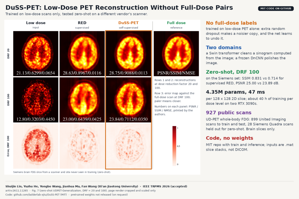
To train a supervised low-dose PET denoiser you need the same patient scanned at low dose and at full dose, and the paper points out that injecting patients twice for a training set raises ethical and safety concerns. Research groups get around it with list-mode data, where a low-dose scan can be simulated by throwing away counts from a full-dose acquisition, but that needs list-mode access and a resampling pipeline at every site. DuSS-PET, from Xi’an Jiaotong University, trains on low-dose images alone. It is described in arXiv:2611.11265, accepted in IEEE Transactions on Radiation and Plasma Medical Sciences, and the code is on GitHub under an MIT licence.
What DuSS-PET does
DuSS-PET takes a reconstructed low-dose PET slice and returns a cleaner slice that should look closer to the full-dose scan. It never sees a full-dose image during training. It builds on Noisier2Noise, a self-supervised denoising idea from Moran and colleagues (CVPR 2020): take a noisy image, corrupt it a second time, and train a network to map the noisier copy back to the noisy one. With the right corruption model, the trained network can be pushed one step further, from noisy towards clean.
The authors add a corruption that resembles low-dose PET and compute the main loss in a projection space as well as on the image.
How it works
The authors started by looking at what lowering the dose does to a PET image. With list-mode data they simulated dose reduction factors (DRF) from 2 to 100 and saw two effects: intensity drops and noise rises overall, and continuous high-uptake regions break up into scattered, hollow-looking patches. Randomly switching pixels off reproduces both, so their corruption, called dose-aware Bernoulli corruption, keeps each pixel with a probability alpha and zeroes the rest. They calibrated alpha against real low-dose data by matching voxel-count histograms in the brain, and ended up with 0.8 for DRF 20, 0.6 for DRF 50 and 0.4 for DRF 100. The paper is clear that this is an empirical proxy and not a physical model of PET acquisition.
The model works in two domains. Each training slice is the low-dose image plus a further-masked copy of it. Both are forward projected with a 2D parallel-beam Radon transform into what the paper calls pseudo-sinograms. A Swin transformer restoration network (SwinIR style, 3.79 million parameters) learns to map the noisier pseudo-sinogram back to the low-dose one. Filtered back-projection turns the result into an image, and a small DnCNN denoiser (0.56 million parameters) refines it.
That DnCNN is pre-trained separately and then frozen. It also learns without full-dose data: its training pairs are two low-dose versions of the same acquisition at different DRFs, for example DRF 100 mapped to DRF 50, both cut from the same list-mode file. A third component, which the paper calls CC-DuAS, adds consistency losses. The refined image is split with complementary random masks, each half is projected, restored by the shared transformer and back-projected, and the two results have to agree with each other and with the refined image.
At inference there is no extra corruption. The paper averages two paths: the denoiser applied to the back-projection of the restored pseudo-sinogram, and the denoiser applied to the back-projection of the untouched one. The total model has 4.35 million parameters and runs in about 47 ms per 2D slice, not counting the projection steps.
What they tested it on
All data come from the public UD-PET collection of whole-body 18F-FDG scans acquired in list-mode. The DnCNN was pre-trained on 664 United Imaging uEXPLORER scans (low-dose images only). The main model was trained and tested on 235 other uEXPLORER subjects, split 188/24/23. A third set, 28 scans from a Siemens Biograph Vision Quadra at a different site (Bern), was held out entirely for zero-shot testing. From each subject the authors took 25 central axial slices of the brain at 128 x 128 pixels. So the reported results are 2D brain slices, even though the source scans are whole-body.
The comparison covers OSEM, two self-supervised methods (N2V and Nr2N) and three supervised ones trained on paired full-dose data: DeepPET, SpachTrans and RED, a 2025 diffusion model that corrects the sinogram. On the uEXPLORER test set, supervised RED keeps a small lead in PSNR (26.39 dB against 25.92 dB at DRF 50), while DuSS-PET has the highest SSIM at DRF 20 and 50 (0.8594 against 0.8389 for RED at DRF 50).
On the Siemens set, trained on United Imaging data and applied without any fine-tuning, DuSS-PET at DRF 100 reached 25.00 dB PSNR and 0.8310 SSIM, against 23.89 dB and 0.7139 for RED and 19.67 dB and 0.6697 for DeepPET. The authors read the drop of the supervised models on unseen data as a weakness of depending on paired data. At DRF 20 the methods were close (28.46 dB for DuSS-PET, 28.49 dB for RED).
The featured image is Fig. 7 of the paper, one Siemens brain slice from that zero-shot test. At DRF 100 the low-dose input is mostly speckle. On this slice the authors’ panel labels give 12.80 dB PSNR for the input, 23.00 dB for RED and 23.84 dB for DuSS-PET, and the error maps below show how far each result sits from the full-dose scan.
In the ablation at DRF 50, the transformer alone lifts PSNR from 19.65 dB (low-dose input) to 22.02 dB. Adding the frozen DnCNN gives the largest single jump, to 25.59 dB, and the consistency losses add the rest, to 25.92 dB.
Why a viewer or clinic team should care
A site that already runs reduced-dose protocols has low-dose images but no matched full-dose scans, and DuSS-PET needs only the former. If you have list-mode data you can also cut several dose levels from one acquisition to pre-train the image prior, still without a full-dose target. For a vendor-neutral platform, the zero-shot Siemens test is the result to read first: at DRF 100, supervised DeepPET went from 22.69 dB on the United Imaging test set to 19.67 dB on Siemens, while DuSS-PET went from 23.08 dB to 25.00 dB.
Inference is fast enough for a post-reconstruction step: about 47 ms per 128 x 128 slice on the authors’ GPUs, plus the projection and back-projection steps. In a viewer you would show it as a derived series next to the original, with the original always available for reading.
Training costs more. The main stage takes about 40 hours per dose level on two RTX 3090 GPUs, and each dose level gets its own transformer. Only the DnCNN is shared across doses.
What it does not tell you yet
- Everything runs on 2D slices at 128 x 128. The authors list 3D extension as future work, likely with slab or patch designs.
- The pseudo-sinograms are computed from reconstructed images with a 2D parallel-beam Radon transform. They do not include attenuation, scatter, randoms, detector response or 3D geometry, and the paper calls this domain a computational consistency space, not a model of the scanner. The method has not been run on real measured sinograms.
- Evaluation is on brain slices only, with FDG only. Body regions, lesions, other tracers and SUV accuracy in lesions are not reported. Metrics are PSNR, SSIM and NMSE; there is no reader study.
- Alpha is fixed per dose level. At DRF 100 the method is sensitive to it: PSNR drops by about 4.5 dB if alpha goes from 0.4 to 0.8. The authors also note that the Bernoulli model matches real low-dose data less well at DRF 100. Real clinical noise varies by patient and protocol, and an adaptive estimate of alpha is listed as future work.
- Low-dose data in the study are retrospectively simulated from list-mode, not separately injected low-dose scans.
How to try it
The repository at github.com/ladderlab-xjtu/DuSS-PET contains the model code (SwinIR-style transformer, DnCNN, a differentiable Radon transform in PyTorch), a training script, an inference script and an evaluation script, with YAML configs for 128 x 128 slices and 180 projection angles. Dependencies are PyTorch 2, timm, scikit-image and SciPy. Inputs are MATLAB .mat files, one per subject, holding an img array of shape (slices, 2 x height, width) where the first half is the low-dose slice and the second half the paired reference used for evaluation. You will need to write your own conversion from DICOM, and for pure inference you would fill the reference half with a placeholder.
Before planning around it, note that no checkpoints are published; the README says weights are expected as external release assets or by request from the authors. There is no script for the DnCNN pre-training stage, only a path to load its weights. The shipped inference script runs only the restored-sinogram path, not the two-path average described in the paper. And no patient data are included, though the UD-PET data are public. For related PET work on the blog, see TB-PET-DDS, a diffusion prior for low-count total-body PET and LesionStudio for tuning PET and SPECT reconstruction.
Source and license
Paper: Shuijin Lin, Yuzhu He, Yongbo Wang, Jianhua Ma and Fan Wang, “DuSS-PET: Self-Supervised Low-Dose PET Reconstruction Using Pseudo-Dual-Domain Consistency Without Paired Data”, arXiv:2611.11265 (cs.CV, 8 November 2026), author accepted manuscript, IEEE Transactions on Radiation and Plasma Medical Sciences, 2026, DOI 10.1109/TRPMS.2026.3696317. Authors are with the School of Life Science and Technology and the School of Mathematics and Statistics at Xi’an Jiaotong University. Code: github.com/ladderlab-xjtu/DuSS-PET, MIT licence. Weights: not released (on request per the README). Data: UD-PET, a public collection, not redistributed in the repo. Featured image: Fig. 7 of the paper, four of the nine columns at DRF 20 and 100, cropped and scaled.