
Portable low-field MRI scanners, the kind that run at 64 mT and can be wheeled to a bedside, are cheap to deploy, but their images are noisy and soft compared with a hospital 3T system. At the other end, 7T research scanners show cortex and white matter in detail that most hospitals will never have. Yiyang Lin, Chenhui Wang, Tianliang Yao, Shuaibo Li, Zijun Sun, Yiming Deng and Yixuan Yuan, from The Chinese University of Hong Kong with co-authors at Fudan University, University Hospital Zurich and Beijing Tiantan Hospital, posted arXiv:2610.22982 on 16 October 2026. Their model, UniField++, takes a brain MRI from the weaker scanner and generates a virtual image of what the stronger one would have shown, for both 64 mT to 3T and 3T to 7T, in one model. It extends the group’s earlier UniField paper from MICCAI 2026.
Why a viewer team should care
Because low-field scanners are affordable and easy to deploy, their images end up in the same PACS and viewer as everything else, and the brain segmentation and volumetry tools in that pipeline were built for 3T. In the paper’s downstream test, SynthSeg run on raw 64 mT images assigned large parts of the brain to the background label. A virtual 3T series, clearly labelled, is a way to get those tools to run. The 3T to 7T direction is more of a research tool, for sharper cortical and hippocampal detail where no 7T scanner exists.
What goes in and what comes out
Input is one brain MRI volume (T1, T2 or FLAIR) from the source scanner plus the target field strength. The authors normalise intensities to the 0.5th to 99.5th percentile with MONAI, resample to 1 mm along the slice axis and resize every volume to 256 x 256 x 160. Pairs that were not already aligned were registered with ANTs. Output is a virtual high-field volume on that same grid, in the same contrast as the input. The model does not change the sequence type; a FLAIR stays a FLAIR.
How it works
UniField++ splits the job between two models. The first is a planner, a Qwen3.5-9B vision-language model fine-tuned with LoRA. It looks at ten slices sampled evenly through the low-field volume and writes out three triplets: a brain region, the problem that region is likely to have after enhancement and a fix. All three come from closed lists. The regions are the frontal, parietal, temporal, occipital and insular lobes, the ventricles and the white matter. The problems include residual blur, over-smoothing, tissue-contrast mismatch, ringing and anatomical inconsistency, and the fixes include detail-preserving denoising, tissue-contrast calibration and conservative local refinement. The planner learned this from radiologists, who compared outputs of earlier enhancement models (UniField and the video super-resolution model FlashVSR) with the matching real high-field scans and annotated where each output went wrong and how it should be fixed.
The second model is the enhancer, a flow-matching network initialised from FlashVSR, with the Wan 2.1 video VAE for the latent space. The authors treat an MRI volume like a short video, since both are stacks of 2D frames. To turn the planner’s region names into pixels, they register a brain atlas to the low-field scan with ANTs and pull out a mask for each named region. A text encoder turns the plans into embeddings, and a small 3D convolutional encoder turns the masks into tokens. Inside the 30-block transformer, a middle group of blocks runs once on low-risk patches and several times on the high-risk patches the planner flagged, with shared weights. Training also weights the loss more heavily inside the flagged regions. A fine-tuned decoder maps the result back to image space.
What the numbers say
Training used five public paired datasets: three for 64 mT to 3T (ULF-EnC, Leiden and KCL, 83 subjects in total) and two for 3T to 7T (UNC and BNU, 30 subjects). The test splits are small, 2 to 10 subjects per dataset. Against six comparison methods across the five task and contrast combinations, UniField++ improves PSNR by 2.74 dB and SSIM by 14.05 percentage points on average (Tables II and III). The downstream result is the one we would look at first. With SynthSeg run on the real high-field scan as reference, the average Macro Dice across T1, T2 and FLAIR rose from 0.566 on the raw 64 mT images to 0.798 on the virtual 3T images (Table VIII). For 3T to 7T the gain is small (T1 Macro Dice 0.829 to 0.841), because the 3T input already segments well.
The external test is a private cohort of 30 patients from Beijing Tiantan Hospital scanned at 50 mT and 3T, a field strength that was never in training. There, UniField++ ranks first or second on most metrics, but the margins are thin. FlashVSR scores a slightly higher T1 PSNR, and the earlier UniField is close on most columns (Table IV).
The figure
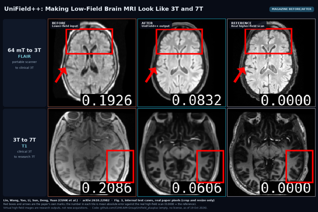
The featured image uses two rows of Fig. 3, both internal test cases. The top row is a 64 mT FLAIR. The input is grainy, the ventricles have soft edges and the frontal cortex inside the red box is barely separable from white matter. The UniField++ output is clean, with crisp ventricles and visible frontal sulci, and its mean absolute error against the real 3T scan drops from 0.193 to 0.083. Next to the real 3T FLAIR it looks smoother and brighter in the white matter, without the fine texture of the real scan. The bottom row is a 3T T1 turned into a virtual 7T T1. The output picks up the 7T contrast, with darker cortex and sharper sulci in the boxed region on the right side of the brain, and its error drops from 0.209 to 0.061. The real 7T still shows small vessels and cortical detail the output does not have.
Limits
The method is brain only, and every training set is small. At least part of the public data comes from healthy volunteers (the Leiden set is described as paired scans of healthy subjects), and the paper does not report any lesion-specific test or reader study. The authors themselves warn that enhancement models can suppress a small lesion or invent a lesion-like structure, and the planner’s seven coarse regions do not cover a tumour, an infarct or a bleed as a region of its own. The atlas masks depend on a nonlinear registration to the low-field scan, which can fail on a brain with large pathology or a strong artifact. The loop count also has a ceiling. In the ablations, quality improves from zero to a small number of extra loops and then gets worse, and more sampling steps trade PSNR for perceptual quality. The compute is heavy. A 9B vision-language model plus a video diffusion transformer is a GPU-server job, the authors trained on four RTX A6000 cards and they do not report inference time.
Code, data and license
The abstract points to CUHK-AIM-Group/UniField_plusplus. As of 19 October 2026 that repository is empty, with no files, no license and no weights, so for now there is nothing to run. The building blocks are open: FlashVSR and Wan 2.1 are both Apache-2.0 on GitHub. The Leiden 64 mT and 3T dataset is on Zenodo under an MIT license. The other datasets (ULF-EnC, KCL, UNC and BNU) come with their own terms, and the Tiantan external cohort is private. Without released planner annotations and weights, reproducing the planner means collecting your own radiologist annotations.
How we would wire it into a viewer
We would run it as a server-side job that writes a new series and never touches the original. When a study comes in from a low-field scanner (the Manufacturer and MagneticFieldStrength tags tell you), convert the series to NIfTI, run the model on a GPU worker, resample the 256 x 256 x 160 output back to the source grid, and write a derived series with ImageType DERIVED\SECONDARY, a series description such as “Virtual 3T (AI, UniField++, not acquired)” and a reference to the source series. In the viewer, hang it next to the original with linked scrolling, keep the acquired scan as the primary image for any read, and put a banner on the virtual viewport. We would also show the planner’s three region, issue and fix lines as a note on the series, so the reader sees where the model expected to go wrong. If the main goal is volumetry, run your segmentation on both series and compare against a real 3T scan of the same patient on your own cases before you trust the numbers. For background, see what a DICOM viewer does, our guides on DICOM to NIfTI and NIfTI back to DICOM, and our earlier post on artifact grading for portable neonatal brain MRI.
Sources
- Lin, Y., Wang, C., Yao, T., Li, S., Sun, Z., Deng, Y., Yuan, Y. UniField++: A Unified Agentic Framework with Loop Reasoning for MRI Field-Strength Enhancement. arXiv:2610.22982, 2026. https://arxiv.org/abs/2610.22982. PDF: https://arxiv.org/pdf/2610.22982.
- Code repository (empty, no license at the time of writing): https://github.com/CUHK-AIM-Group/UniField_plusplus.
- Building blocks (Apache-2.0): FlashVSR, https://github.com/OpenImagingLab/FlashVSR. Wan 2.1, https://github.com/Wan-Video/Wan2.1.
- Leiden paired 64 mT and 3T brain MRI dataset (MIT): https://doi.org/10.5281/zenodo.15862148.
- Featured image: Fig. 3 of arXiv:2610.22982, real paper pixels, cropped and resized only.