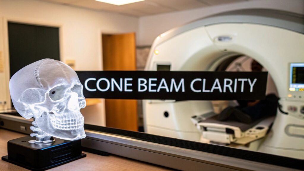
Think about the difference between looking at a flat, two-dimensional map and holding a detailed 3D globe in your hands. That’s the kind of jump we’ve seen in medical imaging, thanks to the power of cone beam images. This technology gives doctors a brilliantly clear, complete view of a patient's anatomy, fundamentally changing how they diagnose and plan treatments.
Seeing Beyond the Surface with Cone Beam Imaging
For years, traditional X-rays were the best tool we had. They gave us valuable, but ultimately flat, pictures of our internal structures. It was like having a single photograph of an intricate building—helpful, certainly, but it couldn't show you the depth, the different dimensions, or how all the internal parts related to one another. Clinicians often had to make educated guesses about what was happening behind or inside the structures they could see.
Then, Cone Beam Computed Tomography (CBCT) came along and changed the game entirely. Instead of a single, flat snapshot, this technology captures an entire volume of data in one quick rotation. A cone-shaped X-ray beam spins around the patient, gathering hundreds of individual images that are then digitally stitched together into a stunningly detailed three-dimensional model.
A New Dimension of Insight
The move from 2D to 3D opens up a whole new world of detail. Suddenly, doctors can virtually fly through a patient's anatomy, looking at teeth, bone, soft tissue, and nerve pathways from any angle imaginable. This kind of insight is absolutely critical in complex fields like:
- Dentistry and Orthodontics: It allows for pinpoint accuracy when placing dental implants or planning intricate orthodontic movements.
- Maxillofacial Surgery: Surgeons get a complete picture of complex facial bone structures before ever making an incision.
- ENT and Sleep Medicine: It’s invaluable for evaluating airways and getting to the root of sinus problems.
The real magic of cone beam images is that they virtually eliminate guesswork. By providing the full anatomical story, CBCT gives healthcare professionals the confidence to create safer, more effective, and deeply personalized treatment plans.
This profound shift is fueling incredible momentum in the industry. The global cone beam CT market, valued at around USD 702.13 million, is on track to soar past USD 1.77 billion by 2034. You can discover more about these market trends and projections to see just how quickly this technology is being adopted.
Here at PYCAD, we take this powerful imaging and make it even more accessible. We at PYCAD, build custom web DICOM viewers and integrate them into medical imaging web platforms, turning these complex 3D scans into interactive tools. You can see some examples of how we bring this data to life in our portfolio.
How Cone Beam Technology Creates 3D Images
To really get a feel for the power of cone beam images, it helps to pull back the curtain and see how these incredible 3D models are born. The whole process is a symphony of efficiency and precision, a world away from older, clunkier imaging methods.
Picture a conventional CT scanner. It works by painstakingly taking one thin, flat "slice" of an image at a time, almost like assembling a photo album page by page to build a full picture. It gets the job done, but it's a slow, methodical approach.
Cone beam technology, on the other hand, is much more direct. Imagine using a powerful, cone-shaped flashlight. In one quick sweep, it illuminates the entire area of interest. That’s exactly how a cone beam CT (CBCT) works—it casts a cone-shaped X-ray beam to capture a whole volume of anatomical data in a single, swift rotation around you. This single difference is what makes it so brilliant.
For the person in the chair, the scan is over in a flash, often in less than a minute. For the clinician, it means getting a massive amount of high-fidelity data almost instantly, all ready to be turned into a digital masterpiece.
The Journey from Scan to Screen
The real magic begins after the scan is complete. Specialized software gets to work, taking the hundreds of individual snapshots captured during that single rotation. Using complex algorithms, it pieces them together, reconstructing them into a single, seamless 3D model. This isn't just a flat picture; it's a complete volumetric dataset.
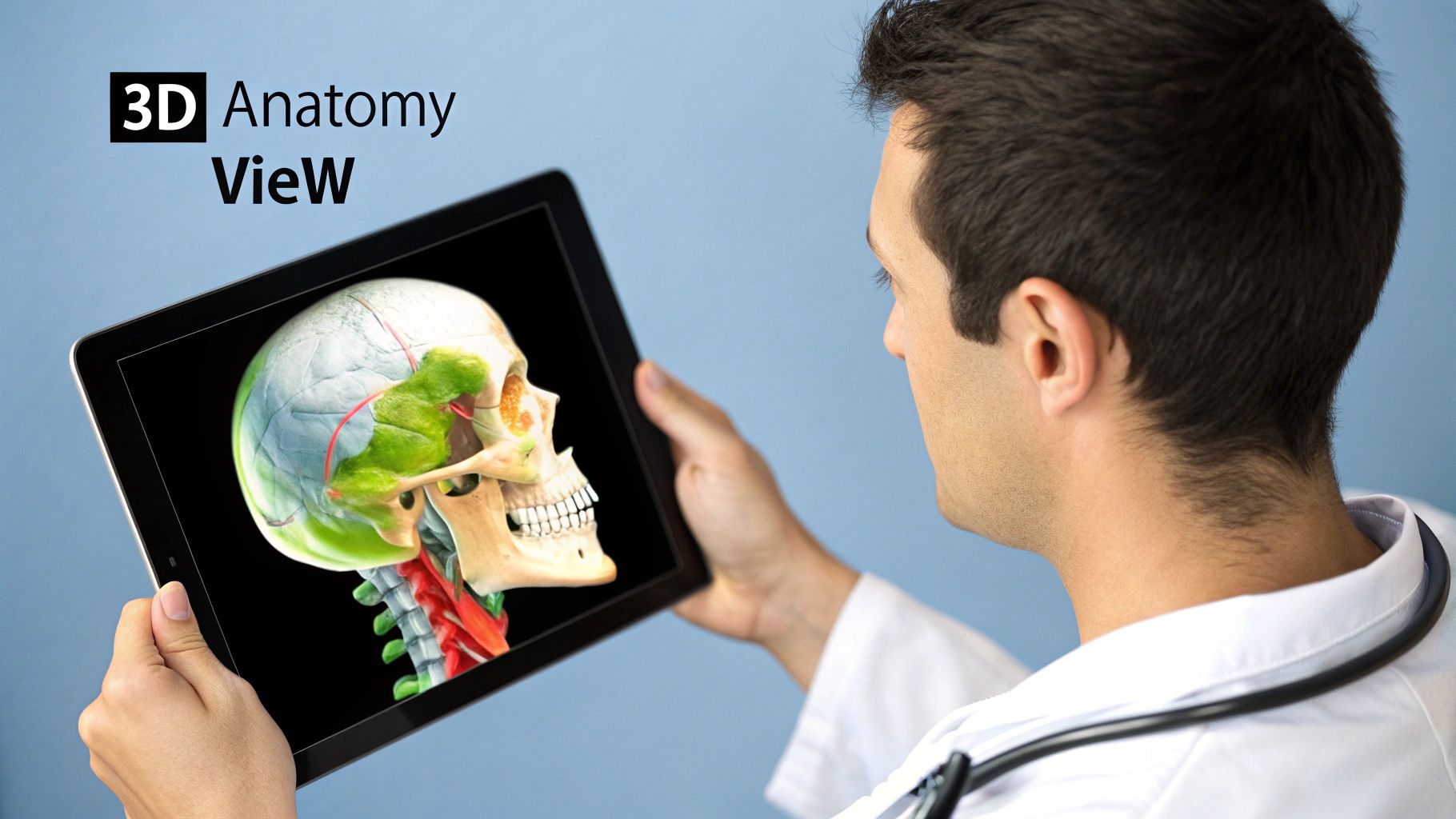
This dataset is built from tiny digital cubes called voxels—think of them as the 3D version of pixels. Each individual voxel holds a value corresponding to the density of the tissue it represents, which allows the software to tell the difference between bone, soft tissue, and air with incredible accuracy. The final product is a stunningly detailed, interactive model of your anatomy that a doctor can spin, slice, and examine from any conceivable angle.
The infographic below walks through this elegant three-step process, from the initial X-ray beam to the final, fully reconstructed 3D model.
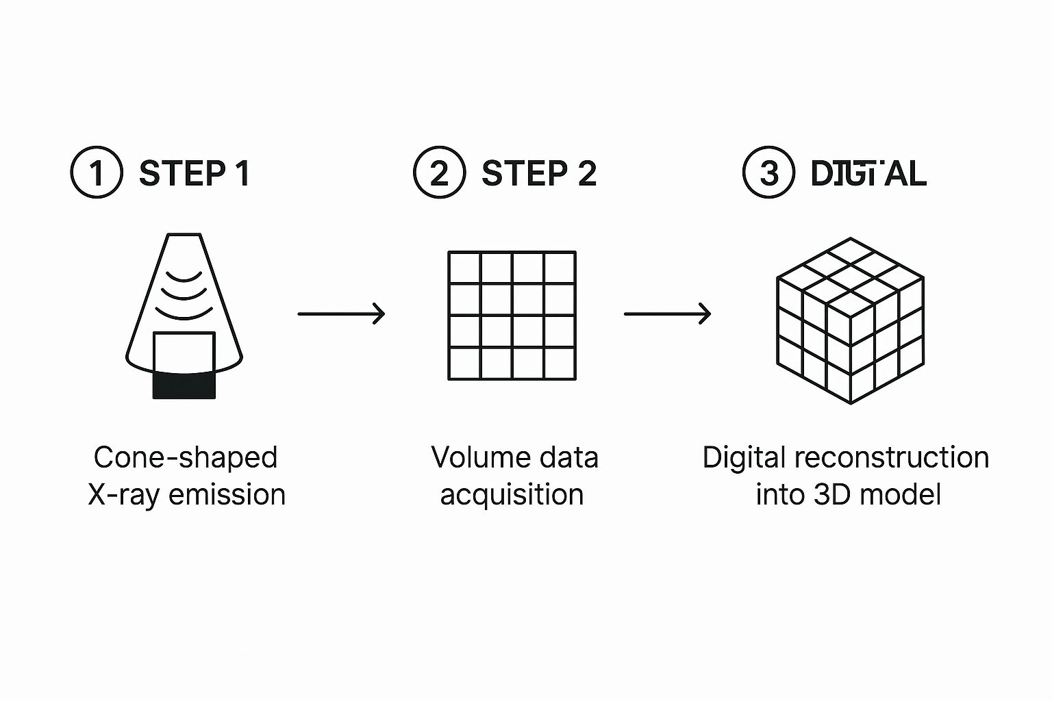
As you can see, that one cone-shaped emission is all it takes to gather every piece of data needed to build the entire volumetric model right away.
Cone Beam CT vs Conventional CT at a Glance
To truly understand the leap forward that CBCT represents, it's helpful to see it side-by-side with the traditional fan-beam CT. While both create detailed internal images, their methods and applications are fundamentally different.
The following table breaks down the key distinctions:
| Feature | Cone Beam CT (CBCT) | Conventional CT |
|---|---|---|
| X-ray Beam Shape | Cone-shaped, captures a large volume at once | Fan-shaped, captures thin, individual slices |
| Scan Motion | Single 360-degree rotation | Multiple rotations (helical/spiral motion) |
| Scan Time | Very fast, typically under 1 minute | Slower, can take several minutes |
| Radiation Dose | Significantly lower | Higher due to multiple passes |
| Image Resolution | Excellent for hard tissues (bone, teeth) | Superior for soft tissue contrast |
| Primary Use | Dentistry, ENT, orthopedics | General medical diagnostics (organs, tumors) |
| Patient Position | Often seated or standing | Lying down on a moving table |
This comparison highlights why CBCT has become the go-to for specific fields like dentistry and otolaryngology. It delivers exactly the information needed, faster and with less radiation exposure for the patient.
Efficiency and Safety: A Winning Combination
This single-rotation method isn't just a neat piece of engineering; it brings huge benefits that directly improve patient care and make clinical workflows smoother.
- Dramatically Lower Radiation: Since the scan is over in one pass, your exposure to radiation is a fraction of what you'd get from a conventional CT—in some cases, it's up to 90% less. This is a massive win for patient safety.
- Quicker Scans, Clearer Images: A full scan is often done in less than a minute. This is not only more comfortable but also dramatically reduces the chances of movement blurring the final image.
The real genius of cone beam technology is its ability to capture everything it needs in one efficient, sweeping scan. This approach doesn't just produce beautiful images; it puts patient safety first by minimizing radiation.
Of course, creating these incredible 3D images is only half the story. You need a powerful and intuitive platform to view, analyze, and truly unlock their clinical value. That's where we come in. At PYCAD, we at PYCAD, build custom web DICOM viewers and integrate them into medical imaging web platforms, turning that raw data into an interactive diagnostic powerhouse. If you're curious to see what this looks like in action, take a look at some of the solutions in our portfolio.
Where Technology Meets Human Healing

This is where the real magic of cone beam images happens—where sophisticated technology directly translates into better patient outcomes. These incredibly detailed 3D scans are far more than just data; they are the architectural blueprints for life-changing treatments.
They give clinicians the confidence to solve complex diagnostic puzzles and restore a patient's health with a level of precision that was once unimaginable. From the local dentist’s office to the advanced surgical suite, CBCT is fundamentally raising the bar for what great care looks like.
A New Standard in Dentistry and Maxillofacial Surgery
The most dramatic impact has been in dentistry, where cone beam technology has become an indispensable part of modern practice. It provides a complete, three-dimensional story of the teeth, jaw, nerves, and surrounding bone, completely changing procedures that previously relied on the flat, limited perspective of 2D X-rays.
Think about planning a dental implant. With a traditional X-ray, a dentist had to make an educated guess about bone depth and the location of critical nerves. Now, with a CBCT scan, they can virtually place the implant before the procedure even begins, measuring bone density to the micrometer and mapping the exact path of the mandibular nerve to sidestep any potential injury.
This leap in clarity is vital for:
- Dental Implants: Ensuring the implant is placed perfectly for long-term stability and safety, which has dramatically boosted success rates.
- Orthodontics: Mapping the precise location of impacted teeth to plan complex tooth movements with absolute certainty.
- Endodontics (Root Canals): Seeing the intricate anatomy of root canals in 3D, revealing hidden canals or fractures that a 2D X-ray would easily miss.
- Maxillofacial Surgery: Creating a complete anatomical guide for reconstructing facial trauma or planning corrective jaw surgery, all before making a single incision.
This growing reliance is fueling incredible adoption. The global cone beam CT market, currently valued around USD 973 million, is projected to skyrocket to nearly USD 2.6 billion in the next decade. This isn't just a trend; it's a testament to its expanding role in day-to-day clinical work. You can read the full research on this market expansion to see the forces driving this growth.
Beyond the Jaw: New Frontiers in Medicine
While its dental roots run deep, the versatility of cone beam imaging extends far beyond the mouth. The same high-resolution 3D views are proving to be invaluable in other specialized medical fields.
In Otolaryngology (ENT), specialists use CBCT to get a crystal-clear look at sinus cavities, middle ear structures, and airway passages. This helps them accurately diagnose chronic sinusitis, assess airway blockages for sleep apnea, and plan delicate ear surgeries. And because the radiation dose is lower, it’s a much safer option for patients who may need repeat scans.
Orthopedics is another exciting frontier, especially for extremities like the hands, wrists, and feet. CBCT delivers exquisite bone detail, allowing surgeons to assess complex fractures, plan joint replacements, and evaluate bone healing with a level of accuracy that simply wasn't possible before. For a closer look at the technology itself, feel free to explore our comprehensive guide on CBCT scans and their many applications.
At its heart, every cone beam scan tells a unique patient story. It provides the crucial details that allow a clinician to turn a complex medical problem into a clear, actionable treatment plan, paving the way for better outcomes and renewed hope.
Bringing this complex data to life is the final, critical step. At PYCAD, this is our expertise. We at PYCAD, build custom web DICOM viewers and integrate them into medical imaging web platforms, turning that raw scan data into interactive, intuitive 3D models that clinicians can truly work with. To see how our solutions help power these clinical applications, take a moment to explore our portfolio.
AI Enhancing the Power of Cone Beam Analysis
The future of medical imaging is already here, and it’s being built on a powerful partnership between artificial intelligence and cone beam technology.
Think of a cone beam image as a remarkably detailed 3D blueprint of a patient’s anatomy. Now, imagine AI as a brilliant co-pilot, helping clinicians read that blueprint with a level of speed and insight we've never seen before.
This isn’t about replacing human expertise; it’s about amplifying it. We're seeing a shift from reactive to proactive healthcare, driven by AI algorithms that can instantly identify anatomical landmarks, flag potential pathologies hidden in complex 3D structures, and segment tissues for surgical planning—all in a fraction of the time it would take a human eye.
AI as a Diagnostic Assistant
AI serves as a tireless assistant, meticulously analyzing every single voxel of data to find patterns that might be too subtle for us to catch.
For instance, an AI can be trained to spot the earliest signs of bone loss around a dental implant or map out intricate root canal structures that are easy to miss during a manual review. This frees up the clinician from the tedious work, allowing them to focus their expertise where it matters most: on the patient and the treatment strategy.
The impact is immediate and profound:
- Sharper Accuracy: By learning from thousands of scans, AI models can spot anomalies with incredible certainty, helping to reduce diagnostic errors.
- Smarter Workflows: Automated segmentation and landmark identification save clinicians a massive amount of time, giving them more capacity to care for patients.
- Predictive Power: By tracking subtle changes over time, AI can help forecast disease progression, opening the door for earlier and more effective intervention.
This fusion of AI and 3D imaging is more than just a technological step up. It represents a genuine leap forward in personalized patient care, setting a new standard where diagnostics are faster, more precise, and profoundly insightful.
Unlocking the Full Potential of 3D Data
A single cone beam scan contains an immense volume of information, and it can be overwhelming to process. This is where AI truly shines, making all that data manageable and, most importantly, actionable.
At PYCAD, this is the world we live in. We at PYCAD, build custom web DICOM viewers and integrate them into medical imaging web platforms. These platforms become exponentially more powerful when infused with AI, turning static images into dynamic, interactive diagnostic tools. You can view our portfolio here to see some of this work in action.
While AI is making huge waves in cone beam analysis, its power is being felt across many medical fields, sparking new possibilities in areas like AI-driven diagnostics in physical therapy.
If you're curious to learn more about how these technologies are being applied across the board, our article on artificial intelligence for radiology is a great place to start. This synergy between advanced imaging and intelligent algorithms is truly paving the way for a new era of proactive and personalized healthcare.
Bringing 3D Cone Beam Images to Your Screen

A powerful 3D scan is only as good as the tools we use to see it. After the cone beam scanner has done its job capturing hundreds of individual images, that raw data needs to be brought to life. It’s the specialized viewing software that works this magic, transforming a massive dataset into a dynamic, interactive model that a clinician can actually work with.
This is where the real power unfolds. The viewing software takes the volumetric data—a collection of digital 3D pixels, or voxels—and makes it completely pliable. Clinicians can spin the model a full 360 degrees, zoom in on the tiniest details, and digitally slice through anatomical structures layer by layer.
It's this interactive quality that makes cone beam images so incredibly valuable in a clinical setting. The freedom to navigate through the data from any angle is crucial for everything from planning a delicate dental implant to assessing an airway for sleep apnea.
The Power of Interactive Visualization
At the heart of the software are core functions that make the 3D data truly come alive. Two of the most critical capabilities are multi-planar reconstruction (MPR) and 3D rendering, which work together to paint a complete diagnostic picture.
-
Multi-Planar Reconstruction (MPR): Imagine being able to create brand new 2D "slices" from the 3D model on the fly. MPR lets a clinician view the anatomy in the three classic planes—axial (top-down), sagittal (side), and coronal (front). Better yet, they can create custom oblique slices to follow the exact path of a nerve or view a tooth root from a very specific, non-traditional angle.
-
3D Rendering: This is the process that builds those stunning, almost photorealistic 3D models we often see. It gives an intuitive, big-picture view of the anatomical structures, making it far easier to grasp spatial relationships and, just as importantly, to explain treatment plans to patients in a way they can instantly understand.
The real breakthrough isn't just seeing a 3D model; it's interacting with it. The software turns clinicians into digital explorers, empowering them to navigate a patient's unique anatomy with a level of precision and clarity that was once unimaginable.
At PYCAD, building these essential tools is what we do. We at PYCAD, build custom web DICOM viewers and integrate them into medical imaging web platforms, making sure that clinicians can access and manipulate powerful cone beam images from anywhere with ease and accuracy. Our mission is to build intuitive, powerful viewing experiences that give healthcare professionals the confidence to make the best decisions. You can check our work on our portfolio page.
A Seamless and Accessible Workflow
Modern viewing platforms are increasingly web-based, which is a huge step forward. This shift eliminates the need for doctors to be tied to a single, high-powered workstation in the office. It opens the door to greater collaboration, allowing specialists in different locations to review the same case simultaneously.
This accessibility provides secure, on-demand access to critical patient data right when it's needed most. You can dive deeper into the technology that drives these platforms in our detailed article about the DICOM viewer. Ultimately, this ease of access is what's making advanced imaging a practical, everyday tool in clinical care, fundamentally changing how we diagnose and plan treatments.
Bringing Your Imaging Data to Life
All the incredible physics and advanced technology behind cone beam imaging mean nothing until you can see the results clearly. The real magic happens when you have a powerful, intuitive viewing platform that turns that raw data into a window into the patient's anatomy.
This is where theory meets practice. It’s about transforming complex scans into a tool that revolutionizes your workflow, sparks collaboration among colleagues, and helps you communicate with patients like never before.
At PYCAD, this is what we do. We at PYCAD, build custom web DICOM viewers and integrate them seamlessly into medical imaging web platforms. We partner with medical and dental practices to create bespoke solutions that finally let them tap into the true power of their imaging data.
Bringing any new medical technology to the clinic involves navigating a complex regulatory environment. The FDA approval process for medical devices is a critical part of ensuring these powerful tools are both safe and effective for patient use.
We invite you to see for yourself what's possible when expert software engineering meets advanced imaging. Visit our portfolio to explore some of the custom viewing platforms we've built for clients around the world.
Let's work together to turn your cone beam images into clear, actionable insights that elevate patient care.
Answering Your Questions About Cone Beam Imaging
It's completely natural to have questions when you're introduced to a new piece of medical technology. When it comes to something as powerful as a cone beam image, getting straight answers is the first step toward feeling confident and empowered in your care. This technology is a huge leap forward, and understanding it helps you see just how valuable it is.
Let's walk through some of the questions we hear most often.
Is a Cone Beam Scan Safe?
This is usually the first thing people ask, and for good reason. The answer is a clear and confident yes. While cone beam imaging does involve X-rays, its design is remarkably efficient. The scanner captures everything it needs in one quick rotation, which means the radiation dose is significantly lower than a traditional medical CT scan—often by as much as 90%.
Your safety is always the priority. The scan is fast, non-invasive, and provides a wealth of diagnostic insight for a very low level of radiation exposure. It’s an incredibly safe and effective tool.
How Should I Prepare for the Scan?
One of the great things about a cone beam scan is how simple it is for the patient. There's almost nothing you need to do beforehand.
- No Fasting Needed: Feel free to eat and drink as you normally would.
- Remove Metal Objects: You'll be asked to take off anything metallic that could get in the way of the image, like jewelry, glasses, or removable dental work.
- Just Stay Still: The most critical part is holding still during the scan itself, which is usually over in less than a minute.
The entire process is designed to be as easy and comfortable as possible, fitting seamlessly into a normal appointment.
How Is It Different from a Regular Dental X-ray?
Imagine the difference between a flat map and a spinning globe. A standard 2D X-ray is like that flat map—it shows you one angle of a place. It's helpful, but things can be hidden or overlap, making it hard to see the full picture.
A cone beam scan, on the other hand, is the globe. It creates a complete, three-dimensional view of your anatomy. Your doctor can look at everything from every angle imaginable—top to bottom, front to back, and side to side. It takes the guesswork out of the equation and reveals details a flat image could never show.
Why Do I Need a Cone Beam Image?
Your doctor will recommend a cone beam scan when they need a level of detail that a standard X-ray simply can't deliver. It has become the gold standard for any procedure where precision is absolutely critical.
This is especially true for:
- Dental Implant Planning: To see exactly how much bone is available and pinpoint the location of nerves.
- Orthodontic Assessment: To find impacted teeth and truly understand the relationship between the jaws.
- Airway Analysis: To check the structure of nasal passages and sinuses, often for conditions like sleep apnea.
- Complex Root Canals: To trace the tiny, complex network of canals inside a tooth.
In the end, a cone beam image provides your clinician with the master blueprint they need to plan your treatment with the highest possible accuracy and safety, which ultimately leads to better results for you.
At PYCAD, our passion is turning this powerful data into something clear, accessible, and intuitive. We at PYCAD, build custom web DICOM viewers and integrate them into medical imaging web platforms, transforming complex scans into interactive models that tell a story.
Ready to see what that looks like? We invite you to explore our portfolio.


