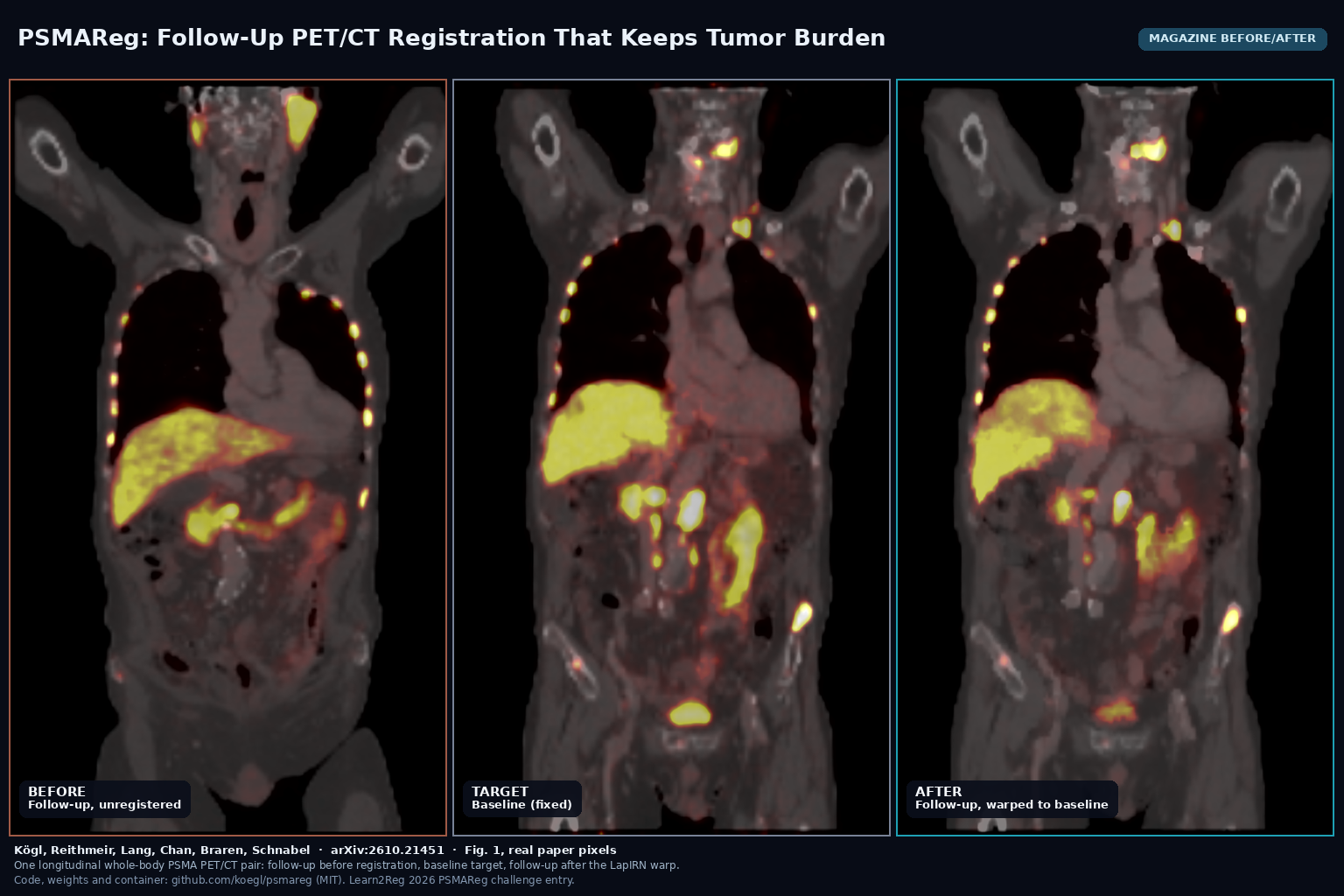
PSMAReg: Follow-Up PET/CT Registration That Keeps Tumor Burden
A LapIRN pipeline from Helmholtz Munich and TUM aligns follow-up PSMA PET/CT to baseline while holding MTV and TLG steady. arXiv:2610.21451, MIT code and weights.

A LapIRN pipeline from Helmholtz Munich and TUM aligns follow-up PSMA PET/CT to baseline while holding MTV and TLG steady. arXiv:2610.21451, MIT code and weights.
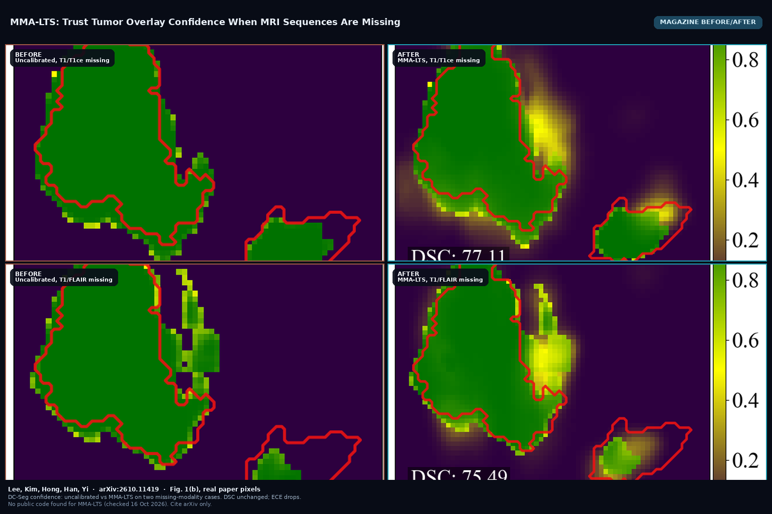
MMA-LTS (arXiv:2610.11419) post-hoc calibrates voxel-wise tumor overlay confidence when MRI sequences are missing, cutting ECE while keeping Dice. KAIST. No public code yet.
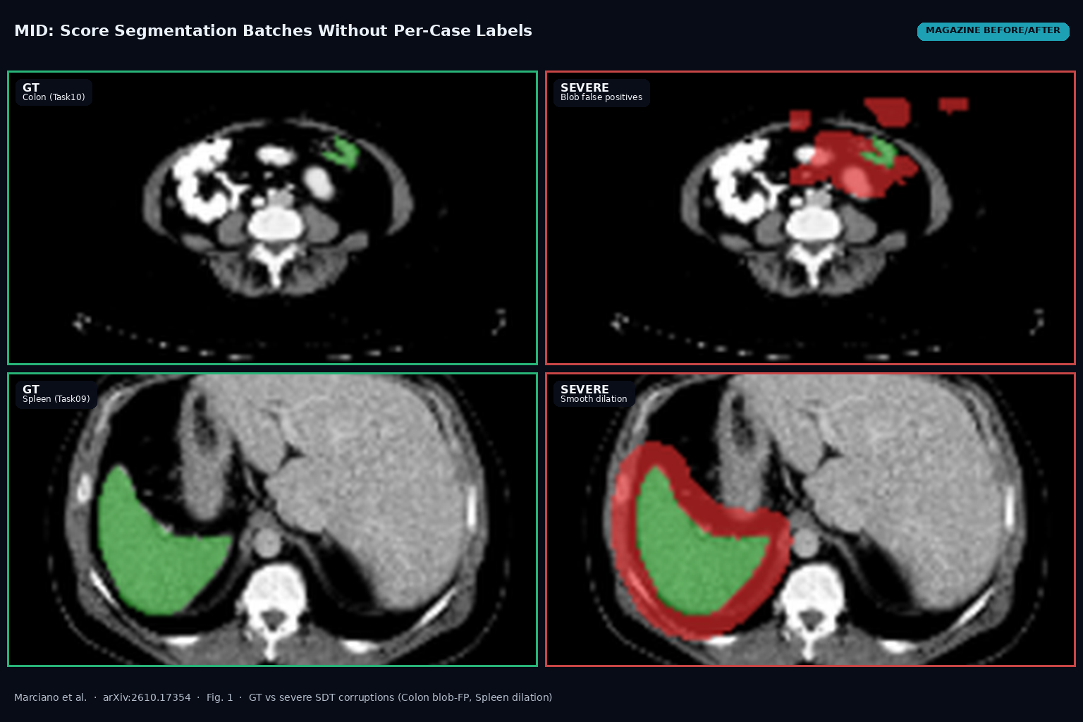
MID (arXiv:2610.17354) scores a batch of predicted segmentations against a labeled reference without per-case GT pairing, via joint MedSigLIP embeddings and a Fréchet / W2^2 distance. Apache 2.0 code on GitHub.
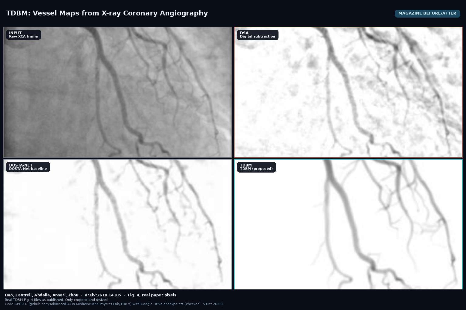
TDBM, by Hao, Cantrell, Abdalla, Ansari and Zhou (arXiv:2610.14105), recovers contrast-preserving coronary vessels from X-ray angiography sequences with a temporal diffusion bridge trained on physics-informed synthetic data, without manual vessel labels. Code is GPL-3.0 on GitHub with Google Drive checkpoints.
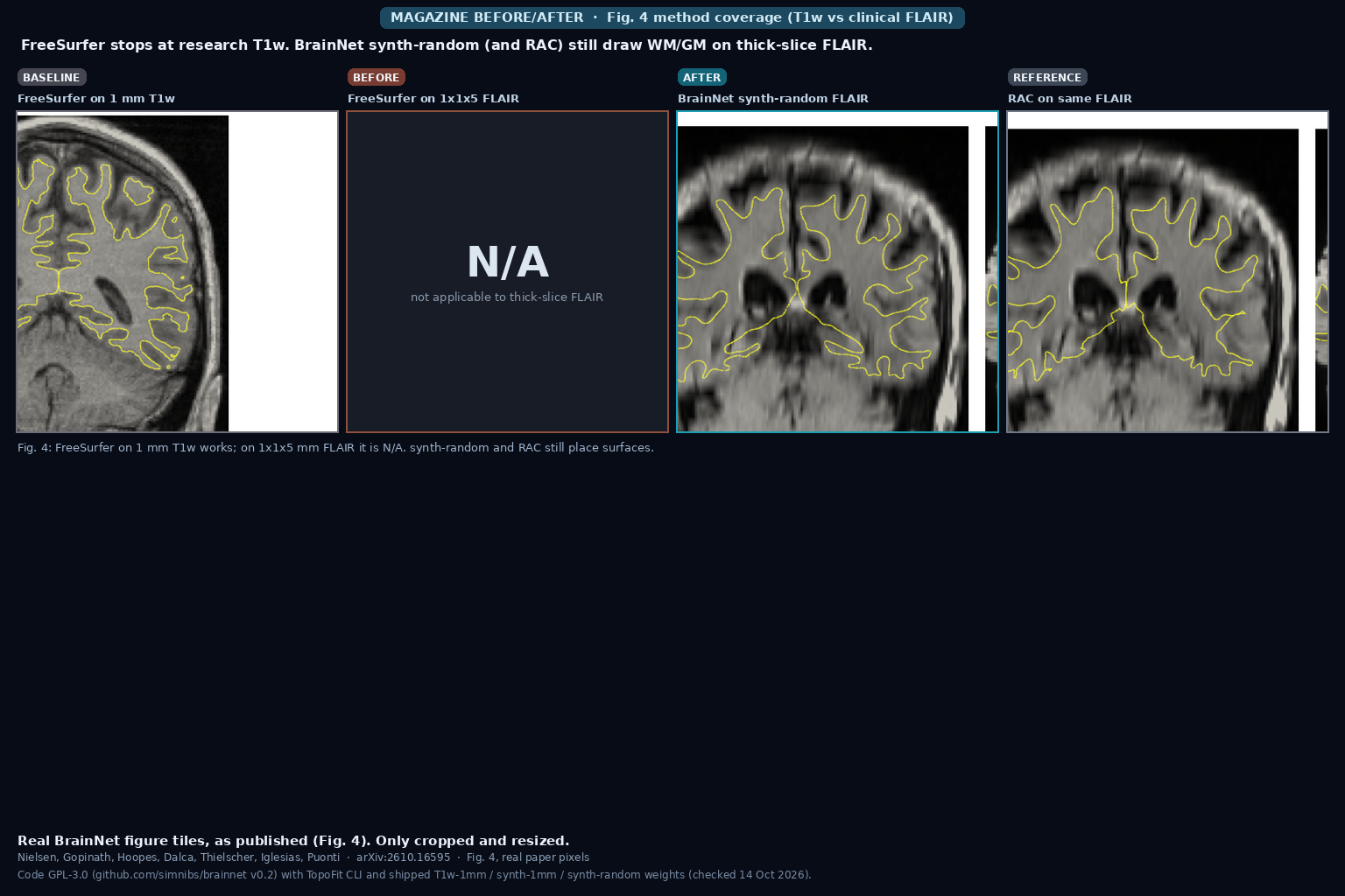
BrainNet, by Nielsen, Gopinath, Hoopes, Dalca, Thielscher, Iglesias and Puonti (arXiv:2610.16595), reconstructs WM and GM cortical surfaces with vertex-wise uncertainty and fsaverage spherical registration from heterogeneous brain MRI, including thick-slice clinical FLAIR. Inference is about 0.6 s on GPU. Code is GPL-3.0 on GitHub with three TopoFit weight variants.
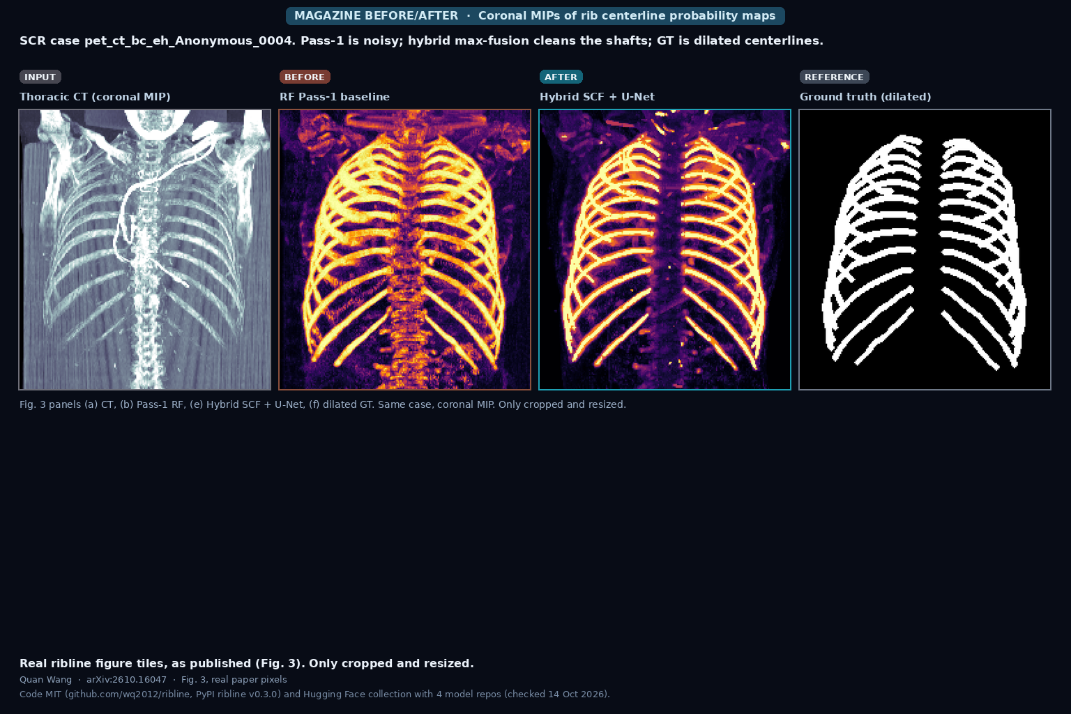
ribline, by Quan Wang (arXiv:2610.16047), traces and numbers all 24 rib centerlines (R01 to R12, L01 to L12) in thoracic CT with a hybrid Semantic Context Forest plus 3D U-Net detector and bilateral Viterbi matching. On the SCR 2 mm benchmark it reports LCD 2.05 mm and 97.2% labeling accuracy on 12 evaluation scans, with honest caveats on Stage-3 tuning and a 5-fold CV at 3.64 mm / 91.8%. Code is MIT on GitHub and PyPI; weights are on Hugging Face.