PYCAD BLOG

Insights on Medical Imaging, AI & DICOM

Guides, project breakdowns, and lessons learned from building medical imaging platforms — written by the PYCAD team.

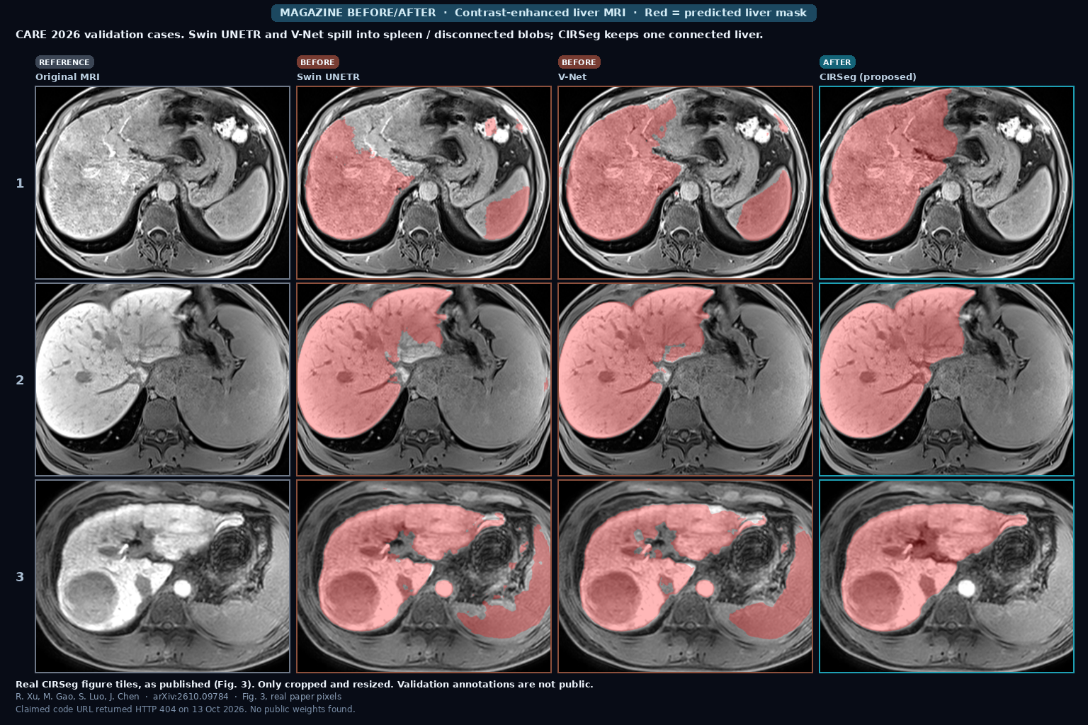
Three CARE 2026 contrast-enhanced liver MRI validation cases with red liver overlays from Swin UNETR and V-Net showing spleen spill and disconnected blobs, beside clean CIRSeg masks and the original images (Xu, Gao, Luo, Chen, arXiv:2610.09784, Fig. 3)

CIRSeg: Liver MRI Masks That Survive Scanner Shift

CIRSeg (Xu, Gao, Luo, Chen) is a cascaded nnU-Netv2 liver segmenter for CARE 2026 contrast-enhanced MRI. It trains with 3D CutMix and Nyul or histogram intensity transfer, refines a coarse prior at full resolution, can adapt at test time without source data, and drops disconnected false positives with LCC filtering. Official CARE eval: Dice 0.9713, HD95 18.31 mm; OOD test 0.9797 / 11.36. Claimed GitHub URL returned HTTP 404 on 13 October 2026. arXiv:2610.09784.
Read More »
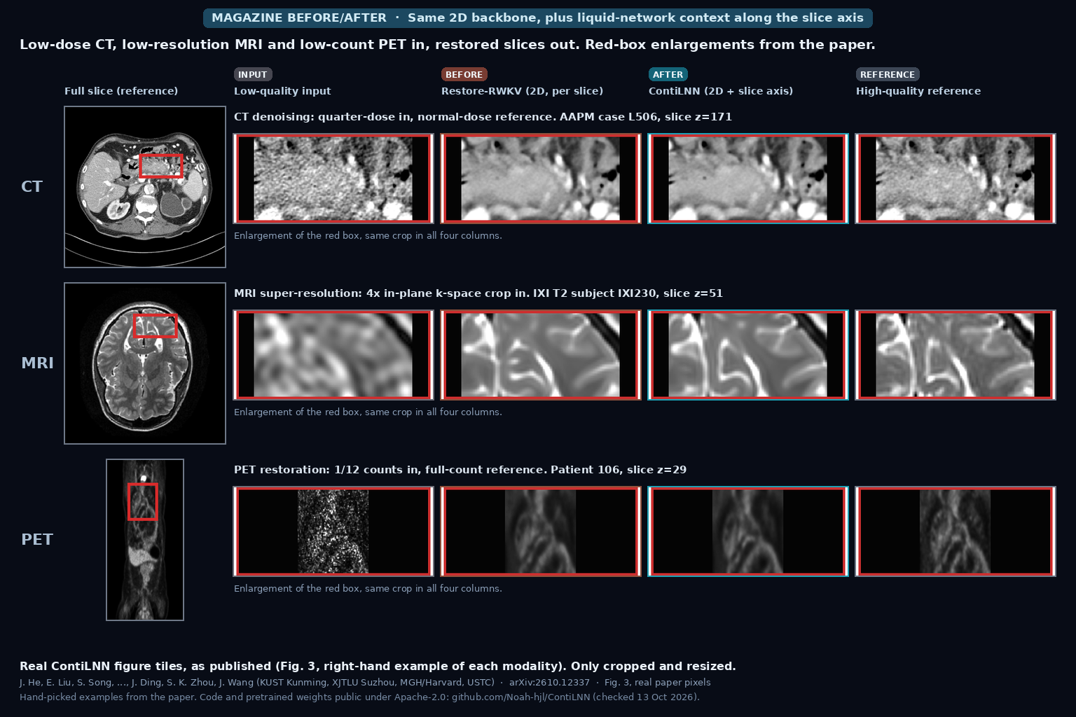
Red-box enlargements of a quarter-dose abdominal CT, a 4x k-space-cropped T2 brain MRI and a 1/12-count PET slice, each shown as low-quality input, Restore-RWKV output, ContiLNN output and high-quality reference, beside the full reference slice (He, Liu, Zhou, Wang et al., arXiv:2610.12337, Fig. 3)

Low-Dose CT, Low-Res MRI or Low-Count PET Slices In, Restored Series Out That Uses Neighboring Slices and Their Gaps, Still Blind to Real DICOM Spacing (ContiLNN)

ContiLNN, from Kunming University of Science and Technology, XJTLU, MGH/Harvard and USTC, adds bidirectional liquid neural network modules along the slice axis of a 2D restoration network, so each restored slice draws on its neighbors and on the index gap to them. Over per-slice Restore-RWKV, mean PSNR rises 1.02 dB for MRI super-resolution and 1.25 dB for low-count PET, the CT gain is small and from one patient, and gaps are counted in slice indices rather than millimeters. Code and weights are public (Apache-2.0). He, Liu, Ding, Zhou, Wang et al., arXiv:2610.12337.

Read More »
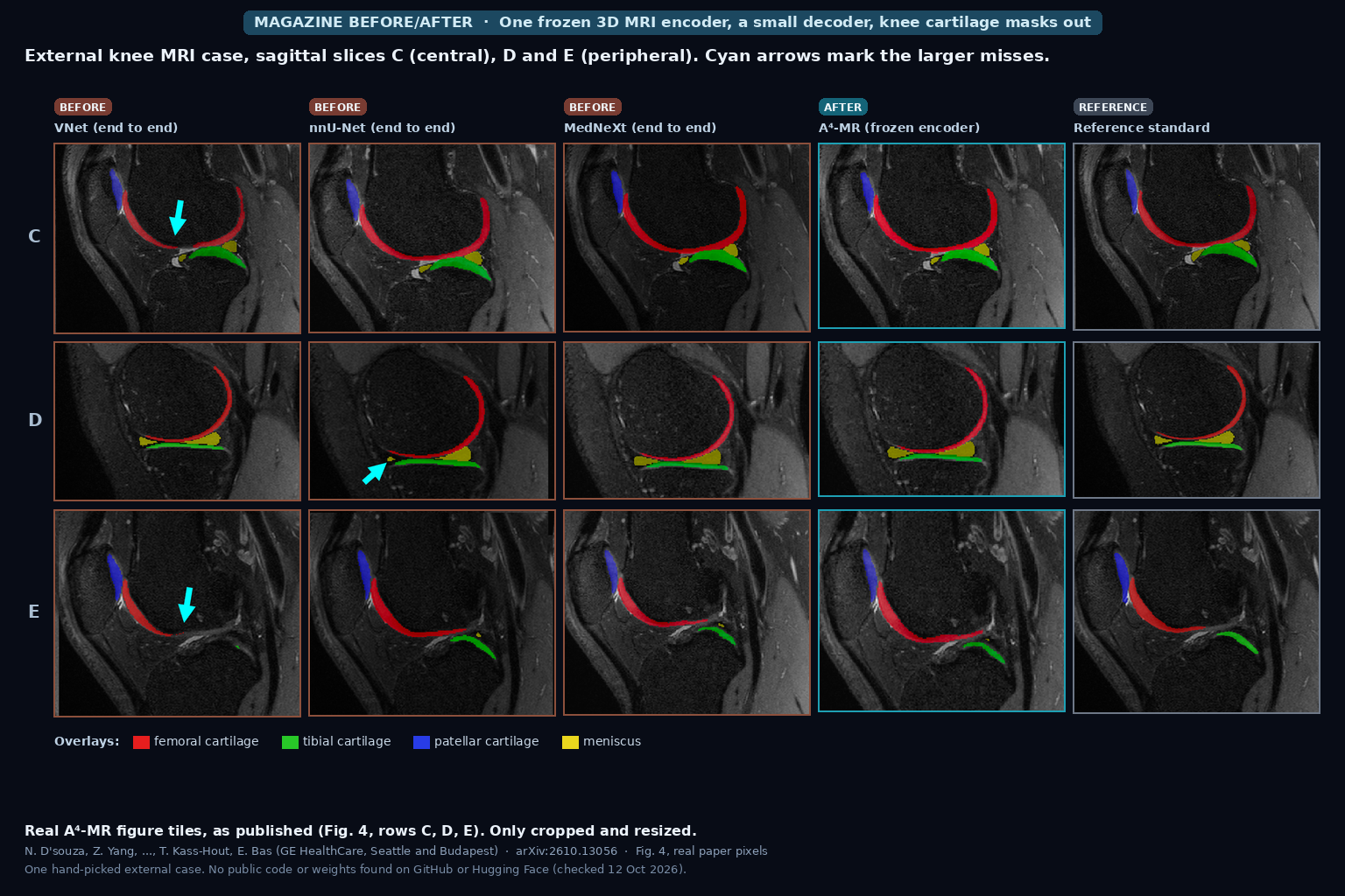
Sagittal knee MRI slices with femoral, tibial, patellar cartilage and meniscus overlays from VNet, nnU-Net, MedNeXt, A4-MR and the reference standard, cyan arrows on larger misses (D'souza, Bas et al., arXiv:2610.13056, Fig. 4)

Whole 3D MRI Series In, Prompt-Free Organ Boxes and Report-Searchable Anatomy Out From One Frozen Encoder, Still Weak on Organs Smaller Than Its 8-Voxel Patches (A4-MR)

A4-MR, a 3D MRI vision-language model from GE HealthCare trained on about 50,000 studies (491,000 series), finds 125 anatomy classes as 3D boxes without prompts, ties each box to report text, and keeps healthy and abnormal organs apart in its embedding. Zero-shot box IoU on AMOS MR is 66.3 against 65.5 for MedSAM2 given a ground-truth box, small organs stay hard, and no code or weights are public. D’souza, Bas et al., arXiv:2610.13056.

Read More »
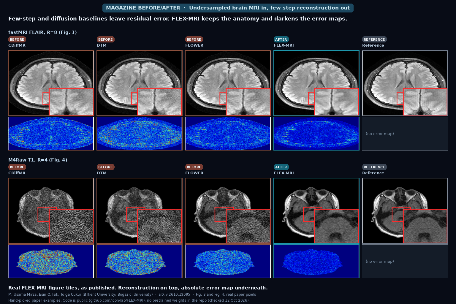
Brain MRI reconstructions at high acceleration: three few-step or diffusion baselines, FLEX-MRI, and the fully sampled reference, each with absolute-error maps (Mirza, Isik, Cukur, arXiv:2610.13095, Fig. 3 and Fig. 4)

Undersampled Brain MRI In, Few-Step Reconstruction Out That Explores Alternate k-Space Paths Without Hundreds of Diffusion Steps

FLEX-MRI takes undersampled multi-coil brain MRI and recovers the image in about ten finite transitions with stochastic k-space exploration and data consistency, without a 1000-step diffusion sampler. On fastMRI at R=8 it reaches 35.8 dB PSNR and 92.7% SSIM, runs in 0.349 s with 1.05 GB on an RTX 4090, and the GitHub repo has training and eval code but no pretrained weights. Mirza, Isik, Cukur, arXiv:2610.13095.

Read More »
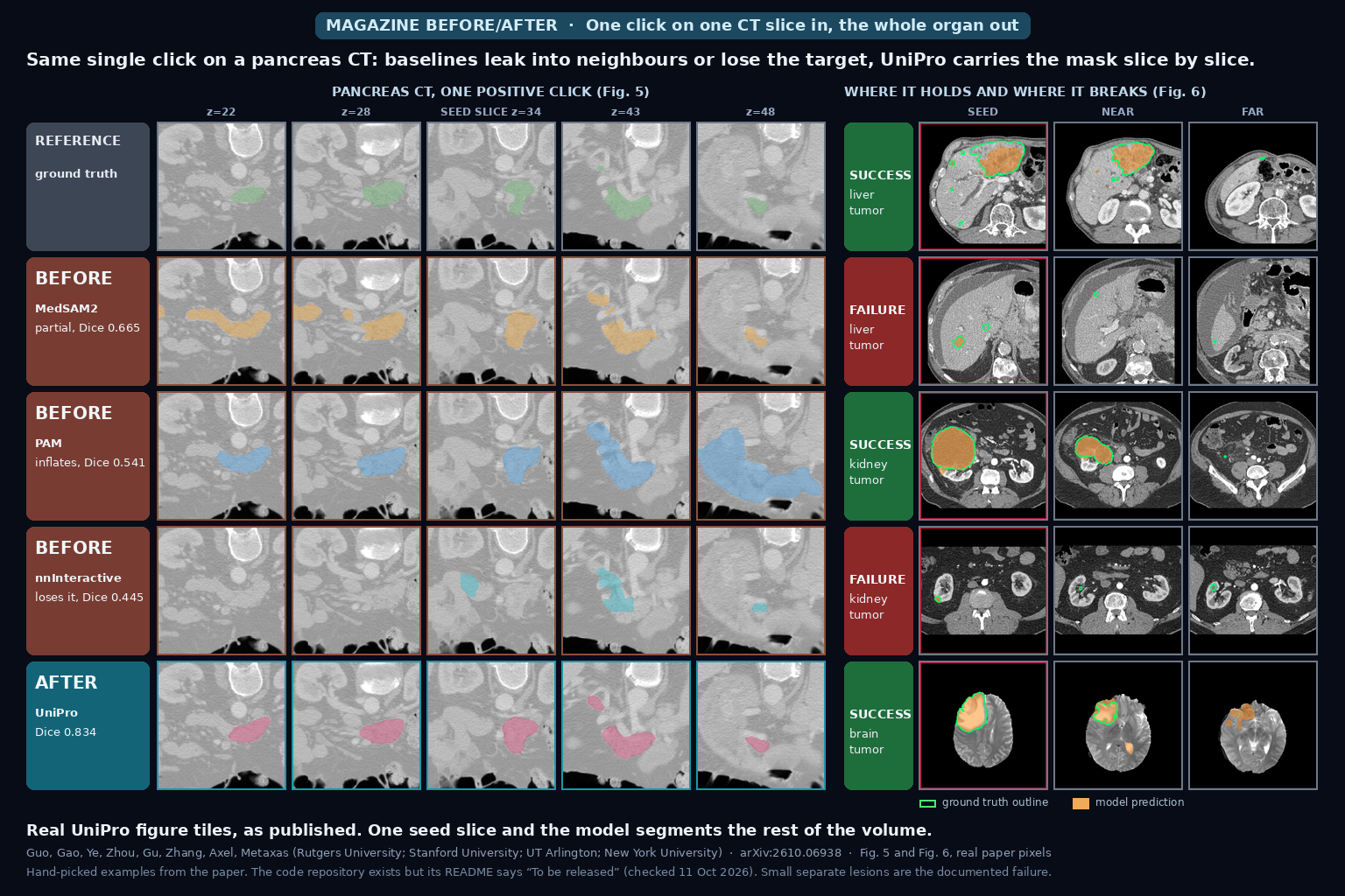
Pancreas CT slices after one click: ground truth, three baseline propagation models and UniPro at five slice positions, with liver, kidney and brain tumor success and failure examples on the right (Guo et al., arXiv:2610.06938, Fig. 5 and Fig. 6)

One Click on One CT Slice In, a Full 3D Organ Mask Out That Is Carried Slice by Slice From the Neighbours and Still Takes Class Prompts, Reference Images and Corrections in the Same Model

Click once on one slice of a CT or MR volume and UniPro carries the mask through the other slices, using the seed slice and the latest predictions as references. The same 2D model also takes a class name, a reference example or corrections. It is best of the compared models on 7 of 8 single-click tests, trails 3D nnU-Net in plain semantic mode, needs 28 seconds per abdominal volume, and the repository README still says “To be released”. Guo et al., arXiv:2610.06938.

Read More »
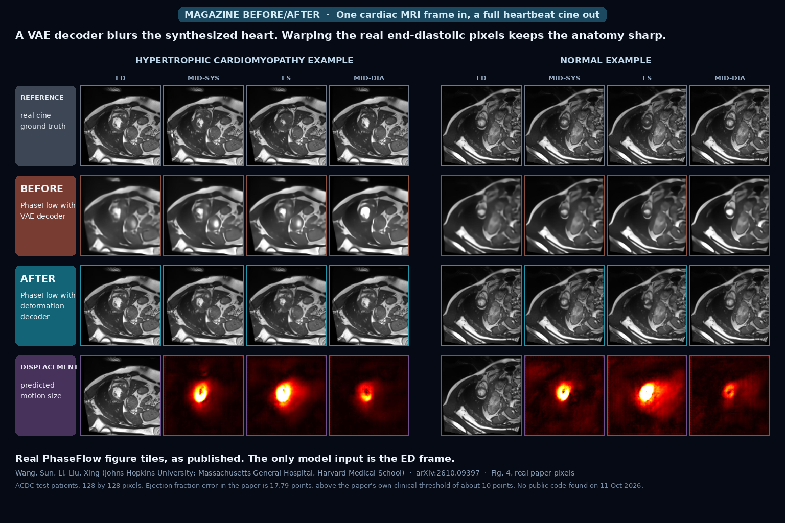
Cardiac MRI cine frames at end-diastole, mid-systole, end-systole and mid-diastole for a hypertrophic cardiomyopathy and a normal example: real cine on top, a blurry VAE-decoder result in the middle, a sharp deformation-decoder result below, and predicted displacement heat maps at the bottom (Wang et al., arXiv:2610.09397, Fig. 4)

One End-Diastole Cardiac MRI Frame In, a Full 30-Frame Beating-Heart Cine Out Without ECG Gating by Warping the Real Pixels With a Phase-Conditioned Flow Model

Give PhaseFlow one end-diastolic cardiac MRI frame and it generates a 30-frame cine of the heartbeat with no ECG gating. It predicts a motion field and warps the real pixels, which keeps the image sharp, and it takes its timing from a mean phase curve for the patient’s pathology group. Tested on ACDC with 30 test patients and one training run, the ejection fraction error is 17.79 points, above the paper’s own clinical threshold of about 10, and I found no public code. Wang et al., arXiv:2610.09397.

Read More »

We build custom medical imaging platforms — advanced DICOM viewers, AI segmentation, and the clinical systems around them.

Copyright © 2026 PYCAD. All Rights Reserved.